Acute polyhydramnios in term pregnancy may be caused by multiple nuchal cord loops
•
1 recomendación•1,235 vistas

1) A 24-year-old pregnant woman presented with severe acute polyhydramnios at 41 weeks gestation, where her amniotic fluid index was 40 cm. 2) An ultrasound revealed multiple loops of the umbilical cord wrapped around the fetus' neck. 3) The fetus was delivered via cesarean section and was found to have the umbilical cord tightly wrapped three times around its neck, limiting neck movement but without signs of strangulation.

Recomendados
Recomendados
Más contenido relacionado
La actualidad más candente
La actualidad más candente (20)
Similar a Acute polyhydramnios in term pregnancy may be caused by multiple nuchal cord loops
Similar a Acute polyhydramnios in term pregnancy may be caused by multiple nuchal cord loops (20)
Más de Asha Reddy
Más de Asha Reddy (20)
Último
Último (20)
Acute polyhydramnios in term pregnancy may be caused by multiple nuchal cord loops
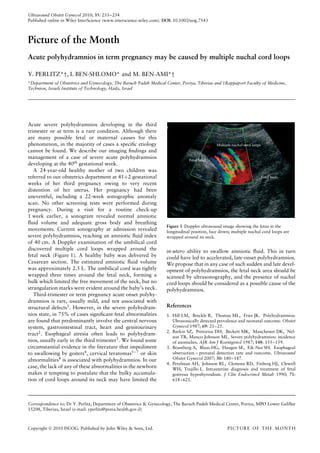
- 1. Ultrasound Obstet Gynecol 2010; 35: 253–254 Published online in Wiley InterScience (www.interscience.wiley.com). DOI: 10.1002/uog.7543 Picture of the Month Acute polyhydramnios in term pregnancy may be caused by multiple nuchal cord loops Y. PERLITZ*†, I. BEN-SHLOMO* and M. BEN-AMI*† *Department of Obstetrics and Gynecology, The Baruch Padeh Medical Center, Poriya, Tiberias and †Rappaport Faculty of Medicine, Technion, Israeli Institute of Technology, Haifa, Israel Acute severe polyhydramnios developing in the third trimester or at term is a rare condition. Although there are many possible fetal or maternal causes for this phenomenon, in the majority of cases a specific etiology Multiple nuchal cord loops cannot be found. We describe our imaging findings and management of a case of severe acute polyhydramnios Fetal head developing at the 40th gestational week. A 24-year-old healthy mother of two children was referred to our obstetrics department at 41+2 gestational Fetal body weeks of her third pregnancy owing to very recent distention of her uterus. Her pregnancy had been uneventful, including a 22-week sonographic anomaly scan. No other screening tests were performed during pregnancy. During a visit for a routine check-up 1 week earlier, a sonogram revealed normal amniotic fluid volume and adequate gross body and breathing Figure 1 Doppler ultrasound image showing the fetus in the movements. Current sonography at admission revealed longitudinal position, face down; multiple nuchal cord loops are severe polyhydramnios, reaching an amniotic fluid index wrapped around its neck. of 40 cm. A Doppler examination of the umbilical cord discovered multiple cord loops wrapped around the in-utero ability to swallow amniotic fluid. This in turn fetal neck (Figure 1). A healthy baby was delivered by could have led to accelerated, late-onset polyhydramnios. Cesarean section. The estimated amniotic fluid volume We propose that in any case of such sudden and late devel- was approximately 2.5 L. The umbilical cord was tightly opment of polyhydramnios, the fetal neck area should be wrapped three times around the fetal neck, forming a scanned by ultrasonography, and the presence of nuchal bulk which limited the free movement of the neck, but no cord loops should be considered as a possible cause of the strangulation marks were evident around the baby’s neck. polyhydramnios. Third-trimester or term pregnancy acute onset polyhy- dramnios is rare, usually mild, and not associated with structural defects1 . However, in the severe polyhydram- References nios state, in 75% of cases significant fetal abnormalities 1. Hill LM, Breckle R, Thomas ML, Fries JK. Polyhydramnios: are found that predominantly involve the central nervous Ultrasonically detected prevalence and neonatal outcome. Obstet system, gastrointestinal tract, heart and genitourinary Gynecol 1987; 69: 21–25. 2. Barkin SZ, Pretorius DH, Beckett MK, Manchester DK, Nel- tract2 . Esophageal atresia often leads to polyhydram- son TR, Manco-Johnson ML. Severe polyhydramnios: incidence nios, usually early in the third trimester3 . We found some of anomalies. AJR Am J Roentgenol 1987; 148: 155–159. circumstantial evidence in the literature that impediment 3. Brantberg A, Blaas HG, Haugen SE, Eik-Nes SH. Esophageal to swallowing by goiters4 , cervical teratomas5 – 7 or skin obstruction – prenatal detection rate and outcome. Ultrasound abnormalities8 is associated with polyhydramnios. In our Obstet Gynecol 2007; 30: 180–187. 4. Perelman AH, Johnson RL, Clemons RD, Finberg HJ, Clewell case, the lack of any of these abnormalities in the newborn WH, Trujillo L. Intrauterine diagnosis and treatment of fetal makes it tempting to postulate that the bulky accumula- goitrous hypothyroidism. J Clin Endocrinol Metab 1990; 71: tion of cord loops around its neck may have limited the 618–621. Correspondence to: Dr Y. Perlitz, Department of Obstetrics & Gynecology, The Baruch Padeh Medical Center, Poriya, MPO Lower Galillee 15208, Tiberias, Israel (e-mail: yperlitz@poria.health.gov.il) Copyright 2010 ISUOG. Published by John Wiley & Sons, Ltd. PICTURE OF THE MONTH
- 2. 254 Perlitz et al. 5. Langer JC, Tabb T, Thompson P, Paes BA, Caco CC. Manage- ´ ˜ 7. Araujo Junior E, Guimaraes Filho HA, Saito M, Pires AB, ment of prenatally diagnosed tracheal obstruction: access to the Pontes AL, Nardozza LM, Moron AF. Prenatal diagnosis of a airway in utero prior to delivery. Fetal Diagn Ther 1992; 7: large fetal cervical teratoma by three-dimensional ultrasonogra- 12–16. phy: a case report. Arch Gynecol Obstet 2007; 275: 141–144. 6. Martino F, Avila LF, Encinas JL, Luis AL, Olivares P, Las- 8. Opitz JM. Pathogenetic analysis of certain developmental and saletta L, Mistral M, Tovar JA. Teratomas of the neck and genetic ectodermal defects. Birth Defects Orig Artic Ser 1988; mediastinum in children. Pediatr Surg Int 2006; 22: 627–634. 24: 75–102. Copyright 2010 ISUOG. Published by John Wiley & Sons, Ltd. Ultrasound Obstet Gynecol 2010; 35: 253–254.