
Recomendados
Más contenido relacionado
Destacado
Similar a final research proposal
Similar a final research proposal (20)
final research proposal
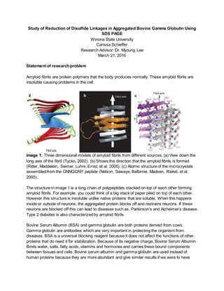
- 1. Study of Reduction of Disulfide Linkages in Aggregated Bovine Gamma Globulin Using SDS PAGE Winona State University Carissa Schieffer Research Advisor: Dr. Myoung Lee March 21, 2016 Statement of research problem Amyloid fibrils are protein polymers that the body produces normally. These amyloid fibrils are insoluble causing problems in the cell. Image 1: Three dimensional models of amyloid fibrils from different sources. (a) View down the long axis of the fibril (Tycko, 2002). (b) Shows the direction that the amyloid fibrils is formed (Ritter, Maddelein, Siemer, Luhrs, Ernst, et al. 2005). (c) Atomic structure of the microcrystals assembled from the GNNQQNY peptide (Nelson, Sawaya, Balbirnie, Madsen, Riekel, et al. 2005). The structure in image 1 is a long chain of polypeptides stacked on top of each other forming amyloid fibrils. For example, you could think of a big stack of paper piled on top of each other. However this structure is insoluble unlike native proteins that are soluble. When this happens inside or outside of neurons, the aggregated protein blocks off and restrains neurons. If these neurons are blocked off this can lead to diseases such as, Parkinson’s and Alzheimer’s disease. Type 2 diabetes is also characterized by amyloid fibrils. Bovine Serum Albumin (BSA) and gamma globulin are both proteins derived from cows. Gamma globulin are antibodies which are very important in protecting the organism from diseases. BSA is a universal blocking reagent because it does not affect the functions of other proteins that do need it for stabilization. Because of its negative charge, Bovine Serum Albumin: Binds water, salts, fatty acids, vitamins and hormones and carries these bound components between tissues and cells. Bovine serum albumin and gamma globulin are used instead of human proteins because they are more abundant and give similar results if we were to have
- 2. used more expensive human proteins. Image 2 shows what the 3 structure of BSA and gamma globulin look like. Image 2: The image on the left shows the 3D model of bovine serum albumin. The pink swirls represent alpha helices in bovine serum albumin (Bujacz, Zielinski, Sekula, 2014).The image on the right shows the 3D model of part of gamma globulin. The yellow arrows indicate beta strands (Wang, Ekiert, Ahmad, et al. 2013). Reducing conditions using betamercaptoethanol breaks up the disulfide linkage in proteins. The reducing and non-reducing conditions change the three dimensional protein structure. Gamma globulin will be broken up into two heavy chains and two light chains under reducing condition. We hypothesize that gamma globulin will be broken up less once amyloid fibril structure is formed because the disulfide linkages will be buried inside the aggregate. We will able to compare the accessibility of the disulfide linkages in gamma globulin before and after aggregation. We hope to apply the knowledge to find agents that break apart the amyloid fibrils that cause diseases such as Parkinson’s, Alzheimer’s, and type two diabetes. Research methodology Bovine serum albumin, is $306.50 (catalog # A7030-50G), will be purchased from Sigma Aldrich. Gamma globulin, is $198.50 (catalog # G7516-10G) and betamercaptoethanol, is $27.80 (catalog # M6250-10mL), will also be purchased from Sigma Aldrich. A 10 pack of PAGE gels will be purchased from BioRad at a cost of $110.00. We will incubate bovine serum albumin and gamma globulin at pH 2.5 at room temperature for 24 hours. We will be using SDS PAGE and native PAGE to analyze the proteins before and after the incubation. However we will mostly be using the SDS PAGE when it comes to investigating the reducing and non-reducing conditions. For example we will be reducing and non-reducing conditions using betamercaptoethanol to break up the disulfide linkage in proteins before and after the aggregate formation. Expected outcomes of project
- 3. We hypothesize that we can change the 3D structure of Bovine Serum Albumin and Gamma Globulin by incubating them at pH 2.5 to form amyloid fibrils. By using reducing conditions using betamercaptoethanol, we expect gamma globulin to break up into two heavy chains and two light chains. Bovine serum albumin will not be affected because it is composed of only one chain and there are no disulfide linkages present. After aggregation of gamma globulin, we expect to see less of heavy and light chains due to the reduced accessibility of the disulfide linkages within the protein aggregate. Schedule and expected completion date: The project will begin as soon as the funding is obtained. First we will prepare four different samples using the bovine serum albumin and gamma Globulin, which will be incubated at different temperatures and combines with different pH buffer. We will be running about 5 different gels in each the Native and SDS solution, or as many as time allows until we are satisfied with our data. After the gels are complete, the next step is to analyze the gels. This experiment is expected to be completed by March 2017. Description of student readiness for proposed project As an undergraduate student at Winona State University, I have completed laboratory courses in principles of chemistry, organic chemistry, physical chemistry, physics, as well as biology. I have worked with many instruments such as the NMR, IR and GC mass spectrum machines. However I have not used the PAGE gel before this experiment, I have started using the PAGE gels and analyzing them in Dr. Lee’s research lab in spring of 2016. I wrote a progress report on using the PAGE gels and what my finding were. I have also read many scholarship articles on the subject and I feel that as a senior chemistry major, I am well trained on the instruments and ono the background knowledge. Statement of where and how results will be presented This research will be followed up by a poster presentation of the results at Judith Ramalay Spring Research Symposium at Winona State University, as well as the possible poster presentation at the ACS National Meeting in March 2017. As well as a poster presentation this research will presented in a final written report. Bibliography 1. Tycko, R., 2002. Biochemistry 42:3151-59 2. Bujacz, A., Zielinski, K., and Sekula, B. 2014. Proteins Proteins: Structure, Function, and Bioinformatics 82, 2199–2208. 3. Wang, F., Ekiert, D. C., Ahmad, I., Yu, W., Zhang, Y., Bazirgan, O., Torkamani, A., Raudsepp, T., Mwangi, W., Criscitiello, M. F., Wilson, I. A., Schultz, P. G., and Smider, V. V. 2013. Cell 153, 1379–1393. 4. Ritter, C., Maddelein, M. L., Siemer, A. B., Luhrs, T., Ernst, M., et al. 2005. Nature 43 5:844- 48 5. Nelson, R., Sawaya, M. R., Balbirnie, M., Madsen, A. O., Riekel, C., et al. 2005. Nature 43 5:773-78