
![[object Object],[object Object],[object Object],[object Object],[object Object],[object Object],[object Object],[object Object],Copyright April 2009 The Pennsylvania State University](data:image/gif;base64,R0lGODlhAQABAIAAAAAAAP///yH5BAEAAAAALAAAAAABAAEAAAIBRAA7)
Recomendados
Más contenido relacionado
La actualidad más candente
La actualidad más candente (20)
Destacado
Destacado (20)
Similar a Nanotech
Similar a Nanotech (20)
Más de LSC-CyFair Library, LIFE Workshops
Más de LSC-CyFair Library, LIFE Workshops (20)
Último
Último (20)
Nanotech
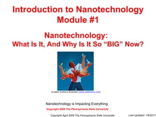
- 1. Introduction to Nanotechnology Module #1 Nanotechnology: What Is It, And Why Is It So “BIG” Now? © patton brothers illustration ( www.pattonbros.com ) Copyright April 2009 The Pennsylvania State University Last Updated: 1/6/2011 Copyright 2009 The Pennsylvania State University Nanotechnology is Impacting Everything
- 7. Where does the Nanometer fit in the length scale? Copyright April 2009 The Pennsylvania State University 1 meter = 3.28 feet 1 / 100 meter = 1 centimeter (cm) 1 / 1000 meter = 1 millimeter (mm) 1 / 1,000,000 meter = 1 micrometer* ( µm) *also called a micron 1 / 1,000,000,000 meter = 1 nanometer (nm) 1 / 1,000,000,000,000 meter = 1 picometer (pm)
- 8. Another way of looking at how small a Nanometer is- ©2009 NanoHorizons Inc. Copyright April 2009 The Pennsylvania State University
- 9. Still another way of looking at how small a Nanometer is- Click on the black box to view Mini Cooper movie Copyright April 2009 The Pennsylvania State University Produced by the Museum of Science, Boston with support from the Nanoscale Informal Science Education Network and the Center for High-rate Nanomanufacturing. © 2007.
- 11. How Do We See Things in These Different Size Ranges? Meter Size Range These are sizes we can see with just our eyes Millimeter Size Range These are sizes we can see with an optical microscope Micrometer Size Range Bigger objects in this range can be seen with an optical microscope . Smaller objects may need an electron microscope Nanometer Size Range Bigger objects can be seen with electron microscopes. Smaller objects require field emission electron or atomic force microscopes MACRO-SCALE NANO-SCALE MICRO-SCALE Copyright April 2009 The Pennsylvania State University
- 12. Let’s look at these size ranges pictorially. Let’s also get some idea of what nature makes and what man makes in these size ranges. Copyright April 2009 The Pennsylvania State University
- 13. Some Small Naturally Occurring and Man-Made Structures 1 mm 100 µm 10 µm 1 µm 100 nm 10 nm 1 nm 100 pm Transistor of 2007 Human hair tissue Bacterium cell Human cell Virus Transistors of 20-30 Years ago Protein Individual atom Drug molecule Quantum dot DNA Nano-scale Micro-scale Macro-scale Copyright April 2009 The Pennsylvania State University Stanford University © 2009 Created by Sean Nash
- 14. Also note from our pictorial representation of scales that the next size range that is smaller than the nano-scale is the pico-scale. Copyright April 2009 The Pennsylvania State University
- 17. What’s After Nanotechnology – Is there a Picotechnology? No, nothing to build at the pico-scale. The elements of the universe are fixed in number (and nicely listed in the periodic table) Copyright April 2009 The Pennsylvania State University
- 19. “ Nanotechnology is the builder’s final frontier.” Richard Smalley 1996 Nobel Laurate in Chemistry, Rice University Smalley Institute for Nanoscale Science & Technology Copyright April 2009 The Pennsylvania State University
- 26. Adapted from Linda Geppert, The Amazing Vanishing Transistor Act, IEEE Spectrum, October 2002, Vol. 39, Number 10, pg. 28-33 Copyright April 2009 The Pennsylvania State University
- 29. Quantum Corral IBM Research Division M.F. Crommie, C.P. Lutz, D.M. Eigler. Confinement of electrons to quantum corrals on a metal surface. Science 262, 218-220 (1993). Copyright April 2009 The Pennsylvania State University
- 31. Introduction to Nanotechnology Module #2 A Brief History of Nanotechnology © patton brothers illustration ( www.pattonbros.com ) Copyright April 2009 The Pennsylvania State University Last Updated: 1/6/2011 Nanotechnology is Impacting Everything
- 36. Around the year 1100. Arab craftsmen made steel swords of legendary strength. Today we know these swords had carbon nanotubes and nanowires in the material. This is the oldest known use of carbon nanotubes and nanowires. These nanostructures may account for the swords’ strength. Carbon nanotubes and carbon nanowires in Damascus steel sword. Copyright April 2009 The Pennsylvania State University Reibold, M., et al. "Carbon Nanotubes in an Ancient Damascus Sabre." Nature 444 (2006).
- 37. Reprinted with permission from Journal of Applied Physics, Vol. 93, Issue 12, P.10058, 2003, American Institute of Physics. 16 th century Renaissance pottery Around the year 1500. The Renaissance Italians used what we now know to be copper and silver nanoparticles to achieve this vibrantly colored pottery. Again, we don’t know the details of how they did it. Copyright April 2009 The Pennsylvania State University
- 42. In 1931. Ruska Develops the Transmission Electron Microscope (TEM)--- The beginnings of being able to really “SEE” at the nanoscale The transmission electron microscope has become a very powerful tool for “seeing” at the nano-scale. Copyright April 2009 The Pennsylvania State University
- 45. In 1959. Richard Feynman gives his famed talk “There is Plenty of Room at the Bottom” “ What I want to talk about is the problem of manipulating and controlling things on a small scale.” In this talk, Feynman said that we have progressed to the point where we can and should manipulate matter at what today we call the nano-scale. Copyright April 2009 The Pennsylvania State University Richard Feynman © 1965
- 46. He uses it to signify machining with tolerances of less than a micron (1000 nanometers). Today the term nanotechnology has evolved to mean making and manipulating “ things” that are much smaller! Today the “things” of nanotechnology have at least one dimension in the range of 1nm to about 100nm. In 1974. Norio Taniguchi coins the word “nanotechnology”. Copyright April 2009 The Pennsylvania State University
- 48. In 1981. Gerd Binnig and Heinrich Rohrer invent the first of the scanning probe tools- -- the scanning tunneling microscope. With this tool, we now could see individual atoms! With the invention of the scanning tunneling microscope , we can now “see” atoms Reprint Courtesy of International Business Machines Corporation copyright © International Business Machines Corporation. Figure: Michael Schmid, TU Wien Copyright April 2009 The Pennsylvania State University
- 50. In 1989. Donald M. Eigler of IBM “writes” for the first time with individual atoms Scanning probe tools (specifically the atomic force microscope (AFM)) can be used to drag atoms into preselected positions. Atoms aren’t blue nor are they cones. The color is computer rendering and shape is artifact of AFM. Copyright April 2009 The Pennsylvania State University Reprint courtesy of International Business Machines Corporation, copyright © 1990 International Business machines Corporation
- 51. In 1986. Eric Drexler publishes: “ Engines of Creation” Science fiction interest in nano begins Copyright April 2009 The Pennsylvania State University
- 52. In 1991. Sumio Iijima of NEC in Tsukubaka, Japan, discovered carbon nanotubes . Another unique carbon bonding configuration tolerated by nature only at the nano-scale . Copyright April 2009 The Pennsylvania State University Image Courtesy of NEC © Queen's University 2007
- 54. In 1998. Cees Dekker’s group at the Delft University of Technology demonstrates a transistor made from a carbon nanotube Courtesy C. Dekker, Delft University Copyright April 2009 The Pennsylvania State University J. Appenzeller, J. Knoch, R. Martel, V. Derycke, S. Wind, Ph. Avouris, Short-channel like effects in Schottky barrier carbon nanotube field-effect transistors, IEEE Technical Digest 2002. p.285. (© 2002 IEEE)
- 55. In 1999. Tour (Rice University) and Reed (Yale University) demonstrate single molecules can act as switches turning electric current on and off, giving experimental support for the idea of “Molecular Electronics” Original arrangement Molecule rearranged The molecule’s physical arrangement depends on the voltage on the contacts. Voltages above a threshold value cause rearrangement of the molecule and thus change the current it can carry. Copyright April 2009 The Pennsylvania State University Courtesy Mark Reed. Yale University. 1997 molecule Contacts
- 61. Copyright April 2009 The Pennsylvania State University Introduction to Nanotechnology Module #3 A Snapshot of Nanotechnology Today Nanotechnology is Impacting Everything © patton brothers illustration ( www.pattonbros.com ) Last Updated: 1/6/2011
- 64. Nanotechnology Investments Government, Corporation, and Venture Capitalist Investments Copyright April 2009 The Pennsylvania State University
- 85. The Product: Quantum dots These are available commercially and used in, for example, medical research for their very strong fluorescence color. The color they give off when excited can be changed simply by buying different sized dots which changes Δ E and thereby the fluorescing color. This picture of vials containing actual quantum dots was captured after the samples were placed in front of a UV hand lamp which excited the electrons from below E 1 to above E 2 . Product Example # 3 (continued) “ Relative Size of a Qdot® Nanocrystal.” Invitrogen Corporation . 2009. Copyright April 2009 The Pennsylvania State University
- 87. A Single-wall Carbon Nanotube The carbon atoms are the balls Copyright April 2009 The Pennsylvania State University
- 89. The product: Easton, a company that makes bicycles, is using carbon nanotubes in bikes they have on the market. Easton has a full carbon-fiber frame bicycle. This frame gives excellent weight savings, ride quality, and strength levels. The frame is a full CNT based material. In fact, the only metal part is the BB thread alloy insert . www.pezcyclingnews.com Product Example # 4 (continued) Copyright April 2009 The Pennsylvania State University
- 90. www.pezcyclingnews.com Copyright April 2009 The Pennsylvania State University
- 92. The very high surface to volume ratio of nanoparticles Lower surface Higher surface to volume ratio to volume ratio Copyright April 2009 The Pennsylvania State University Courtesy of CNEU
- 96. A silver nanoparticle attached to a textile fiber Copyright April 2009 The Pennsylvania State University © 2009 NanoHorizons Inc.
- 97. The Product: A number of companies are manufacturing and selling shoes and clothing containing silver nanoparticles for odor control. Product Example # 5 (continued) Copyright April 2009 The Pennsylvania State University
- 101. Adapted from Linda Geppert, The Amazing Vanishing Transistor Act, IEEE Spectrum, October 2002, Vol. 39, Number 10, pg. 28-33 Copyright April 2009 The Pennsylvania State University
- 104. The Product: Companies are manufacturing microelectronics circuits with more speed and more functionality due to the nanoscale transistors used in these circuits. Product Example # 6 (continued) Copyright April 2009 The Pennsylvania State University Copyright Matco Services Inc. www.materialsforum.com Mick Feuerbacher, December 2005.
- 109. Introduction to Nanotechnology Module #4 The Uniqueness of the Nano-scale © patton brothers illustration ( www.pattonbros.com ) Copyright April 2009 The Pennsylvania State University Last Updated: 1/6/2011 Nanotechnology is Impacting Everything
- 116. An example of an impact of this attribute: Huge Areal Densities The huge number of nano-scale transistors possible per area means today’s state-of-the-art microelectronics circuits with their nano-scale transistors can give more speed and more functionality. Copyright April 2009 The Pennsylvania State University Mick Feuerbacher, December 2005. Copyright Matco Services Inc. www.materialsforum.com
- 117. High surface to volume ratio Ratio = 3/R This ratio is very big when R is very small Ratio = 4 π R 2 _ 4/3 π R 3 Copyright April 2009 The Pennsylvania State University R
- 118. Impact of the Huge Surface to Volume Ratio Percent Surface Atoms Diameter (nm) This figure shows the inverse relationship between particle size and number of surface atoms. As a particle gets smaller, a larger and larger percentage of the atoms that make up the particle are surface atoms. Because the number of atoms or molecules on the surface of a particle influences the particle’s chemical and physical interactions with its environment, this percentage in the figure is key to defining the chemical and biological properties of nanoparticles. Figure 1 from Andre Nel, Tian Xia, Lutz Mädler and Ning Li ., SCIENCE Vol.311. p. 622 (2006). Copyright April 2009 The Pennsylvania State University
- 119. Reprinted figure with permission from Buffat and Borel, “Phys Rev. A” Volume 13, p 2287 (1976). Copyright 1976 by the American Physical Society. An example of an impact of this attribute: Melting Temperature This data is for Gold The melting temperature gets lower as a nanoparticle gets smaller because higher percentage of atoms are on the surface. This makes sense since surface atoms are not bound to each other the same way bulk atoms are. Particle diameter in Angstroms Copyright April 2009 The Pennsylvania State University
- 122. An example of an impact of this attribute: Colloidal Solutions The nano-scale colloidal particles in the solutions seen above will never settle out. (so long as surface forces are not present to cause them to agglomerate. If they agglomerated, this would increase particle volume and give gravity a chance to become important) Copyright April 2009 The Pennsylvania State University “ Electronic Imaging & Signal Processing: Improving biomedical imaging with gold nanocages” by Younan Xia and Sara E. Skrabalak. 12 May 2008, SPIE Newsroom. DOI: 10.1117/2.1200705.1135
- 124. An example of an impact of this attribute: Semiconductor Quantum Dots When excited by light, quantum dots fluoresce (re-emit light). The size and material composition of nano-scale quantum dots dictates the color they re-emit. Copyright April 2009 The Pennsylvania State University Evidenttech.com
- 126. An example of an impact of this attribute: Photonic Crystals Man-made nano-structures can cause light to turn sharp 90 degree corners, as seen below Copyright April 2009 The Pennsylvania State University Reproduced with permission of the MRS Bulletin. www.mrs.org/bulletin
- 127. Butterfly Scales 20,000 x magnification 5000x magnification 220x magnification 1x magnification of wing Another example of an impact of this attribute Nanostructures can cause light of different colors to scatter and diffract differently. This effect is used by nature to give the colors seen in butterfly wings. Militaries Study Animals for Cutting-Edge Camouflage. James Owen in England for National Geographic News March 12, 2003, Proc. R. Soc. Lond. B (1999) 266, 1403-1411 Copyright April 2009 The Pennsylvania State University
- 129. The cell is a complex structure with many compartments and organelles possessing individual and interdependent functions. Many of a cell`s features (e.g., pores) and, of course, DNA and RNA are all in the nano-scale. We can now make structures like these smallest of features in a cell. An example of an impact of this attribute: Copyright April 2009 The Pennsylvania State University Public Library of Science, “The Intersection of Biology and Materials Science” by George M. Whitesides and Amy P. Wong Vol. 31, p. 23.
- 130. Golovchenko, Branton, et. al. (Harvard Nanopore Group) Sequencing DNA using nanopore ionic conductance. Another Example: Structures that can “read” DNA Can now make structures so small that they, for example, can force DNA to line-up single file in order to pass through We can now use electrical signal changes to “read” the DNA as it passes through DNA Copyright April 2009 The Pennsylvania State University
- 131. DNA: millions of atoms long but 2.5 nm wide Nanostructures can have the dimensions seen in macromolecules Copyright April 2009 The Pennsylvania State University The Chaim Weizmann Institute of Chemistry, and the Fritz Haber Research Center for Molecular Dynamics, The Hebrew University of Jerusalem.
- 132. We can now make man-made versions of Nature`s motors like this one. Here Myosin V (blue), a cellular motor protein, carries cargo within cells by moving along actin filaments (red). It takes 37 nanometer steps by placing one “foot” over the other, as revealed by a fluorophore tag (rainbow-colored oval). [Illustration: PrecisionGraphics.com] Ahmet Yildiz, Joseph N. Forkey, Sean A. McKinney, Taekjip Ha, Yale E. Goldman, Paul R. Selvin: Myosin V Walks Hand-Over-Hand: Single Fluorophore Imaging with 1.5-nm Localization . Science. 2003. Vol. 300. p.2061 An example of an impact of this attribute: Copyright April 2009 The Pennsylvania State University
- 133. Unique chemical bonding configurations possible Carbon nanotubes Copyright April 2009 The Pennsylvania State University Odom et al, J. Phys. Chem. B104, 2794 (2000). Copyright Wiley-VCH Verlag GmbH & Co. KGaA. Reproduced with permission.
- 134. An example of an impact of this attribute Superior strength concrete for construction made with CNTs Carbon nanotubes distributed on small cement grains Copyright April 2009 The Pennsylvania State University S Bulletin, Vol. 31. P. 23, January 2006 Makar, J. M Beaudoin, J.J/NRCC-46618 http://irc.nrc-cnrc.gc.ca/ircpubs . Reproduced with the permission of the Minister of Public Works and Government Services Canada, 2009.
- 135. Molecular Self-Assembly An example: a mixture of two polymeric molecules can be made to self-assemble, under the influence of heat, into a structure with one phase made up only of molecules of type 1 and another phase made up only of molecules of type 2 White phase (regions) is made up only of molecules of type 1 Black phase (regions) is made up only of molecules of type 2 Copyright April 2009 The Pennsylvania State University Courtesy of CNEU
- 136. New Ways of Seeing things The nano-scale tips on scanning probe microscopes (SPMs) allow us to even “see” atoms Actual Atoms! IBM Research Division M.F. Crommie, C.P. Lutz, D.M. Eigler. Confinement of electrons to quantum corrals on a metal surface. Science 262, 218-220 (1993). Copyright April 2009 The Pennsylvania State University
- 138. Introduction to Nanotechnology Module #5 How Do We “See” Things at the Nano-scale: An Introduction to Characterization Techniques Nanotechnology is Impacting Everything © patton brothers illustration ( www.pattonbros.com ) Copyright April 2009 The Pennsylvania State University Last Updated: 1/6/2011
- 145. Schematic of a TEM or FE-TEM JEOL 2010F Semiconductor Material and Device Characterization, 3 rd ed. Dieter K. Schroder, John Wiley & Sons, Inc. p 647. 1999 Copyright April 2009 The Pennsylvania State University
- 146. Size, Shape, and Structure Observations using a TEM Here a silver nanowire is seen at various levels of magnification and finally, on the right, at a magnification that resolves the individual atoms (The Ag atoms appear as the white “dots”) Copyright April 2009 The Pennsylvania State University © 2005 College of Engineering at The University of Texas at Austin. Nanoscale Materials: Metal Nanowires
- 148. SEM Operation Click on the image to view the movie Copyright April 2009 The Pennsylvania State University SEM Images and Text Courtesy of the Museum of Science, Boston
- 149. A Size and Shape Observation using an SEM A carbon nanotube Copyright April 2009 The Pennsylvania State University Copyright 2006-2009 JEOL Ltd.
- 153. A Size, Shape, and Composition Observations using X-rays Imaging Technology Group, Beckman Institute of Advanced Science and Technology, University of Illinois Copyright April 2009 The Pennsylvania State University Ni Colors assigned to different elements C Ca Na SEM Image of a grain of sand showing well defined regions Composition mapping (of the regions seen in the SEM micrograph) using X-rays
- 156. SPM Tools All Use a Probe with a Nano-scale Sized Tip Copyright April 2009 The Pennsylvania State University Image courtesy: Greg McCarty tip sample
- 159. Deflection of the cantilever due to varying forces between the nano-scale tip and the atoms of the surface is picked up by changes in the laser beam reflection and converted by a computer into a picture. Copyright April 2009 The Pennsylvania State University
- 160. AFM Operation Movie courtesy of Veeco Instruments Inc. Tapping mode Click on the black box to view the movie Copyright April 2009 The Pennsylvania State University
- 161. Size and Shape Observations using an AFM Seeing DNA using an AFM Copyright April 2009 The Pennsylvania State University Courtesy of SPMage. http://www.icmm.csic.es/spmage/ "Toroidally supercoiled DNA” Dr. Jozef Adamcik. Ecole Polytechnique Federale de Lausanne (EPFL). S witzerland
- 162. AFM Probes can be used to move Nano-particles Copyright April 2009 The Pennsylvania State University Courtesy of CNEU
- 164. Picture of the Atoms on a Silicon Surface Imaged using STM Atoms on a silicon surface. Note that you can see that Nature has made some mistakes and there are defect sites where atoms are missing. Scanning tunneling microscopes allow surfaces to be imaged at the atomic-scale Schematic of the scanning tunneling microscope Image courtesy: Greg McCarty Copyright April 2009 The Pennsylvania State University
- 165. STM Probes can also be used to move Individual Atoms or Molecules Using Voltages applied between the Tip and the selected Atom or Molecule Copyright April 2009 The Pennsylvania State University Courtesy of CNEU
- 166. Here atoms on a surface are being arranged by an STM to form a corral Don Eigler and co-workers at IBM published these greyscale STM images showing the moving of atoms across a surface to form a corral as the corral was being constructed. The atoms were moved by the STM also by applying voltages to the probe that attracted the atom during the moving process. The atoms that form the surface itself can be seen in these STM images lying below those atoms in the process of being moved. Some Corral Atoms are in Position Atoms in various Stages of Being Moved Atoms of the Underlying Surface Finished Corral Copyright April 2009 The Pennsylvania State University Science 1993
- 167. A Quantum Corral As Seen By STM The STM tunneling current has been turned by a computer into this false color STM image of the Quantum Corral. The computer also tilted the image for us. Copyright April 2009 The Pennsylvania State University Science 1993
- 172. Introduction to Nanotechnology Module #6 How Do You Make Things So Small? An Introduction to Nanofabrication © patton brothers illustration ( www.pattonbros.com ) Copyright April 2009 The Pennsylvania State University Last Updated: 1/6/2011 Nanotechnology is Impacting Everything
- 174. Making nano-scale “things” is called Nanofabrication Copyright April 2009 The Pennsylvania State University
- 187. Top-down Nanofabrication is like Sculpting Start with a material supported on a substrate Add some new material according to a pattern (lithography) Copyright April 2009 The Pennsylvania State University Image courtesy of Bruce Hirst Image courtesy of Bruce Hirst
- 188. Top-down Nanofabrication is like Sculpting Subtract some of the material according to a pattern (Process order is not important; can subtract before or after adding) Repeat the adding/subtracting as needed following the pattern Copyright April 2009 The Pennsylvania State University Image courtesy of Bruce Hirst Image courtesy of Bruce Hirst
- 189. Bottom-up Nanofabrication is like putting blocks together The building blocks can go together in some inherent pattern dictated by shape or they can go together randomly. The building blocks can be atoms, molecules, or nanoparticles Copyright April 2009 The Pennsylvania State University
- 195. Etching Lithography Depositing or Growing Material Modification The Top-down Fabrication Methodology Copyright April 2009 The Pennsylvania State University Courtesy of CNEU
- 197. An Example of a Top-Down Nanofabrication Processing Sequence Copyright April 2009 The Pennsylvania State University Film Grown by Chemical Reaction of Ambient species with the Substrate Thin Film Substrate Photoresist PLASMA ETCH + IONS + IONS + IONS LITHOGRAPHY ETCHING Chemistry Chemistry Chemistry Spin on Photoresist Align Photomask Expose with Light Chemical Bonds are Altered in Exposed Areas Dissolve Exposed Photoresist in Liquid Developer Remove the Photoresist (Etch/Ion Implantation) Barrier (Negative Bias) Pattern Transfer and Substrate Modification Complete HEAT Substrate THIN FILM GROWTH OR DEPOSITION Oxygen SURFACE MODIFICATION + + + + + + + Ion Implantation Thermal Anneal HEAT Mask
- 202. Building Block Fabrication Self Assembly The Bottom-up Fabrication Methodology Copyright April 2009 The Pennsylvania State University Courtesy of CNEU
- 204. An Example of a Bottom-Up Nanofabrication Processing Sequence Functionalize the Nanoparticle Link with Antibodies Antigen Attachment Synthesize Nanoparticle Copyright April 2009 The Pennsylvania State University Courtesy of CNEU
- 206. Introduction to Nanotechnology Module #7 How Do You Build Things So Small: Top-Down Nanofabrication Copyright April 2009 The Pennsylvania State University © patton brothers illustration ( www.pattonbros.com Last Updated: 1/6/2011 Nanotechnology is Impacting Everything
- 208. Top-down Nanofabrication is like Sculpting Start with a material supported on a substrate (or just start with the substrate) Add some new material according to a pattern (lithography; i.e., pattern transfer) Copyright April 2009 The Pennsylvania State University Image courtesy of Bruce Hirst Image courtesy of Bruce Hirst
- 209. Top-down Nanofabrication is like Sculpting Subtract some of the material according to a pattern (Process order is not important; can subtract before or after adding) Repeat the adding/subtracting as needed following the pattern Copyright April 2009 The Pennsylvania State University Image courtesy of Bruce Hirst Image courtesy of Bruce Hirst
- 212. Etching Lithography Depositing or Growing Material Modification The Basic Steps of Top-down nanofabrication. These are used in any sequence. Copyright April 2009 The Pennsylvania State University Courtesy of CNEU
- 213. An Example of a Top-Down Nanofabrication Processing Sequence Copyright April 2009 The Pennsylvania State University Film Grown by Chemical Reaction of Ambient species with the Substrate Thin Film Substrate Photoresist PLASMA ETCH + IONS + IONS + IONS LITHOGRAPHY ETCHING Chemistry Chemistry Chemistry Spin on Photoresist Align Photomask Expose with Light Chemical Bonds are Altered in Exposed Areas Dissolve Exposed Photoresist in Liquid Developer Remove the Photoresist (Etch/Ion Implantation) Barrier (Negative Bias) Pattern Transfer and Substrate Modification Complete HEAT Substrate THIN FILM GROWTH OR DEPOSITION Oxygen SURFACE MODIFICATION + + + + + + + Ion Implantation Thermal Anneal HEAT Mask
- 214. In the preceding cartoon sequence, all 4 steps were used. Sometimes, one or more of these steps is not needed and is omitted in nanofabrication. Copyright April 2009 The Pennsylvania State University
- 218. Material modification Deposition or growth of films/layers Lithography (pattern transfer) Etching (material removal) The Top-down nanofabrication methodology Deposition or Growth Step Copyright April 2009 The Pennsylvania State University Courtesy of CNEU
- 220. Growth by Chemical Reaction Thin Film Substrate HEAT Substrate (This Example Shows Oxidation) Film Grown by Chemical Reaction of Ambient species with the Substrate Oxygen Copyright April 2009 The Pennsylvania State University Courtesy of CNEU
- 221. Growth by chemical reaction differs from physical application, physical vapor deposition, and chemical vapor deposition in that part of the layer is used up (chemically reacted) in any growth process. Copyright April 2009 The Pennsylvania State University
- 222. Physical Application There are many types of physical application processes; e.g., dipping, spraying, and spin-on. Here we see layer spin-on in cartoon form. Thin Film Substrate Layer Substrate Copyright April 2009 The Pennsylvania State University Courtesy of CNEU
- 223. Physical Vapor Deposition (PVD) In this example of PVD called sputtering , a film (purple) is being deposited on the substrate by argon ions (green). These ions act as hammers knocking film atoms (yellow) off the target (yellow too). A negative voltage attracts the Ar ions to the target . Copyright April 2009 The Pennsylvania State University Courtesy of CNEU
- 224. Chemical Vapor Deposition (CVD) In this example of CVD, gas molecules (the precursor) are broken apart by a plasma. The radicals, ions, and electrons produced result in a chemical reaction on the substrate producing the creation of a film as shown . Copyright April 2009 The Pennsylvania State University Courtesy of CNEU Ω ~ Impedance Match AC Power Source Throttle Valve Silane Plasma Si Amorphous silicon film growing H Gas Inlet #1 Gas Inlet #2
- 226. Material modification Deposition or growth of films/layers Lithography (pattern transfer) Etching (material removal) The top-down nanofabrication methodology Lithography (pattern transfer) Lithography Step Copyright April 2009 The Pennsylvania State University Courtesy of CNEU
- 229. The controlling pattern that is “written” to guide the fabrication processes permanently resides in a “ mask ”, in a mold , or in an computer data file , depending on the type of lithography. A mask is a plate of transparent material (e.g., glass) on which a pattern resides. A mold is a plate in which a pattern resides Copyright April 2009 The Pennsylvania State University
- 230. Basic Terms used in Lithography Lithography – The transferring (writing) of a pattern-usually to a “resist” Resist – Medium into which pattern on a mask, on a mold, or in computer file is transferred. Used in most types of lithography Developer – Needed in some types of lithography to bring out the pattern written in the resist Copyright April 2009 The Pennsylvania State University
- 231. There are many types of lithography Copyright April 2009 The Pennsylvania State University Type of Lithography Initial Location of Pattern “ Pencil” Doing the Writing Resist Used? Developer Used? Photo-lithography Mask Flood of light (photons) Yes Yes Electron Beam Lithography Data File Beam of Electrons Yes Yes Ion Beam Lithography Data File Beam of Ions Yes Yes Dip Pen Lithography Data File Physical Contact No No Embossing Lithography Mold Physical Contact Yes, usually If resist used Flash Mold Physical Contact Yes etching Stamp Lithography Mold Physical Contact Can be If resist used Molding Lithography Mold Deposited Material Can be If resist used Self-Assembly Lithography Mask, Mold, or Data File Deposited Material Can be If resist used
- 233. Photo (or Optical ) Lithography Copyright April 2009 The Pennsylvania State University
- 234. Photo (or Optical) Lithography Mask (pattern is on the mask) Resist Substrate Visible or UV light Resist, which has been coated onto the substrate, is exposed to light—but only in regions allowed by the mask Copyright April 2009 The Pennsylvania State University Courtesy of CNEU
- 235. Photo Lithography (continued) Exposed photo-resist has been designed to have its chemical bonding changed in the regions exposed to light Copyright April 2009 The Pennsylvania State University Courtesy of CNEU
- 236. Photo Lithography (continued) These changed regions are then chemically attacked by a developer and removed. Copyright April 2009 The Pennsylvania State University Courtesy of CNEU
- 237. Photo Lithography (continued) The result of the chemical attack by the developer is that the pattern is now “written” in the resist Copyright April 2009 The Pennsylvania State University Courtesy of CNEU
- 239. Photo Lithography (continued) The pattern is put into the substrate by using the resist as a chemical-attack barrier and etching the substrate. After etching, the resist is removed and the pattern is now in the substrate. Copyright April 2009 The Pennsylvania State University Courtesy of CNEU
- 240. Dip Pen Lithography Another Example of a Type of Lithography Copyright April 2009 The Pennsylvania State University
- 241. Dip Pen Nanolithography - An AFM probe tip is used to write alkanethiols (a type of molecule) onto a surface -Writes sub – 100 nm features - Moving the probe tip can be slow. Manufacturing through-put problem? S. Hong and C.A. Mirkin, Northwestern University Center for Nanofabrication and Molecular Assembly Copyright April 2009 The Pennsylvania State University
- 242. Embossing (or Nano-imprinting ) Lithography Another Example of a Type of Lithography Copyright April 2009 The Pennsylvania State University
- 243. Copyright April 2009 The Pennsylvania State University Courtesy of CNEU
- 244. Copyright April 2009 The Pennsylvania State University Courtesy of CNEU
- 245. Structures Produced using Patterns Created by Embossing Lithography Followed by Etching Copyright April 2009 The Pennsylvania State University "Current Status of Nanonex Nanoimprint Solutions," Hua Tan, Linshu Kong, Mingtao Li, Colby Steer and Larry Koecher, SPIE, (2004) "Four-inch Photo-Curable Nanoimprint Lithography Using NX-2000 Nanoimprint," Mingtao Li, Hua Tan, Linshu Kong, and Larry Koecher, SPIE, (2004)
- 246. Another example of
Notas del editor
- There is a glossary that comes along with this set of modules. Each term in blue print in every module is defined in this glossary.
- The boundaries between these various size ranges are not rigid but are blurry. For example, some engineers and scientists might say 1/5 of a micrometer is still in the nano-scale. Engineering and Science are very precise except to something like this where the boundaries between these size scales can vary somewhat.
- The glossary goes into the terms in blue italics. We don`t directly “see” with tools like electron microscopes and tunneling microscopes. These tools take a response and use computer processing to create an image which the human eye can decipher. This table just shows some of the imaging tools available
- The pico-scale is the size range of an individual atom. The individual atom types found in nature are seen in the periodic table on this viewgraph. These are the basic legos everything else is made of. There`s nothing to make at this size range unless you consider the man-made atoms (also listed in the periodic table of this viewgraph) that are made in accelerators and that live for fractions of a second. There in one technology based in the pico scale---nuclear technology (reactors, bombs, etc)—but that`s the only one. Nanotechnology, on the other hand, impacts and covers many technologies.
- At the nanoscale, mild thermal vibrations are equivalent to massive earthquakes, and an atom is not able to stand still to have its picture taken. In the cases of the 35 xenon atoms spelling “IBM” in 1990 and the 48 iron atoms defining the quantum corral in 1993, it was necessary for Donald Eigler and his co-workers at the IBM Almaden Research Center in California to reduce the temperature within their scanning tunnelling microscope (STM) chamber to just 4 Kelvin. In both cases, Eigler and co-workers included a sequence of images to show the process of creating the final picture. In the case of the quantum corral, there is a panel of four images showing the construction of the quantum corral, which was reported in Science in 1993.
- The corral is made by dragging atoms around using an Scanning Tunneling Microscope (STM) tip. The system was under ultra high vacuum and cooled to 4K. The original image used for this viewgraph was taken with an STM in 1993. The image shown in this viewgraph is a later modification where color is added and the image is rotated. In forming the corral, the atoms remain in contact with the surface but are pulled by the STM tip to the desired location by increasing the tunneling current. They move at a rate of about 4 Angstroms per second and never leave the surface. The tip can be withdrawn by decreasing the tunneling current and the attractive force is eliminated. The 3rd paragraph on page 525 of the second article noted below goes into good detail about this process. Background reading on the moving and imaging of atoms can be found at the following: (1) Confinement of Electrons to Quantum Corrals on a Metal Surface M. F. Crommie; C. P. Lutz; D. M. Eigler Science , New Series, Vol. 262, No. 5131. (Oct. 8, 1993), pp. 218-220. (2) Positioning Single Atoms with a Scanning Tunneling Microscope , D. Eigler and E. Schweizer, Nature 344 , 524, 1990.
- There is a glossary that comes along with this set of modules. Each term in blue print in every module is defined in this glossary.
- For more information, see http://www.thebritishmuseum.ac.uk/science/lycurguscup/sr-lycugus-p1.html
- For more information, see http://www.thebritishmuseum.ac.uk/science/lycurguscup/sr-lycugus-p1.html And read: (1) Paul Mulvaney, Not all That’s Gold Does Glitter, MRS Bulletin, December 2001, pg.’s 1009-1013 and (2) Barber, D J and Freestone, I C, An investigation of the origin of the colour of the Lycurgus Cup by analytical transmission electron microscopy, Archaeometry , 32 (1), 33-45, 1990.
- Please note the references for further reading: References: (1) Paul Mulvaney, Not all That’s Gold Does Glitter, MRS Bulletin, December 2001, pg.’s 1009-1013 and (2) Barber, D J and Freestone, I C, An investigation of the origin of the colour of the Lycurgus Cup by analytical transmission electron microscopy, Archaeometry , 32 (1), 33-45, 1990.
- See the article Reibold, M., et al. "Carbon Nanotubes in an Ancient Damascus Sabre." Nature 444 (2006).
- This work was done in one of Einstein`s famous 1905 papers. He produced three amazingly insightful papers that year: two on relativity and one on what we`ve come to call nanotechnology.
- His book is Zsigmondy, R. "Colloids and the Ultramicroscope", J.Wiley and Sons, NY, (1914). It is available today in paperback.
- Students can get a “feel” for what it is like to operate a TEM by going to http://nobelprize.org/educational_games/physics/microscopes/tem/tem.html
- Scanning tunnelinng miicroscopes are now one of many scanning probe tools. One such tool is the atomic force microscope (AFM). Your students can operate an AFM over the web by going to
- Your students can operate an AFM over the web by contact us and setting up a time slot.
- There is a glossary that comes along with this set of modules. Each term in blue print in every module is defined in this glossary.
- For an up-date on nanotechnology products on the market for sale, see the Woodrow Wilson Center for Scholars web site at www.nanotechproject.org. Specifically go to http://www.nanotechproject.org/index.php?id=44 Another excellent web site is http://www.nano.gov/ A general podcast series called SmallTalk is run by Dr. Stephanie Chasteen, of the Exploratorium’s Teacher Institute. One of the podcasts in this series covers nanotechnology products. See http://www.nisenet.org/publicbeta/podcasts/index.php .
- Nanoparticles are so small that they can stay in solution forever (so long as there`s no interaction)—so small that gravity can`t make them settle out. These particles are made of gold but not gold in color. We saw already in our module on the history of nanotechnology that the Romans used the fact that nano-scale gold is not “gold” in color to get colored glass.
- Clicking on words in blue takes you to the glossary for the module set
- The individual atoms of the nanoparticle are see as the red, green, and blue balls. The drug molecules are attached onto the nanoparticle that has been “functionalized” to accept them. Such functionlization may be achieved, for example, by using a linker molecule which links to the drug molecule and to the nanoparticle. The actual linking can be accomplished with chemical bonding.
- Human albumin is actually used so that the human antibody system is not set off by the presence of the nanoparticles.
- As a type of semiconductor nanparticle is made smaller, the Δ E gets bigger and the light given off shifts more to blue.
- Specific uses of quantum dots are given in the module on medical applications of nanotechnology
- They can kill bacteria, fungi, and algae depending on the metal
- Have the students decide which products use which nanoscale attributes.
- There is a glossary that comes along with this set of modules. Each term in blue print in every module is defined in this glossary.
- Bigger energy differences between allowed energies as R gets smaller means larger energy photons are emitted when electrons relax from the higher (excited) energy band to the lower (ground state) energy band. Higher energy photons means shorter wavelength light.
- We have already seen that Nature has a number of ways to create colors. In these modules we have so far seen (1) metal nanoparticle plasmom behavior which gives rise to color, (2) single electron transitions in semiconductor nanoparticles which gives rise to color, and now (3) geometric interference effects which also can give rise to color.
- CNTs = carbon nanotubes
- Tools and the size range they can “see” at
- There is a glossary that comes along with this set of modules. Each term in blue print in every module is defined in this glossary.
- The incident beam originates at a source. The electron source is heated to boil-off the electrons (thermionic emission) or it is designed so that the electrons can quantum mechanically tunnel out. The latter is called a field emission (an old name for tunneling) source and it is better because the electrons are more mono-energetic and therefore easier to manipulate to get better images. The electrons that come from a thermal emission source have a variety of energies because they have picked up some thermal energy and are therefore harder to manipulate..
- The electron source is heated to boil-off the electrons (thermionic emission) or it is designed so that the electrons can tunnel out. The latter is called a field emission (an old name for tunneling) source and it is better because the electrons are more mono-energetic.The electrons that come from a thermal emission source have a variety of energies because they have picked up some thermal energy. When an electron beam from the source impinges on a specimen, electrons can make it through the specimen if it is thinner than ~100nm. Using different apertures, bright field, dark field, and diffraction images can be obtained from these transmitted electrons. In all these variations, one thing is common: the number and distribution of emerging electrons is turned by a computer program into an image. See, for example, Dieter K. Schroder, Semiconductor Material and Device Characterization, 2nd ed., John Wiley & Sons, Inc.
- The white line on the lower left of the right-hand side picture shows how wide 2 nm is. This beautiful, repetitive pattern of atoms is the signature of a crystalline solid. Pictures like these are called micrographs.
- The white line at the bottom of this SEM picture (micrograph) shows how wide 100nm is in this picture. A demonstration of using an SEM for seeing can be set-up using our web based capabilities. Contact us.
- Composition here refers to the elemental composition
- A demonstration using the x-rays produced by an SEM to map elemental composition can be set-up using our web access capability. Contact us.
- Thermal energy gives atoms energy and the move and vibrate. At the nanoscale, all this motion is equivalent to massive earthquakes and an atom is not able to stand still at (or anywhere near) room temperature to have its picture taken. In the case the 48 iron atoms defining the quantum corral, it was necessary for Donald Eigler and his co-workers at the IBM Almaden Research Center in California to reduce the temperature within their scanning tunnelling microscope (STM) chamber to just 4 Kelvin to get the atomic motion to an acceptable level.
- An STM was used to both move the atoms (using the forces created by the application of voltages) and image the surface.
- This is a false color version of the image seen in the lower right of the previous viewgraph. The computer also titled the image for us to give some perspective. The blue things that look like “Hershey`s Kisses” are the atoms. Of course, atoms are blue and aren`t shaped like “Hershey`s Kisses”. That shape is an artifact of tunneling. The color blue was the computer programer`s choice. The red wave (color choice due to programmer) seen is the distribution of the corral atoms` electrons that have been penned-up in the “Corral”. This structure is called a quantum corral because the “red” wave seen is the quantum mechanical probability density for these electrons as predicted by Shcroedinger`s Wave Equation of quantum mechanics. As is clear, this probability density can actually be seen!
- There is a glossary that comes along with this set of modules. Each term in blue print in every module is defined in this glossary.
- Following this sculpting analogy, the modification step could be coloring the eyes blue.
- Following this sculpting analogy, the modification step could be coloring the eyes blue.
- The instructor may want to break this module into two lectures. There’s a lot of material here. There is a glossary that comes along with this set of modules. Each term in blue print in every module is defined in this glossary.
- The instructor may want to break this module into two lectures. There`s a lot of material here.
- Following this sculpting analogy, the modification step could be coloring the eyes blue.
- The instructor may want to break this module into two lectures. There`s a lot of material here.
- There are hundreds of such steps used in the manufacture of a state-of-the-art chip.
- The instructor may want to break this module into two lectures. There`s a lot of material here.
- There are hundreds of such steps used in the manufacture of a state-of-the-art chip.
- The instructor may want to break this module into two lectures. There`s a lot of material here.
- Materials for which the bonding is weakened where light impinges are called a positive resist. There is also negative resists for which the bonding gets stronger where light hits. The developer for these resists removes regions where light did not impinge.
- The instructor may want to break this module into two lectures. There`s a lot of material here.
- The instructor may want to break this module into two lectures. There`s a lot of material here.
- The instructor may want to break this module into two lectures. There`s a lot of material here.
- There is a glossary that comes along with this set of modules. Each term in blue print in every module is defined in this glossary.
- The instructor may want to break this module into two lectures. There`s a lot of material here.
- Following this sculpting analogy, the modification step could be coloring the eyes blue.
- The instructor may want to break this module into two lectures. There`s a lot of material here.
- The instructor may want to break this module into two lectures. There`s a lot of material here.
- The instructor may want to break this module into two lectures. There`s a lot of material here.
- There is a glossary that comes along with this set of modules. Each term in blue print in every module is defined in this glossary.
- This article “The ‘right”size in nanobiotechnology”, G. M. Whitesides NATURE BIOTECHNOLOGY, Volume 21, Number 10, October 2003 is good reading.
- There are lots of structures and organells in a cell. Many are at the nano-scale. An exampleis the microtubule structure seen in the lower left part of the cell.
- Fluorescing molecules (fluorophores) have been used for a while in biology to visualize structures. Here they have been attached to the microtubules and actin in a cell so we can see those structures.
- Quantum dots are semiconductor nano-particles whose fluorescing color is controlled by the diameter of the nano-particle. The smaller the particle, the more blue shifted (i.e., the shorter the wavelength of the emitted light) is the fluorescence.
- Everyone has heard about MRI scans so it’s a good example of the impact of nanotechnology—even in the local hospital.
- MRI versus CAT scans: A computed tomography (CT) scanner uses the scattering and attenuation of high energy X-ray photons, a type of ionizing radiation, to acquire its images, making it a good tool for examining tissue composed of elements of a relatively higher atomic number than the tissue surrounding them, such as bone and calcifications (calcium based). It caan be used for parts of the the body (carbon based flesh), or for structures (vessels, bowel) which have been artificially enhanced with contrast agents (iodine, barium) containing elements of a higher atomic number than the surrounding flesh. MRI, on the other hand, uses non-ionizing radio frequency signals to acquire its images using the quantum mechanical concept of nuclear spin. MRI is best suited for non-calcified tissue.
- Interesting for the students to learn that a quantum mechanical concept is being used to get those MRI pictures so basic today to medicine. Protons, along with neutrons, are found in the nucleus of an atom, and so, in its early years. MRI imaging was referred to as nuclear magnetic resonance imaging (NMRI), but the word nuclear has been associated by the public with ionizing radiation exposure, which is not used in an MRI. So, to prevent patients from making a flase and negative association between MRI and ionizing radiation, the word nuclear has been almost universally removed. Scientists still us the term NMRI when discussing non-medical imaging operating on the same principles. Common magnetic field strengths range from 0.3 to 3 T, although field strengths as high as 9.4 T or higher are used in research scanners [2] and research instruments for animals or only small test tubes range as high as 20 T. Commercial suppliers are investing in 7 T platforms. For comparison, the Earth's magnetic field averages around 50 μT, less than 1/100,000 times the field strength of a typical MRI. Go to the web site http://www.simplyphysics.com for a nice discussion of the physics of MRI.
- Water and fat account for approximately 63 per cent of the body's hydrogen atoms. Water is composed of two hydrogens and one oxygen. The hydrogen nuclei each have one proton---no neutrons. Medical MRI most frequently relies on the relaxation properties of excited hydrogen nuclear spins in water and lipids (fat).
- The contrast in MRI images is altered by contrast agents due to their influence on the relaxation times of hydrogen nuclei in tissue. Nanoparticles are promising candidates for molecular imaging enhancement because they convey the possibility to high relaxivity per molecular binding site. Using nanoparticles with a ligand specific for a certain tissue, will enhance the local contrast owing to the high relaxivity of each particle. For many applications a positive contrast agent that increases the signal intensity would be advantageous. Gadolinium-containing nanoparticles, for example, give excellent contrast enhancement properties.
- These nanoparticles have molecules attached to them (they are functionalized) allowing them to bond only to specific places; for example to just to cancer cells
- These dots were irradiated with the exciting light and the near-infra-red fluorescence was captured by a sensor to give the picture. A computer convered the image to a color the human eye can see
- Attaching ligands onto nanoparticles can be done to make them target only certain cells, to regulate their solubility, or both.
- One approach: use of liposome nanoparticles for carrying drugs. Extensive development of liposome delivery systems is already underway. The advantage of this approach is the diversity of cargoes (genes, drugs, proteins) that can be delivered to the cytosol of cells by liposome delivery vehicles.
- Until recently, cancer researchers have taken a shotgun approach to killing cancer – administering drug that goes everywhere in the body with the hope that enough reaches malignant cells to kill them. The results of this approach are often serious side effects and suboptimal therapeutic response. But in order to develop more effective chemotherapeutics and imaging agents, scientists need to improve their aim—switching to a sniper’s rifle to deliver agents more accurately to tumors. Nanoparticles, with the ability to store large payloads within their cores and with “targeting” molecules on their surfaces, would seem ideally suited to the “sniper” role.
- Docetaxel is the anti-cancer drug. PEG adjusts hydrophilicity for solubility control while RNA attaches to targeted tumor.
- To test the effectiveness of these new drug-delivery devices, the investigators conducted experiments on mice bearing human prostate tumors. After waiting until the tumors had become large (to simulate the stage at which most prostate cancers are detected in humans), the researchers injected the nanoparticles directly into the tumors of the cancer-laden animals. The shrinkage in tumors after approximately three months was dramatic. Moreover, all of the treated mice survived the study. In contrast, only 57 percent of the animals treated with untargeted nanoparticles survived for the duration of the study, and only 14 percent of the animals treated with docetaxel alone survived. These results indicated that the encapsulated drug did not move throughout the body, but rather stayed anchored to the tumor and delivered docetaxel to only cancer cells. Weight loss and white cell counts also confirmed lower toxicity of the treatment in mice that received the targeting nanoparticles versus those treated with untargeted ones. "A single injection of our nanoparticles completely eradicated the tumors in five of the seven treated animals, and the remaining animals also had significant tumor reduction, compared to the controls
- Stamping is covered in detail in the nanofabrication discussions.
- There is a glossary that comes along with this set of modules. Each term in blue print in every module is defined in this glossary.
- The instructor may want to break this module into two lectures. There`s a lot of material here.
- Vertical axis is number of transistors on a chip.
- The instructor may want to break this module into two lectures. There`s a lot of material here.
- The instructor may want to break this module into two lectures. There`s a lot of material here.
- These a cross-sections of MOSFET transistors. They are made by cutting a chip and looking in edge-wise at the cross-sections of the transistors using FESEM. These are lab transistors. The smallest in production today is 45nm.
- Schematic cross-section. Note that the transistor look as they really do in the earlier FESEM micrograph.
- Moore’s Second Law is that the cost of the factories (or fabs) for fabricating electronic devices has also been increasing exponentially with time. This is because the requirements on the size and positioning of features of features on a chip have been getting much more stringent, as has the complexity of the chips and the necessity to maintain a clean environment. In the past, the cost of the fabrication facility was small compared to all other costs, which have been increasing essentially linearly. However, now the cost of the fab is becoming a dominant cost, and the rate of its increase is significantly larger than the rate at with the market is growing. Thus, many experts believe that the economic issues illustrated by Moore’s Second Law will bring an end to the First.
- The instructor may want to break this module into two lectures. There`s a lot of material here.
- The instructor may want to break this module into two lectures. There`s a lot of material here.
- The term “TFT” used here means thin film transistor.
- Diode behavior is seen from these nanowires in the picture. Vertically oriented nanowires are p-type; horizontally oriented nanowires are n-type. Where they cross forms a p/n junction.
- The term SiNW means silicon nanowire.
- Many SiNW MOSFET transistors on a chip.
- The instructor may want to break this module into two lectures. There`s a lot of material here.