SlideShare una empresa de Scribd logo
1 de 3
Illustration and Evaluation of the Patient Pathway for Suspected Non-Massive Pulmonary Embolism
Introduction
National guidelines for suspected non-massive pulmonary embolism (PE) recommend pre-
test probability (PTP) assessment and D-dimer assays before imaging (Bayes et al, 2011).
This poster illustrates and evaluates the patient pathway for presentations of suspected
non-massive PE at the authors Trust, which employs the algorithm from NICE clinical
guidance 144 (fig. 6). The value of following the various steps of the pathway will be
discussed, and their applications justified alongside alternative techniques.
Discussion
Common risk factors for a PE include malignancy, previous emboli, and immobilisation or
surgery, and presenting symptoms are non-specific (Shujaat, Shapiro, and Eden, 2013).
Clinical assessment alone is not sufficient, yet performing imaging on all suspected cases
is not justifiable (Bayes et al, 2011). Being that as few as 10% of suspected PEs are
positive, and since they can be excluded by other means, utilising the guidance can save a
quarter of patients from imaging studies (Shujaat, Shapiro, and Eden, 2013).
CT pulmonary angiogram (CTPA) has replaced other techniques as the gold standard
(Shujaat, Shapiro, and Eden, 2013). With specificity and sensitivity at 100% (Subramaniam
et al, 2006), the rapid acquisition of high contrast images, the availability, and non-
invasive nature, CT is safe to exclude a PE as a sole imaging test. Experts agree that it has
fulfilled conditions to replace conventional angiography as the reference standard, and
has demonstrated low numbers of inadequate studies varying from 0.9-3% in large
cohorts (Huisman and Klok, 2009). Fig.1 and fig.2: positive PE visualised on CTPA
3. Multi detector CT (MDCT) advances have dramatically
increased PE diagnoses (Remy-Jardin et al, 2007), and the
PTP Score
D-Dimer
CTPA
1. The two level simplified and dichotomised Wells score
(Wells et al, 2000) utilised at the authors Trust (fig. 5)
categorises a PE as being ‘likely’ or ‘unlikely’, based on
clinical features and risk factors (Huisman and Klok,
2009). With a result of 4 points or greater proving that a
PE is likely, immediate imaging should be offered. Those
with an unlikely result should first have a D-dimer assay
performed (Yin et al, 2012).
Other PTP scoring tools: see appendix 1.
2. D-dimer is a fibrin degradation product detected by
blood test. It is elevated in pregnancy, infection, liver
complications, malignancy, advanced age, thromboembolic
disease and post-surgery or trauma (Yin et al, 2012). Due to
being detected in multiple instances, its specificity is
reported to be 34%, however, with sensitivity of 90%, a
negative D-dimer and low PTP excludes a PE (Shujaat,
Shapiro, and Eden, 2013). The cut off for serum D-dimer is
500mg/l, below which a PE can be excluded (Vossen et al,
2012). In cases with a raised D-dimer, imaging should be
performed, as evidence shows a clear correlation between
elevated D-dimer, and PEs confirmed radiologically (Shah
et al, 2013).
Adjusting D-dimer assay for age: see appendix 2.
The Diagnostic Pathway Imaging Considerations
7. Availability of Imaging: In the case of a likely PTP or raised D-dimer when CT or
VQ scanning is not immediately available, parenteral anticoagulant therapy should
be given, followed by imaging, once it becomes available. (NICE, 2012).
9. Overuse of Imaging: Finding clinically insignificant emboli or incidental diagnoses
demonstrate that imaging is being adopted without consideration of the risks (Costa
et al, 2014). An increase in the number of deaths due to contrast induced
nephropathy, radiation-induced cancers, and anticoagulation of false-positive results
is well reported, with such aggressive testing shifting the risk for a few patients with
PE to many without (Shah et al, 2013).
8. Alternative Diagnoses: While an alternative or incidental diagnosis may be found
on a CTPA, it is not its intended application, and VQ scans are not useful for this
purpose (Costa et al, 2014). A limited CTPA which just images the main pulmonary
arteries, and a simple report stating ‘PE present’, ‘PE absent’ or ‘Non-diagnostic’, is
employed at the authors Trust for all CTPAs. This acts as a deterrent for requests for
alternative diagnoses, and to encourage the correct use of the algorithm, ultimately
saving healthy patients from irradiation (Chandra et al, 2013).
10. Dose Concerns: Radiosensitive organs such as the thyroid, breasts, and lungs are
irradiated during a CTPA. A 20 year old woman is at a 68% greater risk of breast
cancer manifestation by 35 years following a CTPA, than a 20 year old woman who is
not scanned, with a dose of 20mGy being delivered to each breast. However, at the
authors Trust, CTPA is preferred over VQ scans in pregnancy which give a dose of
0.29mGy, as while the mother receives a greater dose from the CTPA than the VQ,
the opposite is true for the foetus. (Huisman and Klok, 2009).
4. Ventilation/Perfusion (VQ) Scan: The VQ scan is used as an alternative for patients
with contraindications to contrast media (Shujaat, Shapiro, and Eden, 2013) and due it
its overall lower dose, it may be favourable in pregnancy (Bayes et al, 2011). It has a
positive predictive value of 85-90%, but has non diagnostic readings of 28-46%
(Huisman and Klok, 2009). Guidance recommends VQ for patients not suited for CTPA
due to compromised renal function (estimated glomerular filtration rate at the
authors trust must be >45), or for limiting radiation dose. (NICE, 2012). Fig.3 ‘V’ scan
and fig. 4 ‘Q’ scan demonstrating a PE.
5. Compression Ultrasound (CUS) of lower limbs: CUS is performed if the VQ scan is
non-diagnostic or a CTPA was performed on single slice scanner. A normal CUS with an
unlikely PTP rules out a PE, and with a normal CUS and a likely PTP, a D-dimer assay
should be performed. If this is normal, then PE is ruled out. A raised D-dimer should
have repeat CUS in 7 days (Huisman and Klok, 2009).
Other imaging techniques: see appendix 3.
6. Investigations for Malignancy: At the authors Trust, patients who have an
unprovoked PE are recommended to have further investigations for malignancy, which
involves physical examination, a chest radiograph, various blood tests and urinalysis. In
patients over 40, a venous abdomen pelvis CT scan (with an arterial chest added if the
apices and bases were omitted in the CTPA) is offered, and a mammogram in females.
This recommendation is supported in the NICE guidance 144 (NICE, 2012)
Clinical Feature Points
Clinical symptoms/signs of DVT
(leg swelling and pain on palpation of
deep veins)
3
Alternative diagnosis less likely than
PE
3
HR > 100bpm 1.5 1.5
Immobilisation (>3 days) or surgery in
past 4 wks
1.5
Previous DVT/PE 1.5
Haemoptysis 1
Malignancy (on treatment, treated in
last 6m or palliative)
1
Conclusion
This poster illustrated and evaluated the patient pathway for presentations of suspected
non-massive PE at the authors Trust, which utilises clinical guidance from NICE. It has
been discovered that while imaging is invaluable for PE exclusion or diagnosis when
necessary, it is being overused when non-imaging tests can be conclusive. Evidence has
shown that following the guidance on PTP and D-dimer algorithms can safely exclude the
diagnoses, saving a quarter of patients from unnecessary imaging. It has been discussed
that patients with unprovoked PE should be followed up with investigations for
malignancy, utilising recommendations from NICE.
Fig. 1 Fig. 2
Fig. 3 Fig. 4
Fig. 5 Wells PTP Score
(Wells et al, 2000)
Axial and coronal CTPA slices from the authors Trust showing left and right sided PEs (yellow circles)
Images courtesy of the authors Trust
Ventilation (V) scan and Perfusion (Q) scan showing images of a PE
Images courtesy of the authors Trust
Fig. 6 PE Diagnostic Algorithm
Courtesy of the authors Trust
040044947
U
N
L
I
K
E
L
Y
R
A
I
S
E
D
References
Bayes, H.K., O'Dowd, C.A., Glassford, N.J., McKay, A. & Davidson, S. 2011, "D-dimer assays - a help or hindrance in suspected pulmonary
thromboembolism assessment?” Journal of postgraduate medicine, vol. 57, no. 2, pp. 109-114.
Chandra, S., Sarkar, P.K., Chandra, D., Ginsberg, N.E. & Cohen, R.I. 2013, "Finding an alternative diagnosis does not justify increased use
of CT-pulmonary angiography", BMC Pulmonary Medicine, vol. 13, pp. 9-9.
Costa, A., Basseri, H., Sheikh, A., Stiell, I. & Dennie, C. 2014, "The yield of CT pulmonary angiograms to exclude acute pulmonary
embolism", Emergency Radiology, vol. 21, no. 2, pp. 133-141.
Huisman, M.V. & Klok, F.A. 2009, "Diagnostic management of clinically suspected acute pulmonary embolism", Journal of Thrombosis &
Haemostasis, vol. 7, pp. 312-317.
National Clinical Guideline Centre 2012, "Venous thromboembolic diseases: the management of venous thromboembolic diseases and
the role of thrombophilia testing.
Clinical Guideline Methods, evidence and recommendations", National Institute for Health and Clinical Excellence.
Remy-Jardin, M., Pistolesi, M., Goodman, L.R., Gefter, W.B., Gottschalk, A., Mayo, J.R. & Sostman, H.D. 2007, "Management of suspected
acute pulmonary embolism in the era of CT angiography: a statement from the Fleischner Society", Radiology, vol. 245, no. 2, pp.
315-329.
Shah, K., Quaas, J., Rolston, D., Bansal, S., Bania, T., Newman, D., Wiener, D. & Lee, J. 2013, "Magnitude of D-dimer matters for
diagnosing pulmonary embolus", The American Journal of Emergency Medicine, vol. 31, no. 6, pp. 942-945.
Shujaat, A., Shapiro, J.M. & Eden, E. 2013, "Utilization of CT pulmonary angiography in suspected pulmonary embolism in a major urban
emergency department", Pulmonary Medicine, , pp. 1-6.
Subramaniam, R.M., Chou, T., Swarbrick, M. & Karalus, N. 2006, "Pulmonary embolism: accuracy and safety of a negative CT pulmonary
angiogram and value of a negative D-dimer assay to exclude CT pulmonary angiogram-detectable pulmonary embolism",
Australasian Radiology, vol. 50, no. 5, pp. 424-428.
Vossen, J.A., Albrektson, J., Sensarma, A. & Williams, S.C. 2012, "Clinical usefulness of adjusted D-dimer cut-off values to exclude
pulmonary embolism in a community hospital emergency department patient population", Acta radiologica, vol. 53, no. 7, pp. 765-
768.
Wells PS, Anderson DR, Rodger M, Ginsberg JS, Kearon C, Gent M et al. Derivation of a simple clinical model to categorize patients
probability of pulmonary embolism: increasing the models utility with the SimpliRED D-dimer. Thrombosis & Haemostasis. 2000;
83(3):416-420.
Yin, F., Wilson, T., Della Fave, A., Larsen, M., Yoon, J., Nugusie, B., Freeland, H. & Chow, R.D. 2012, "Inappropriate use of D-dimer assay
and pulmonary CT angiography in the evaluation of suspected acute pulmonary embolism", American Journal of Medical Quality,
vol. 27, no. 1, pp. 74-79.
Bounameaux, H. 2010, "Contemporary management of pulmonary embolism: the answers to ten questions", Journal of internal
medicine, vol. 268, no. 3, pp. 218-231.
Bowman, A.W., Johns, G.S. & Karstaedt, N. 2011, "Markedly elevated plasma D-dimer and the prevalence of acute pulmonary embolus",
Acta Medica Academica, vol. 40, no. 2, pp. 152-158.
Bibliography
Appendices:
Appendix 2: Adjusting D-dimer for age
Typically, the cut off value for serum D-dimer is 500 mg/L, below which the diagnosis of PE can be ruled out. However one study concluded that this could be increased
to 1000 mg/L without affecting its sensitivity, safely eliminating the diagnosis in more patients, and further reducing the use of imaging (Bowman, Johns and Karstaedt,
2011). A PE is more likely to occur in the elderly, and multiple comorbidities associated with aging can mimic a PE clinically. Hence, the argument exists regarding the
usefulness of this value for the elderly, with evidence showing that a PE may be excluded in as few as 5% of patients over 80, compared with 60% in those under 40,
using this D-dimer value (Bounameaux, 2010).
Appendix 1: Other PTP scoring tools
“Geneva score (original and revised) - The original Geneva score is based on seven clinical factors and required interpretation of the findings on chest X-ray and arterial
blood gases. The revised Geneva score covers eight parameters in 3 clinical areas: risk factors, symptoms and clinical signs. Each of these is given 1 to 5 points
accordingly. This gives a possible score range of 0 to 25. A score of 11 or higher is classified as ‘high risk’ of PE; a score of 4 to 10 as ‘intermediate risk’ and a score of 0
to 3 as ‘low risk’.
Charlotte rule - Patients with suspected PE (based on empiric clinical assessment) are stratified into low-risk and high-risk (pre-test probability groups). Patients are
classified as high risk if they have at least two of the following:
• Age greater than 50
• Heart rate greater than systolic blood pressure
• Surgery in the past month
• Unilateral leg swelling
• Haemoptysis
• Unexplained oxygen saturation less than 95% on room air.”
(National Clinical Guideline Centre 2012, p 69.)
Appendix 3: Other imaging techniques
Pulmonary Angiogram (PA)
The PA was the traditional gold standard (Yin et al, 2012) reference imaging method (Huisman and Klok, 2009). However it is Invasive and resource demanding and no
longer routinely performed (Bounameaux, 2010).
Magnetic Resonance Angiography (MRA)
MRA has potential as an alternative for those unsuited to iodinated CM (Huisman and Klok, 2009), due to shortened acquisition times and improved spatial resolution.
Combined perfusion and non-contrast/contrast scans have a specificity of 93% (Remy-Jardin et al, 2007), however, 25% of scans are inadequate (Bounameaux, 2010).

Más contenido relacionado

La actualidad más candente

What is the place of CT coronary angiography in ED chest pain?
What is the place of CT coronary angiography in ED chest pain?What is the place of CT coronary angiography in ED chest pain?
What is the place of CT coronary angiography in ED chest pain?kellyam18
 
Comparative study of Balthazar Computed Tomography Severity Index and Modifie...
Comparative study of Balthazar Computed Tomography Severity Index and Modifie...Comparative study of Balthazar Computed Tomography Severity Index and Modifie...
Comparative study of Balthazar Computed Tomography Severity Index and Modifie...Apollo Hospitals
 
BALKAN MCO 2011 - R. Curca - Palliative and supportive care in lung cancer
BALKAN MCO 2011 - R. Curca - Palliative and supportive care in lung cancer BALKAN MCO 2011 - R. Curca - Palliative and supportive care in lung cancer
BALKAN MCO 2011 - R. Curca - Palliative and supportive care in lung cancer European School of Oncology
 
Evaluation of subclavian, thoracic aorta and innominate artery injuries in bl...
Evaluation of subclavian, thoracic aorta and innominate artery injuries in bl...Evaluation of subclavian, thoracic aorta and innominate artery injuries in bl...
Evaluation of subclavian, thoracic aorta and innominate artery injuries in bl...Clinical Surgery Research Communications
 
Manual of Geriatric Anesthesia
Manual of Geriatric AnesthesiaManual of Geriatric Anesthesia
Manual of Geriatric AnesthesiaSpringer
 
MCQs in evidence based practice
MCQs in evidence based practiceMCQs in evidence based practice
MCQs in evidence based practiceSamir Haffar
 
Clinical application and efficacy analysis of 3 d navigation module in the tr...
Clinical application and efficacy analysis of 3 d navigation module in the tr...Clinical application and efficacy analysis of 3 d navigation module in the tr...
Clinical application and efficacy analysis of 3 d navigation module in the tr...Clinical Surgery Research Communications
 
Samir rafla real life outcome of atrial fibrillation ablation-cardio alex 2014
Samir rafla  real life outcome of atrial fibrillation ablation-cardio alex 2014Samir rafla  real life outcome of atrial fibrillation ablation-cardio alex 2014
Samir rafla real life outcome of atrial fibrillation ablation-cardio alex 2014Alexandria University, Egypt
 
CT coronary angiography in ED chest pain patients
CT coronary angiography in ED chest pain patientsCT coronary angiography in ED chest pain patients
CT coronary angiography in ED chest pain patientskellyam18
 
Wh fecho criteriafordxrhdnrcardio.2012.7
Wh fecho criteriafordxrhdnrcardio.2012.7Wh fecho criteriafordxrhdnrcardio.2012.7
Wh fecho criteriafordxrhdnrcardio.2012.7gisa_legal
 
Pulmonary review - Chest World Congress Madrid 2014
Pulmonary review - Chest World Congress Madrid 2014Pulmonary review - Chest World Congress Madrid 2014
Pulmonary review - Chest World Congress Madrid 2014Dr. Josep Morera Prat
 

La actualidad más candente (20)

Pancholy S - AIMRADIAL 2013 - Radiation exposure
Pancholy S - AIMRADIAL 2013 - Radiation exposurePancholy S - AIMRADIAL 2013 - Radiation exposure
Pancholy S - AIMRADIAL 2013 - Radiation exposure
 
What is the place of CT coronary angiography in ED chest pain?
What is the place of CT coronary angiography in ED chest pain?What is the place of CT coronary angiography in ED chest pain?
What is the place of CT coronary angiography in ED chest pain?
 
Open Journal of Surgery
Open Journal of SurgeryOpen Journal of Surgery
Open Journal of Surgery
 
Comparative study of Balthazar Computed Tomography Severity Index and Modifie...
Comparative study of Balthazar Computed Tomography Severity Index and Modifie...Comparative study of Balthazar Computed Tomography Severity Index and Modifie...
Comparative study of Balthazar Computed Tomography Severity Index and Modifie...
 
Roberts J - AIMRADIAL 2015 - Ultrasound guidance
Roberts J - AIMRADIAL 2015 - Ultrasound guidanceRoberts J - AIMRADIAL 2015 - Ultrasound guidance
Roberts J - AIMRADIAL 2015 - Ultrasound guidance
 
BALKAN MCO 2011 - R. Curca - Palliative and supportive care in lung cancer
BALKAN MCO 2011 - R. Curca - Palliative and supportive care in lung cancer BALKAN MCO 2011 - R. Curca - Palliative and supportive care in lung cancer
BALKAN MCO 2011 - R. Curca - Palliative and supportive care in lung cancer
 
Evaluation of subclavian, thoracic aorta and innominate artery injuries in bl...
Evaluation of subclavian, thoracic aorta and innominate artery injuries in bl...Evaluation of subclavian, thoracic aorta and innominate artery injuries in bl...
Evaluation of subclavian, thoracic aorta and innominate artery injuries in bl...
 
Manual of Geriatric Anesthesia
Manual of Geriatric AnesthesiaManual of Geriatric Anesthesia
Manual of Geriatric Anesthesia
 
Jolly S - Radiation exposure and transradial - 201507
Jolly S - Radiation exposure and transradial - 201507Jolly S - Radiation exposure and transradial - 201507
Jolly S - Radiation exposure and transradial - 201507
 
Antithrombotic pp
Antithrombotic ppAntithrombotic pp
Antithrombotic pp
 
15 aimradial2016 fri A Amin
15 aimradial2016 fri A Amin15 aimradial2016 fri A Amin
15 aimradial2016 fri A Amin
 
MCQs in evidence based practice
MCQs in evidence based practiceMCQs in evidence based practice
MCQs in evidence based practice
 
Clinical application and efficacy analysis of 3 d navigation module in the tr...
Clinical application and efficacy analysis of 3 d navigation module in the tr...Clinical application and efficacy analysis of 3 d navigation module in the tr...
Clinical application and efficacy analysis of 3 d navigation module in the tr...
 
Samir rafla real life outcome of atrial fibrillation ablation-cardio alex 2014
Samir rafla  real life outcome of atrial fibrillation ablation-cardio alex 2014Samir rafla  real life outcome of atrial fibrillation ablation-cardio alex 2014
Samir rafla real life outcome of atrial fibrillation ablation-cardio alex 2014
 
Sciahbasi A - AIMRADIAL 2013 - Radiation exposure
Sciahbasi A - AIMRADIAL 2013 - Radiation exposureSciahbasi A - AIMRADIAL 2013 - Radiation exposure
Sciahbasi A - AIMRADIAL 2013 - Radiation exposure
 
CT coronary angiography in ED chest pain patients
CT coronary angiography in ED chest pain patientsCT coronary angiography in ED chest pain patients
CT coronary angiography in ED chest pain patients
 
Wh fecho criteriafordxrhdnrcardio.2012.7
Wh fecho criteriafordxrhdnrcardio.2012.7Wh fecho criteriafordxrhdnrcardio.2012.7
Wh fecho criteriafordxrhdnrcardio.2012.7
 
Cardoso C - AIMRADIAL 2014 - Radial operators and femoral
Cardoso C - AIMRADIAL 2014 - Radial operators and femoralCardoso C - AIMRADIAL 2014 - Radial operators and femoral
Cardoso C - AIMRADIAL 2014 - Radial operators and femoral
 
Pulmonary review - Chest World Congress Madrid 2014
Pulmonary review - Chest World Congress Madrid 2014Pulmonary review - Chest World Congress Madrid 2014
Pulmonary review - Chest World Congress Madrid 2014
 
02 aimradial2016 fri2 EM Vegh
02 aimradial2016 fri2 EM Vegh02 aimradial2016 fri2 EM Vegh
02 aimradial2016 fri2 EM Vegh
 

Similar a 040044947

076 advances in pulmonary imaging
076 advances in pulmonary imaging076 advances in pulmonary imaging
076 advances in pulmonary imagingmuhammed Yasar
 
DIFFERENT IMAGING MODALITIES USED FOR THE DETECTION OF PROSTATE CANCER – A RE...
DIFFERENT IMAGING MODALITIES USED FOR THE DETECTION OF PROSTATE CANCER – A RE...DIFFERENT IMAGING MODALITIES USED FOR THE DETECTION OF PROSTATE CANCER – A RE...
DIFFERENT IMAGING MODALITIES USED FOR THE DETECTION OF PROSTATE CANCER – A RE...IRJET Journal
 
Health Surveillance of asbestos-exposed workers at Helsinki Asbestos 2014
Health Surveillance of asbestos-exposed workers at Helsinki Asbestos 2014Health Surveillance of asbestos-exposed workers at Helsinki Asbestos 2014
Health Surveillance of asbestos-exposed workers at Helsinki Asbestos 2014Työterveyslaitos
 
Atypical Presentations of lung cancers.pdf
Atypical Presentations of lung cancers.pdfAtypical Presentations of lung cancers.pdf
Atypical Presentations of lung cancers.pdfKimberly Pulley
 
Imaging of Pulmonary Embolism
Imaging of Pulmonary Embolism  Imaging of Pulmonary Embolism
Imaging of Pulmonary Embolism Gamal Agmy
 
Pruebas radiologicas a evitar American College of Radiology.
Pruebas radiologicas a evitar American College of Radiology. Pruebas radiologicas a evitar American College of Radiology.
Pruebas radiologicas a evitar American College of Radiology. Cristobal Buñuel
 
kellygarcia.positionpaperdraft
kellygarcia.positionpaperdraftkellygarcia.positionpaperdraft
kellygarcia.positionpaperdraftKelly Garcia
 
Whole body screening – risks and benefits
Whole body screening – risks and benefitsWhole body screening – risks and benefits
Whole body screening – risks and benefitsWan Najwa Zaini
 
Co-relation of multidetector CT scan based preoperative staging with intra-op...
Co-relation of multidetector CT scan based preoperative staging with intra-op...Co-relation of multidetector CT scan based preoperative staging with intra-op...
Co-relation of multidetector CT scan based preoperative staging with intra-op...Apollo Hospitals
 
i.a.Preoperative ovarian cancer diagnosis using neuro fuzzy approach
i.a.Preoperative ovarian cancer diagnosis using neuro fuzzy approachi.a.Preoperative ovarian cancer diagnosis using neuro fuzzy approach
i.a.Preoperative ovarian cancer diagnosis using neuro fuzzy approachJonathan Josue Cid Galiot
 
Secondary Malignancy after Treatment of Prostate Cancer. Radical Prostatectom...
Secondary Malignancy after Treatment of Prostate Cancer. Radical Prostatectom...Secondary Malignancy after Treatment of Prostate Cancer. Radical Prostatectom...
Secondary Malignancy after Treatment of Prostate Cancer. Radical Prostatectom...asclepiuspdfs
 
ROLE OF HIGH-RESOLUTION COMPUTER TOMOGRAPHY IN SPUTUM POSITIVE.pptx
ROLE OF HIGH-RESOLUTION COMPUTER TOMOGRAPHY IN SPUTUM POSITIVE.pptxROLE OF HIGH-RESOLUTION COMPUTER TOMOGRAPHY IN SPUTUM POSITIVE.pptx
ROLE OF HIGH-RESOLUTION COMPUTER TOMOGRAPHY IN SPUTUM POSITIVE.pptxNagasai Pelala
 

Similar a 040044947 (20)

Ultrasound Diagnosis Of Appendicitis
Ultrasound Diagnosis Of Appendicitis  Ultrasound Diagnosis Of Appendicitis
Ultrasound Diagnosis Of Appendicitis
 
076 advances in pulmonary imaging
076 advances in pulmonary imaging076 advances in pulmonary imaging
076 advances in pulmonary imaging
 
Imagen Torácica
Imagen TorácicaImagen Torácica
Imagen Torácica
 
DIFFERENT IMAGING MODALITIES USED FOR THE DETECTION OF PROSTATE CANCER – A RE...
DIFFERENT IMAGING MODALITIES USED FOR THE DETECTION OF PROSTATE CANCER – A RE...DIFFERENT IMAGING MODALITIES USED FOR THE DETECTION OF PROSTATE CANCER – A RE...
DIFFERENT IMAGING MODALITIES USED FOR THE DETECTION OF PROSTATE CANCER – A RE...
 
Clots and Zealots
Clots and ZealotsClots and Zealots
Clots and Zealots
 
Health Surveillance of asbestos-exposed workers at Helsinki Asbestos 2014
Health Surveillance of asbestos-exposed workers at Helsinki Asbestos 2014Health Surveillance of asbestos-exposed workers at Helsinki Asbestos 2014
Health Surveillance of asbestos-exposed workers at Helsinki Asbestos 2014
 
Atypical Presentations of lung cancers.pdf
Atypical Presentations of lung cancers.pdfAtypical Presentations of lung cancers.pdf
Atypical Presentations of lung cancers.pdf
 
pe.pptx
pe.pptxpe.pptx
pe.pptx
 
Imaging of Pulmonary Embolism
Imaging of Pulmonary Embolism  Imaging of Pulmonary Embolism
Imaging of Pulmonary Embolism
 
Nódulos pulmonares
Nódulos pulmonares Nódulos pulmonares
Nódulos pulmonares
 
Pruebas radiologicas a evitar American College of Radiology.
Pruebas radiologicas a evitar American College of Radiology. Pruebas radiologicas a evitar American College of Radiology.
Pruebas radiologicas a evitar American College of Radiology.
 
Factors determining outcomes in grown up patients operated
Factors determining outcomes in grown up patients operatedFactors determining outcomes in grown up patients operated
Factors determining outcomes in grown up patients operated
 
kellygarcia.positionpaperdraft
kellygarcia.positionpaperdraftkellygarcia.positionpaperdraft
kellygarcia.positionpaperdraft
 
Whole body screening – risks and benefits
Whole body screening – risks and benefitsWhole body screening – risks and benefits
Whole body screening – risks and benefits
 
Co-relation of multidetector CT scan based preoperative staging with intra-op...
Co-relation of multidetector CT scan based preoperative staging with intra-op...Co-relation of multidetector CT scan based preoperative staging with intra-op...
Co-relation of multidetector CT scan based preoperative staging with intra-op...
 
i.a.Preoperative ovarian cancer diagnosis using neuro fuzzy approach
i.a.Preoperative ovarian cancer diagnosis using neuro fuzzy approachi.a.Preoperative ovarian cancer diagnosis using neuro fuzzy approach
i.a.Preoperative ovarian cancer diagnosis using neuro fuzzy approach
 
10 pulmonary
10 pulmonary10 pulmonary
10 pulmonary
 
Pulmonary embolism
Pulmonary embolismPulmonary embolism
Pulmonary embolism
 
Secondary Malignancy after Treatment of Prostate Cancer. Radical Prostatectom...
Secondary Malignancy after Treatment of Prostate Cancer. Radical Prostatectom...Secondary Malignancy after Treatment of Prostate Cancer. Radical Prostatectom...
Secondary Malignancy after Treatment of Prostate Cancer. Radical Prostatectom...
 
ROLE OF HIGH-RESOLUTION COMPUTER TOMOGRAPHY IN SPUTUM POSITIVE.pptx
ROLE OF HIGH-RESOLUTION COMPUTER TOMOGRAPHY IN SPUTUM POSITIVE.pptxROLE OF HIGH-RESOLUTION COMPUTER TOMOGRAPHY IN SPUTUM POSITIVE.pptx
ROLE OF HIGH-RESOLUTION COMPUTER TOMOGRAPHY IN SPUTUM POSITIVE.pptx
 

040044947

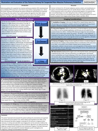
  • 1. Illustration and Evaluation of the Patient Pathway for Suspected Non-Massive Pulmonary Embolism Introduction National guidelines for suspected non-massive pulmonary embolism (PE) recommend pre- test probability (PTP) assessment and D-dimer assays before imaging (Bayes et al, 2011). This poster illustrates and evaluates the patient pathway for presentations of suspected non-massive PE at the authors Trust, which employs the algorithm from NICE clinical guidance 144 (fig. 6). The value of following the various steps of the pathway will be discussed, and their applications justified alongside alternative techniques. Discussion Common risk factors for a PE include malignancy, previous emboli, and immobilisation or surgery, and presenting symptoms are non-specific (Shujaat, Shapiro, and Eden, 2013). Clinical assessment alone is not sufficient, yet performing imaging on all suspected cases is not justifiable (Bayes et al, 2011). Being that as few as 10% of suspected PEs are positive, and since they can be excluded by other means, utilising the guidance can save a quarter of patients from imaging studies (Shujaat, Shapiro, and Eden, 2013). CT pulmonary angiogram (CTPA) has replaced other techniques as the gold standard (Shujaat, Shapiro, and Eden, 2013). With specificity and sensitivity at 100% (Subramaniam et al, 2006), the rapid acquisition of high contrast images, the availability, and non- invasive nature, CT is safe to exclude a PE as a sole imaging test. Experts agree that it has fulfilled conditions to replace conventional angiography as the reference standard, and has demonstrated low numbers of inadequate studies varying from 0.9-3% in large cohorts (Huisman and Klok, 2009). Fig.1 and fig.2: positive PE visualised on CTPA 3. Multi detector CT (MDCT) advances have dramatically increased PE diagnoses (Remy-Jardin et al, 2007), and the PTP Score D-Dimer CTPA 1. The two level simplified and dichotomised Wells score (Wells et al, 2000) utilised at the authors Trust (fig. 5) categorises a PE as being ‘likely’ or ‘unlikely’, based on clinical features and risk factors (Huisman and Klok, 2009). With a result of 4 points or greater proving that a PE is likely, immediate imaging should be offered. Those with an unlikely result should first have a D-dimer assay performed (Yin et al, 2012). Other PTP scoring tools: see appendix 1. 2. D-dimer is a fibrin degradation product detected by blood test. It is elevated in pregnancy, infection, liver complications, malignancy, advanced age, thromboembolic disease and post-surgery or trauma (Yin et al, 2012). Due to being detected in multiple instances, its specificity is reported to be 34%, however, with sensitivity of 90%, a negative D-dimer and low PTP excludes a PE (Shujaat, Shapiro, and Eden, 2013). The cut off for serum D-dimer is 500mg/l, below which a PE can be excluded (Vossen et al, 2012). In cases with a raised D-dimer, imaging should be performed, as evidence shows a clear correlation between elevated D-dimer, and PEs confirmed radiologically (Shah et al, 2013). Adjusting D-dimer assay for age: see appendix 2. The Diagnostic Pathway Imaging Considerations 7. Availability of Imaging: In the case of a likely PTP or raised D-dimer when CT or VQ scanning is not immediately available, parenteral anticoagulant therapy should be given, followed by imaging, once it becomes available. (NICE, 2012). 9. Overuse of Imaging: Finding clinically insignificant emboli or incidental diagnoses demonstrate that imaging is being adopted without consideration of the risks (Costa et al, 2014). An increase in the number of deaths due to contrast induced nephropathy, radiation-induced cancers, and anticoagulation of false-positive results is well reported, with such aggressive testing shifting the risk for a few patients with PE to many without (Shah et al, 2013). 8. Alternative Diagnoses: While an alternative or incidental diagnosis may be found on a CTPA, it is not its intended application, and VQ scans are not useful for this purpose (Costa et al, 2014). A limited CTPA which just images the main pulmonary arteries, and a simple report stating ‘PE present’, ‘PE absent’ or ‘Non-diagnostic’, is employed at the authors Trust for all CTPAs. This acts as a deterrent for requests for alternative diagnoses, and to encourage the correct use of the algorithm, ultimately saving healthy patients from irradiation (Chandra et al, 2013). 10. Dose Concerns: Radiosensitive organs such as the thyroid, breasts, and lungs are irradiated during a CTPA. A 20 year old woman is at a 68% greater risk of breast cancer manifestation by 35 years following a CTPA, than a 20 year old woman who is not scanned, with a dose of 20mGy being delivered to each breast. However, at the authors Trust, CTPA is preferred over VQ scans in pregnancy which give a dose of 0.29mGy, as while the mother receives a greater dose from the CTPA than the VQ, the opposite is true for the foetus. (Huisman and Klok, 2009). 4. Ventilation/Perfusion (VQ) Scan: The VQ scan is used as an alternative for patients with contraindications to contrast media (Shujaat, Shapiro, and Eden, 2013) and due it its overall lower dose, it may be favourable in pregnancy (Bayes et al, 2011). It has a positive predictive value of 85-90%, but has non diagnostic readings of 28-46% (Huisman and Klok, 2009). Guidance recommends VQ for patients not suited for CTPA due to compromised renal function (estimated glomerular filtration rate at the authors trust must be >45), or for limiting radiation dose. (NICE, 2012). Fig.3 ‘V’ scan and fig. 4 ‘Q’ scan demonstrating a PE. 5. Compression Ultrasound (CUS) of lower limbs: CUS is performed if the VQ scan is non-diagnostic or a CTPA was performed on single slice scanner. A normal CUS with an unlikely PTP rules out a PE, and with a normal CUS and a likely PTP, a D-dimer assay should be performed. If this is normal, then PE is ruled out. A raised D-dimer should have repeat CUS in 7 days (Huisman and Klok, 2009). Other imaging techniques: see appendix 3. 6. Investigations for Malignancy: At the authors Trust, patients who have an unprovoked PE are recommended to have further investigations for malignancy, which involves physical examination, a chest radiograph, various blood tests and urinalysis. In patients over 40, a venous abdomen pelvis CT scan (with an arterial chest added if the apices and bases were omitted in the CTPA) is offered, and a mammogram in females. This recommendation is supported in the NICE guidance 144 (NICE, 2012) Clinical Feature Points Clinical symptoms/signs of DVT (leg swelling and pain on palpation of deep veins) 3 Alternative diagnosis less likely than PE 3 HR > 100bpm 1.5 1.5 Immobilisation (>3 days) or surgery in past 4 wks 1.5 Previous DVT/PE 1.5 Haemoptysis 1 Malignancy (on treatment, treated in last 6m or palliative) 1 Conclusion This poster illustrated and evaluated the patient pathway for presentations of suspected non-massive PE at the authors Trust, which utilises clinical guidance from NICE. It has been discovered that while imaging is invaluable for PE exclusion or diagnosis when necessary, it is being overused when non-imaging tests can be conclusive. Evidence has shown that following the guidance on PTP and D-dimer algorithms can safely exclude the diagnoses, saving a quarter of patients from unnecessary imaging. It has been discussed that patients with unprovoked PE should be followed up with investigations for malignancy, utilising recommendations from NICE. Fig. 1 Fig. 2 Fig. 3 Fig. 4 Fig. 5 Wells PTP Score (Wells et al, 2000) Axial and coronal CTPA slices from the authors Trust showing left and right sided PEs (yellow circles) Images courtesy of the authors Trust Ventilation (V) scan and Perfusion (Q) scan showing images of a PE Images courtesy of the authors Trust Fig. 6 PE Diagnostic Algorithm Courtesy of the authors Trust 040044947 U N L I K E L Y R A I S E D
  • 2. References Bayes, H.K., O'Dowd, C.A., Glassford, N.J., McKay, A. & Davidson, S. 2011, "D-dimer assays - a help or hindrance in suspected pulmonary thromboembolism assessment?” Journal of postgraduate medicine, vol. 57, no. 2, pp. 109-114. Chandra, S., Sarkar, P.K., Chandra, D., Ginsberg, N.E. & Cohen, R.I. 2013, "Finding an alternative diagnosis does not justify increased use of CT-pulmonary angiography", BMC Pulmonary Medicine, vol. 13, pp. 9-9. Costa, A., Basseri, H., Sheikh, A., Stiell, I. & Dennie, C. 2014, "The yield of CT pulmonary angiograms to exclude acute pulmonary embolism", Emergency Radiology, vol. 21, no. 2, pp. 133-141. Huisman, M.V. & Klok, F.A. 2009, "Diagnostic management of clinically suspected acute pulmonary embolism", Journal of Thrombosis & Haemostasis, vol. 7, pp. 312-317. National Clinical Guideline Centre 2012, "Venous thromboembolic diseases: the management of venous thromboembolic diseases and the role of thrombophilia testing. Clinical Guideline Methods, evidence and recommendations", National Institute for Health and Clinical Excellence. Remy-Jardin, M., Pistolesi, M., Goodman, L.R., Gefter, W.B., Gottschalk, A., Mayo, J.R. & Sostman, H.D. 2007, "Management of suspected acute pulmonary embolism in the era of CT angiography: a statement from the Fleischner Society", Radiology, vol. 245, no. 2, pp. 315-329. Shah, K., Quaas, J., Rolston, D., Bansal, S., Bania, T., Newman, D., Wiener, D. & Lee, J. 2013, "Magnitude of D-dimer matters for diagnosing pulmonary embolus", The American Journal of Emergency Medicine, vol. 31, no. 6, pp. 942-945. Shujaat, A., Shapiro, J.M. & Eden, E. 2013, "Utilization of CT pulmonary angiography in suspected pulmonary embolism in a major urban emergency department", Pulmonary Medicine, , pp. 1-6. Subramaniam, R.M., Chou, T., Swarbrick, M. & Karalus, N. 2006, "Pulmonary embolism: accuracy and safety of a negative CT pulmonary angiogram and value of a negative D-dimer assay to exclude CT pulmonary angiogram-detectable pulmonary embolism", Australasian Radiology, vol. 50, no. 5, pp. 424-428. Vossen, J.A., Albrektson, J., Sensarma, A. & Williams, S.C. 2012, "Clinical usefulness of adjusted D-dimer cut-off values to exclude pulmonary embolism in a community hospital emergency department patient population", Acta radiologica, vol. 53, no. 7, pp. 765- 768. Wells PS, Anderson DR, Rodger M, Ginsberg JS, Kearon C, Gent M et al. Derivation of a simple clinical model to categorize patients probability of pulmonary embolism: increasing the models utility with the SimpliRED D-dimer. Thrombosis & Haemostasis. 2000; 83(3):416-420. Yin, F., Wilson, T., Della Fave, A., Larsen, M., Yoon, J., Nugusie, B., Freeland, H. & Chow, R.D. 2012, "Inappropriate use of D-dimer assay and pulmonary CT angiography in the evaluation of suspected acute pulmonary embolism", American Journal of Medical Quality, vol. 27, no. 1, pp. 74-79. Bounameaux, H. 2010, "Contemporary management of pulmonary embolism: the answers to ten questions", Journal of internal medicine, vol. 268, no. 3, pp. 218-231. Bowman, A.W., Johns, G.S. & Karstaedt, N. 2011, "Markedly elevated plasma D-dimer and the prevalence of acute pulmonary embolus", Acta Medica Academica, vol. 40, no. 2, pp. 152-158. Bibliography
  • 3. Appendices: Appendix 2: Adjusting D-dimer for age Typically, the cut off value for serum D-dimer is 500 mg/L, below which the diagnosis of PE can be ruled out. However one study concluded that this could be increased to 1000 mg/L without affecting its sensitivity, safely eliminating the diagnosis in more patients, and further reducing the use of imaging (Bowman, Johns and Karstaedt, 2011). A PE is more likely to occur in the elderly, and multiple comorbidities associated with aging can mimic a PE clinically. Hence, the argument exists regarding the usefulness of this value for the elderly, with evidence showing that a PE may be excluded in as few as 5% of patients over 80, compared with 60% in those under 40, using this D-dimer value (Bounameaux, 2010). Appendix 1: Other PTP scoring tools “Geneva score (original and revised) - The original Geneva score is based on seven clinical factors and required interpretation of the findings on chest X-ray and arterial blood gases. The revised Geneva score covers eight parameters in 3 clinical areas: risk factors, symptoms and clinical signs. Each of these is given 1 to 5 points accordingly. This gives a possible score range of 0 to 25. A score of 11 or higher is classified as ‘high risk’ of PE; a score of 4 to 10 as ‘intermediate risk’ and a score of 0 to 3 as ‘low risk’. Charlotte rule - Patients with suspected PE (based on empiric clinical assessment) are stratified into low-risk and high-risk (pre-test probability groups). Patients are classified as high risk if they have at least two of the following: • Age greater than 50 • Heart rate greater than systolic blood pressure • Surgery in the past month • Unilateral leg swelling • Haemoptysis • Unexplained oxygen saturation less than 95% on room air.” (National Clinical Guideline Centre 2012, p 69.) Appendix 3: Other imaging techniques Pulmonary Angiogram (PA) The PA was the traditional gold standard (Yin et al, 2012) reference imaging method (Huisman and Klok, 2009). However it is Invasive and resource demanding and no longer routinely performed (Bounameaux, 2010). Magnetic Resonance Angiography (MRA) MRA has potential as an alternative for those unsuited to iodinated CM (Huisman and Klok, 2009), due to shortened acquisition times and improved spatial resolution. Combined perfusion and non-contrast/contrast scans have a specificity of 93% (Remy-Jardin et al, 2007), however, 25% of scans are inadequate (Bounameaux, 2010).