❤️Amritsar Escorts Service☎️9815674956☎️ Call Girl service in Amritsar☎️ Amri...
Ch07
1. “For there was never yet a philosopher who could endure the
toothache patiently.”
–William Shakespeare,
Much Ado About Nothing, Act V, Scene 1
Orofacial pain is a major public health problem. This
fact was recently emphasized by a report from the US
National Center for Health Statistics (NCHS).
Although their figures and estimates apply only to the
United States, they may generally be extrapolated
worldwide.
The NCHS interviewed “45,711 households in the
US civilian population.” This was the 1989 National
Health Interview Survey (NHIS). Using the statistics
developed from this survey, the National Institute of
Dental Research reported in 1993 that “about 39 mil-
lion people or about 22% of the U.S. population 18
years of age or older are estimated to have experienced
at least one of five types of orofacial pain more than
once during the past six months.”1
This alarming cohort of 39 million can be further
broken down into 22 million toothaches (12.2%), 15
million oral sores (8.4%), 9.5 million “jaw joint” pains
(1.4%), and 1.3 million burning mouth pains (0.7%).
The total adds up to more than 39 million because some
respondents suffered from more than one type of orofa-
cial pain.1 Of the 45,711 households interviewed, 9,072
people reported orofacial pain. These figures, extrapolat-
ed nationwide, may actually be too low because the
Armed Forces, institutions (prisons, etc), and children
under age 18 were not included in the statistics.
The shocking revelation that 22 million people in
the United States suffer from a toothache within a
6-month period is an overall prevalence rate of 12,261
persons per 100,000 population. The prevalence for
African Americans (14,584) and Hispanics (14,226)
was even higher.1
By all odds, the most frequently seen “pain” patient
will be experiencing acute, true intraoral pain,
toothache and its sequelae being the most common.
Pain accompanying intraoral lesions and infections is
the next most commonly seen. After that, the field thins
somewhat, with top priority going to the acute pains of
everyday general, endodontic, and oral surgery practice.
Less commonly seen, but a good deal more baffling,
are the chronic pains found in and around the mouth,
a number of them referred there from faraway sites.
These are the craniofacial pains to be discussed in detail
in Chapter 8. Because these two types of oral and peri-
oral pain are so different in diagnosis and manage-
ment, they will be dealt with separately, even though
there is often a confusing overlap.
The first premise in diagnosis is to “play the percent-
ages,” thinking first of the most commonly occurring.
As the old saying goes, “If you hear hoofbeats, think of
horses, not zebras.”If an obvious diagnosis is not imme-
diately apparent, a good diagnostician begins with the
most frequent cause of this type of pain, not some
obscure, seldom seen syndrome. Systematically working
down through the pain roster, from most common to
least common, a logical diagnosis is finally arrived at.
A well-trained dentist may render many services that
gain him a deep personal satisfaction. Foremost among
these are the diagnosis and relief of excruciating or
long-lasting pain. These are the occasions patients
remember most vividly: “the night you came back at
midnight and relieved my terrible toothache,” or
“…you diagnosed the constant pain I had for 3 years,
Doctor, and after everyone else had given up.”
Solving these problems is rarely rewarding financial-
ly, but they are the few moments we as a profession
enjoy that set us apart from the lay public. These prob-
lems also try one’s patience and ingenuity; only the
skilled are successful in diagnosing and managing the
really difficult cases.
Chapter 7
DIFFERENTIAL DIAGNOSIS AND
TREATMENT OF DENTAL PAIN
John I. Ingle and Dudley H. Glick
2. Diagnosis is a personal and cognitive experience;
therefore, many of the qualities of a good diagnostician
are of an interpersonal nature and based on knowledge,
experience, and diagnostic tools. Diagnosing orofacial
disease is similar to medical diagnosis. The pulp test,
radiographs, percussion, palpation, and other tests and
procedures can facilitate the diagnosing of facial disease,
just as the electrocardiograph, electroencephalograph,
liver and kidney function tests, echocardiograph, com-
puted axial tomographic scanners, and a host of other
radiographs can facilitate medical diagnosis.
REQUIREMENTS OF A DIAGNOSTICIAN
A dentist can develop a number of assets to become a
successful diagnostician. Again, the most important of
these are knowledge, interest, intuition, curiosity, and
patience. The successful diagnostician must also have
acute senses and the necessary tools for diagnosis. For a
detailed discussion of these assets, see Chapter 6.
HISTORY, EXAMINATION, DIAGNOSTIC TESTS,
AND CONSULTATIONS
The important steps leading to a diagnosis and estab-
lishing a plan of treatment have been dealt with in
depth in the previous chapter. Additional information,
necessary to establish a diagnosis in chronic extracra-
nial and intracranial pain complexes, will be detailed in
the next chapter dealing with craniofacial pain.
PULP PAIN
Pulp pain, or pulpalgia, is by far the most commonly
experienced pain in and near the oral cavity and may
be classified according to the degree of severity and the
pathologic process present:
1. Hyperreactive pulpalgia
a. Dentinal hypersensitivity
b. Hyperemia
2. Acute pulpalgia
a. Incipient
b. Moderate
c. Advanced
3. Chronic pulpalgia
a. Barodontalgia
4. Hyperplastic pulpitis
5. Necrotic pulp
6. Internal resorption
7. Traumatic occlusion
8. Incomplete fracture
The mildest pulp discomfort, experienced when no
inflammation is present, is hyperreactive pulpalgia.
260 Endodontics
Hyperreactive Pulpalgia
Hyperreactive pulpalgia is characterized by a short,
sharp, shock—that is, “pain” best described as a sensa-
tion of sudden shock. The sensation is as sharp as it is
sudden and must be elicited by some exciting factor. It
is never spontaneous. The pain is of short duration,
lasting only slightly longer than the time during which
the irritating element is in contact with the tooth. In
some manner, the odontoblastic cellular body within
the dentin must be excited by a noxious stimulus, either
hot or cold, sweet or sour, or touch. Excitation of the
odontoblast conducts the excitation to the pulp nerves.
These “dentinal receptors have the characteristics of
slow adaptation.”3
It is difficult to explain to a patient that the severe
pain experienced when eating ice cream—a blinding
pain that extends upward through the eye and into the
forehead—is really normal and not pathologic. In “lay
terms,” one can only tell the patient that the cold
“excites”the nerve in the tooth, and the pain is so severe
it is referred upward through the eye.
The dentist, however, requires a more sophisticated
explanation. Although this is hard to come by, the best
explanation revolves around thinking of the fluid in the
dentinal tubules, along with the odontoblast cells, as a
“pump”—a hydrodynamic theory, if you will, that pro-
poses that the fluid moves back and forth to stretch,
compress, and excite the pulp nerves.4
Brännström pointed out that “the displacement of
tubule contents, if the movement occurs rapidly
enough, may produce deformation of nerve fibers in
the pulp or predentin or damage to the cells; both of
these effects may be capable of producing pain.”4 Such
a mechanical transmission of the stimulus would
account for the hitherto inexplicably great sensitivity of
the dentin to pain, in spite of the apparent absence of
nerve fibers in this tissue.4
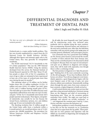
Brännström further confirmed the damage and pain
generated by blowing air over exposed dentin.5 A short
air blast evaporates from 0.1 to 0.3 mm of fluid from
the dentinal tubule. This results in immediate capillary
fluid replacement from the pulp’s blood supply, suck-
ing the odontoblasts and nerve fibers up into the
tubule. The nerves are stretched or even torn off, elicit-
ing pain (Figure 7-1).
On continued exposure to an air blast, however, a
plug of fluid protein builds up in the tubule, preventing
fluid outflow. This plug “closes the pump” and leads to
dentin insensitivity. When water is applied to the
dentin surface, however, the plug “melts” and sensitivi-
ty returns (see Figure 7-1).
3. Differential Diagnosis and Treatment of Dental Pain 261
The same phenomenon is produced by the dental
drill, the frictional heat and surface pressure displacing
the tubule fluid and causing pain. Scraping or chiseling
the dentin produces similar pressure and pain.
Pulp pain produced by cold is a similar phenome-
non. When cold is applied, the fluid in the tubules con-
tracts, thus redirecting the fluid volume. Fluid contrac-
tion within the tubules again produces fluid outflow
owing to the normal pulp pressure, and the nerves are
once again stretched into the tubules along with the
odontoblasts (Figure 7-2).5 Beveridge measured a fall
in intrapulp pressure when cold was applied to a tooth6
(Figure 7-3). Researchers in Israel found this to be an
“interstitial” pressure fall, whereas the “arterial trans-
mural” pressure rose.7
Hyperreactive pulpalgia owing to the application of
heat is more easily explained. Again, Beveridge easily
demonstrated a true increase in intrapulp pressure
when heat was applied to the tooth (Figure 7-4). An
increase of pressure within the pulp serves to excite the
sensory pulp nerves.6
Three different types of response to heat have also
been recorded from pulp nerve fibers: (1) a transient
type of response when pulp nerve fiber was excited by
heat over 43˚C (the response ceased as soon as the tem-
perature fell below the firing points), (2) a long-lasting
type of response that started at over 45˚C and contin-
ued even after the temperature had returned to the ini-
tial level for a few minutes, and (3) a pulsating type of
response in which the discharge of the fiber was syn-
chronized with the heartbeat.8
Brännström added to the understanding of the
“pumping” excitation by dentinal fluid when he point-
ed out, “Fluids have a considerably greater coefficient
of expansion than solids—a sudden rise in temperature
of 20˚C at the outer one-third of the closed tubule
Figure 7-1 Pain produced by air blast. A, Air evaporates dentinal
fluid, causing rapid outflow (arrows) owing to capillary pressure
from the pulp’s vessels. B, Odontoblast and accompanying nerve
fiber aspirated into tubule, stretching nerve and causing pain. C,
Prolonged air blast caused a protein plug to form in the tubule, pre-
venting outward flow. Redrawn with permission from Brännström
M. Dentin and pulp in restorative dentistry. Nacka (Sweden):
Dental Therapeutics AB; 1981. p. 15.
Figure 7-2 The effect of cold stimulus on the pulp. A, Cold is
applied to the tooth, causing a contraction of fluid in tubule. B,
Pulp capillary pressure forces replacement fluid into the tubule
along with the odontoblast and afferent nerve. Stretching of nerve
(arrow) produces intense pain. Redrawn with permission from
Brännström M. Dentin and pulp in restorative dentistry. Nacka
(Sweden): Dental Therapeutics AB; 1981. p. 17.
Figure 7-3 Effect of intrapulp pressure by application of cold
ethyl chloride spray to anesthetized mandibular premolar in a
13-year-old boy. Within 8 seconds, intrapulp pressure had dropped
from 30 mm Hg to nearly zero. After the irritant was removed, pres-
sure returned to initial baseline within 1 minute. Reproduced with
permission from Beveridge EE, and Brown AC.14
4. might result in an immediate pulpward movement of
about 5 micra of the content of the tubules,”9 once
again stretching the afferent nerves.
Hyperreactive pulpalgia is common following the
placement of a new restoration. Patients also complain
after root planing and curettage or following periodon-
tal surgery, which exposes the root surface. Hyper-
reactive pulpalgia also may be present in the tooth with
a carious lesion. Teeth traumatized by bruxism or
incompletely fractured teeth are generally more hyper-
reactive, as are the maxillary teeth involved in maxillary
sinusitis.
It is possible, with our present level of knowledge, to
divide the sensations of hyperreactive pulpalgia into
hypersensitivity and hyperemia.
Dentinal Hypersensitivity
The exciting factors of a hypersensitive pulp are usual-
ly cold food or drink or cold air, contact of two dissim-
ilar metals that will yield a galvanic shock, or stimula-
tion of the exposed dentin on the root surface by cold,
sweet or sour substances, vegetable or fruit acid, salt, or
glycerine, or often just touching the surface with a fin-
gernail, a toothbrush, an interdental stimulator, or an
explorer. One should not be surprised at this latter
reaction when the microanatomy of the dentin and
pulp is reviewed. The cementum covering the gingival
root dentin frequently is missing or has been removed
by curettage or brushing, exposing the dentinal
tubules. It has also been reported that the use of the
new “calculus-removing” toothpastes leads to an
increased dentinal hypersensitivity. Evidently, these
agents remove the surface smear layer and open the
dentinal tubuli orifices (Palm Springs Seminars, Palm
Springs, California, 1990, Lecture, Ingle JI.).
262 Endodontics
When one considers that “one square millimeter of
dentin contains approximately 30,000 tubules*10 and that
“approximately 25% of the volume of the dentin is occu-
pied by fluid, most of which is in the dentinal tubules,”11
one must be struck by the capacity for fluid dynamic
hypersensitivity emanating from exposed dentin.
This is a frequent complaint following periodontal
surgery when whole areas of root are exposed by the
apically repositioned gingiva. Add to this the careful
root preparation that removes most of the cementum
covering the dentin. The problem is then compounded
by two other avenues leading to irritation: the use of
citric acid on the root surface to remove the smear layer
that may “plug” the tubuli and the formation of dental
plaque on the root surfaces.12 The acid-releasing bacte-
ria in the plaque set up a steady barrage of irritation
into the dentinal tubules.
This is a “catch-22” equation: the hypersensitive
dentin is “painful” to brush and floss and therefore is
avoided by the patient. The bacterial plaque that then
forms causes greater sensitivity, so the area is avoided
all the more during home care, which, in turn, leads to
more plaque and greater sensitivity. Relieving the sen-
sitivity is the only solution to the problem.
One is led to conjecture about the pulp sensation stim-
ulated by sour substances, fruit juices, sugar, salt, and dis-
similar metals, which may be described as an electric cur-
rent flowing between the oral cavity and the pulp. Sicher
postulated that the oral cavity is positively charged and
the pulp is negatively charged (personal communication,
1958). Any electrolyte, such as salt or fruit acid, upsets
this ionic balance, and the resultant current stimulates
the nerve endings to the odontoblasts. The sensation dis-
appears as soon as the electrolyte is diluted away or metal
(such as aluminum foil) is removed. In addition to the
current flow theory, Anderson believes that “pain can be
evoked from dentin by applying to it solutions which
exert high osmotic pressure.”13 Brännström pursued this
idea further, although he believes it is not simply a ques-
tion of osmosis but relates again to the hydrodynamic
“pump”—concentrated solutions of sugar, salt, etc dehy-
drate the tubule contents,causing their rapid outflow and
deformation of the nerves within the tubule.4,5
Brännström also pointed out that a similar mechanism is
operant when cracks develop in the dentin—that is, as a
cusp flexes with biting pressure, the fluid in the tubules is
pumped back and forth (especially on release), which
stimulates the pain response.5
*This figure varies between 10,000 and 30,000 depending on the
location in the tooth—crown or root.
Figure 7-4 Effect on intrapulp pressure by application of hot
gutta-percha to an anesthetized maxillary premolar in a 12-year-old
girl. Within 15 seconds, the intrapulp pressure had more than dou-
bled. After heat was removed, the intrapulp pressure dropped pre-
cipitously but not to the original baseline. Reproduced with per-
mission from Beveridge EE, and Brown AC.14
5. Differential Diagnosis and Treatment of Dental Pain 263
Hyperemia
All minor pulp sensations were once thought to be
associated with hyperemia, an increased blood flow in
the pulp. The investigations of Beveridge and Brown
demonstrated, however, that an increase in intrapulp
tissue pressure is produced only when heat is applied to
the tooth, not when cold is applied.6,14 The increased
pressure against the sensory nerve endings in the pulp
might well produce the sensation associated with
hyperemia. Quite possibly, this will explain why the
pain appears to be of a different intensity and character
with applications of cold or heat, the cold producing a
sharp hypersensitivity response and the heat producing
true transient hyperemia and a dull pain.
This difference in the character of the painful
response between cold and hot might well be explained
by the difference in the nerve fibers supplying the pulp:
The pulp contains both myelinated A nerve fibers
and unmyelinated C nerve fibers. The former [A]
are fast-conducting and have a low response
threshold, whereas the latter [C] are slow-con-
ducting with a higher activation threshold.
Activation of the A fibers…will cause a sharp
localized response, whereas activation of C fibers
will cause a dull, poorly localized response.10
Cold stimulates the fast-conducting A fibers, pro-
ducing the sharp, localized pain. Continued heat appli-
cation, on the other hand, will more likely stimulate the
slower-conducting C fibers, deeper in the pulp, with a
resulting dull pain of longer duration, the pain also
experienced with early pulpitis.10 Trowbridge con-
curred in reviewing the action of the A and C fibers and
pointed out that approximately 25% of the dentinal
tubules contain nerve fibers.11
The converse of “pain from pressure” also appears to
be true. Beveridge and Brown have shown the effect of
pain on intrapulp pressure.14 Paradoxically, pulp pain
causes first a fall and then, when removed, a rise in
intrapulp tissue pressure (Figure 7-5): “This again rais-
es the question of the role of neural control in the reg-
ulation of intrapulp pressure.”14 It was also discovered
that intrapulp pressure decreased when the patient fell
asleep and increased when she awakened.
Examination
Determining which tooth is hyperreactive by examina-
tion is not always as simple a step as it might seem. A
patient may complain of the symptoms of hypersensi-
tivity on taking cold water into the mouth. On the
other hand, ice on the suspected tooth during exami-
nation may not elicit an unusual reaction. In this case,
the entire tooth must be surrounded by cold for the
pulp to react. This particular condition is best checked
by isolating the teeth adjacent to the suspected tooth
behind a heavy rubber dam and then playing a stream
of ice water onto the tooth being examined.
If the tooth has had a recent restoration, it usually
responds to applications of ice, carbon dioxide “ice,” or
Fluori-Methane (Gebauer Chemical Co., Cleveland,
Ohio) or ethyl chloride sprayed on a large cotton pellet.
Cervical dentin exposed by scratching with an explorer
may also elicit a pain response.
Hyperreactive teeth are also said to be more sensitive
to the pulp tester; that is, “they require lower levels of
electrical stimulation to produce a response.”10
“Electrical stimulation is different from other types of
stimuli in that it does not cause movement of the fluid
within the dentinal tubules.”10 The sensation derived
from electrical stimulation has been described as a
“prepain” sensation—“tingling, hot, sharp or warm;
rarely is it described as painful.”10
Some of the fast-conducting A fibers are initially
stimulated by electricity and are described by Nahri as
A beta fibers with conduction velocities well beyond
the A delta fibers stimulated by tubule fluid movement.
At higher levels of electrical stimulation, the slower C
fibers “kick in” so that the summation of A and C fibers
produces the painful response “associated with higher
electrical stimulation.”15
Treatment
Grossman has stated, “The best treatment for hyper-
emia lies in its prevention.”16 This is sound advice.
Application of the new resin adhesives or placement of
Figure 7-5 Effect on a partially anesthetized pulp from pain elicit-
ed by cavity preparation on the maxillary premolar of an
11-year-old girl. Intrapulp pressure dropped about 10 mm Hg
within 25 seconds, during which the patient experienced pain.
When drilling ceased, intrapulp pressure climbed slowly to a level
slightly higher than the original baseline. Reproduced with permis-
sion from Beveridge EE, and Brown AC.14
6. an insulating base under metallic restorations will
materially reduce most hypersensitivity. Moreover, this
sensation usually diminishes gradually as irritation
dentin builds to protect the dental pulp.
There is, however, another source of continuing
irritation often overlooked—microleakage.17 Virtually
every restoration placed—amalgam, resin, cemented
restorations—will share some degree of microleakage
around and under the filling. The bacteria collected
here will again produce acidic irritants that could
affect the pulp through the dentinal fluid. The result-
ing degree of sensitivity will, in great measure, depend
on the presence or absence of a smear layer that
obstructs the tubuli.17 Removal of the smear layer
(which is very fragile to acids such as those found in
soda drinks and fruit juices) and its replacement with
one of the new resin bonding agents will materially
overcome the problem of microleakage. These adhe-
sives have been shown to be a substitute for an insu-
lating cement base.
Since a true hyperreactive pulp is not a pathologic
condition, it may continue to be sensitive for years, act-
ing as a distress signal, warning against insult to a partic-
ular tooth. The patient learns to avoid the involved tooth
and often becomes a unilateral masticator in the process.
The pulp seems well able to accept constant insult, and
the statement that long-standing“hyperemia”eventually
terminates in pulp inflammation and death is patently
false. Apparently, something more than hypersensitivity
or hyperemia must be present to lead to necrosis. One
would suspect inflammation and/or infection.
Recent interest in eliminating dentinal hypersensi-
tivity has stimulated the revival or development of a
number of modalities—physiologic, chemical, or
mechanical in nature. Physiologic methods are rem-
ineralization of the dentinal tubulii from the “calcium
phosphate-carbohydrate-protein complex”in the saliva
and/or from the formation of irritation dentin from
the pulp. Both of these techniques can take place natu-
rally over long periods of time, but artificially stimulat-
ing salivary flow and/or pulp activity are too time con-
suming and painful to be practical.18
For chemical/mechanical obstruction, “the ideal
desensitizing agent should be non-irritating to the pulp;
be relatively painless on application; be easily applied; be
rapid in action; have long-term or permanent effective-
ness; and produce no staining.”19 Krauser pointed out
the obverse, “that an agent may be effective (1) in one
individual but not in another, (2) on one tooth but not
on others, and (3) against one stimuli but not others.”20
“Various agents have been used in attempts to seal
the peripheral ends of tubules in sensitive dentin.”18
264 Endodontics
Agents that have been tried and found wanting are cal-
cium hydroxide, formalin, and silver nitrate.
Tubule-sealing agents that have proved successful are
potassium oxalate, strontium chloride, sodium and
stannous fluoride, and the resins, including the new
adhesives. Another approach, using potassium nitrate,
blocks sensory nerve activity at the pulpal end of the
tubules by altering the excitability of the nerves.
Potassium oxalate as a desensitizing agent was
developed by Greenhill and Pashley.21 It is sold com-
mercially as PROTECT (John O. Butler Co., Chicago,
Ill.). Applying potassium oxalate to the dentin surface,
which, in turn, produces “calcium oxalate crystals of
different particle sizes within the dentinal tubules, is a
means of obstructing the tubules’ apertures (Figure 7-
6).” “Calcium oxalate is poorly soluble and is formed
when the potassium oxalate contacts the calcium ions
in the dentinal fluid.”18 A single-dose applicator per-
mits pinpoint delivery, to the sensitive area, of
monopotassium-monohydrogen oxalate. Although the
degree and duration of relief will vary from patient to
patient, the effectiveness of a single application by the
dentist can last up to 6 months.
One rather crude study was less than enthusiastic
about oxalate dentin desensitization after 3 months
using a monopotassium-monohydrogen oxalate
agent,22 whereas a more sophisticated American and
two Japanese reports conveyed a good impression of
the oxalate solution for densensitization.23–25
Strontium chloride is contained in two toothpastes
on the market, Sensodyne (Block Drug Co., Jersey City,
N.J.) and Thermadent (Mentholatum Co., Buffalo,
N.Y.). Strontium combines “with phosphate in the
dentinal fluid and exchanging for calcium in the
hydroxyapatite of the dentinal tubule walls may pro-
duce strontium phosphate crystals and dentinal tubules
closure.”18 Goodman believes that the strontium ion
alters neural transmission, which may account for the
immediate improvement in relieving sensitivity.26
Strontium may also stimulate the formation of irrita-
tion dentin, and it has been reported “as well to bind to
the matrix of the tubule, thus reducing its radius.”27
The fluorides, sodium and stannous, have been used
as desensitizing agents longer than any of the other min-
eral salts. Initially, sodium fluoride was used in paste
form (33%) and burnished into the sensitive areas.
Repeated applications were necessary. Neither stannous
nor sodium fluoride anticaries rinses or toothpastes,
however, are particularly effective desensitizers.
Goodman has stated that, “Fluoride is thought to
work by reaction between the fluoride ion and ionized
calcium in the tubular fluid to form an insoluble calci-
7. Differential Diagnosis and Treatment of Dental Pain 265
um fluoride precipitate.”26 It may also stimulate the for-
mation of irritation dentin.
Stannous fluoride with carboxylmethylcelluose in a
glycerine gel was found to be significantly more effec-
tive than a placebo gel in reducing hypersensitivity,18
and an acidulated sodium fluoride solution decreased
conduction in the tubuli by 24.5%. If sodium fluoride
was applied by iontophoresis, hydraulic conduction in
the dentinal tubules was decreased by 33%.21
Fluoride iontophoresis has been recognized for
years as a consistently successful treatment for dentinal
hypersensitivity. Gangerosa is credited with populariz-
ing this treatment when he introduced the Electro
Applicator. The Desensitron (Parkell Products,
Farmingdale, N.Y.) has also proved effective.
To use these battery-powered devices, the patient
holds the positive electrode in his hand and the dentist,
using the negative electrode, applies a 2% solution of
sodium fluoride to the sensitive areas of the teeth.
Using this technique, Simmons reported 94 to 99%
reduction in hypersensitivity.28 According to a report
from India,29 a comparative evaluation of the desensi-
tizing effects of the topical application of 33.3% sodi-
um fluoride paste, of iontophoresis with a 1% solution
of sodium fluoride, and of iontophoresis with the
patient’s own saliva was made. Iontophoresis with
sodium fluoride produced immediate relief after one
application, whereas topical application required two
to three applications. The authors concluded that “ion-
tophoresis with 1% sodium fluoride is the method of
choice for the treatment of hypersensitive dentin, as it
meets all the requirements of an ideal desensitizing
agent except permanency of effect, which requires fur-
ther investigation” (Table 7-1).29
Gangerosa reported very similar results as well as
recommending the iontophoretic application of the
fluoride in a tray to desensitize a number of teeth.30–33
Carlo and colleagues reported 100% desensitization
after two iontophoretic fluoride treatments 73.9% of
the time.34 Brough et al., on the other hand, found one
Figure 7-6 A, Smear layer–covered dentin treated with 3%
monopotassium-monohydrogen oxalate for 30 seconds (original
×1,000 magnification). Enlarged inset (×10,000 original magnifi-
cation) reveals a crack over the tubule. Much of the surface is
angular crystals of calcium oxalate. (Courtesty of David H.
Pashley.) B, Smear layer treated with neutral 30% dipotassium
oxalate. Note large crystals growing out of the smear layer. C,
Surface of B treated with 3% monopotassium-monohydrogen
oxalate, pH 2.0, acid etches the smear layer away but reacts with
calcium from tubular fluid to release a host of finer crystals effec-
tively plugging the tubules. B and C reproduced with permission
from Pashley DH, Galloway SE. Arch Oral Biol 1985;30:731.
A B
C
8. application of 2% sodium fluoride by iontophoresis to
be no more effective to cold response than a similar
application with distilled water or 2% sodium fluoride
without iontophoresis.35
Potassium nitrate as a desensitizing agent was devel-
oped by Hodash, who reported the use of saturated
solutions and pastes to be used for home care that
contain up to 5% potassium nitrate.36 These pastes are
sold over the counter as Promise and Sensodyne Fresh
Mint (Block Drug Co., Jersey City, N.J.) and Denquel
(Vicks Oral Health Group, Wilton, Conn.).
Hodash reported that “Relief of hypersensitivity
was notable and rapid in most instances,” and that
“Potassium nitrate is also an extremely safe chemi-
cal.”36 Goodman has shown some impressive clinical
results using dentifrices containing potassium
nitrate.26 He suggested that desensitization may occur
either by the oxidizing nature of potassium nitrate or
by crystallization, which blocks the tubules, or both.26
Pashley, on the other hand, believes that potassium
nitrate does not block the tubules but instead reduces
the sensitivity of the mechanoreceptor nerves to the
movement of dentinal fluid in the tubuli, which nor-
mally would produce painful stimuli. Although the
fluid still shifts, according to Pashley, “the nerves
would not fire because they would be rendered unex-
citable.”37 Goodman also believes that the “potassium
ion depolarizes the nerve fiber membrane…in which
few or no action potentials can be evoked.”26 Patients
should be encouraged to use the desensitizing denti-
frices frequently.
Composite resins and bonding adhesives have also
been used very successfully to reduce or eliminate denti-
nal hypersensitivity. Early on, isobutyl cyanoacrylate was
found to be effective by blocking the dentinal tubules.
Bahram stated that “Cyanoacrylate should be repeated
after 6 weeks.”38
266 Endodontics
In another study, a light-curing dentin bonding
agent, Scotchbond (3-M Co., St. Paul, Minn.), was
painted onto sensitive areas of exposed dentin and
light-cured for 20 seconds.27 After one treatment, all
sensitivity was eliminated in 89% of the extremely sen-
sitive surfaces and in 97% of the moderately sensitive
surfaces.“After 6 months, 85 percent of these teeth were
[still] without sensitivity and only 15 percent exhibited
any sensitivity at all.”27 In contrast, a “control” group
treated with a sodium fluoride/strontium chloride
solution received virtually no relief.27 More recently,
Amalgambond (Parkell Products, Farmingdale, N.Y.), a
4-META bonding agent, was tested: “Initially all teeth
treated had an immediate response of no sensitivity.”At
6 months, 18 of 19 treated teeth showed decreased sen-
sitivity, 15 of those showing no sensitivity. All control
teeth remained sensitive.39 A similar study, in which
“several coats of a dentin primer (NPG-GMA, BPDM)
were applied to root sensitive teeth, achieved similar six
months results—all patients symptomless except one
who was slightly sensitive to ice.”40
The authors of this study are very positive that the
success of resin adhesive therapy depends on careful
preparation of the root surface and application of the
resin before curing. If or when the resin wears away and
sensitivity returns, additional application should elim-
inate the discomfort once again. Many of the manufac-
turers of dental adhesives are making extended claims
for the effectiveness of their product to reduce hyper-
sensitivity, and this appears justified.
In conclusion, there are a number of alternative
modalities that will desensitize hypersensitive dentin.
It “boils down” to what works best in the dentist’s
and/or the patient’s hands. One must remember that
the placebo effect is always present and that at least
30% of the time, anything that is done will achieve a
result, for example, the relief achieved over 3 months
using only water.35
It should also be remembered that “molars are usu-
ally less sensitive than cuspids or premolars, which are,
in turn, usually less sensitive than incisors…older teeth
are less sensitive than younger teeth.”18 Also, dental
plaque elimination should be the first priority before
any treatment is undertaken.
Acute Pulpalgia
True pulpalgia begins with the development of pulp
inflammation or pulpitis. Beveridge and Brown have
shown that an increased intrapulp tissue pressure is
possible.14 It may be postulated that this pressure
might well be the stimulus that is applied to the senso-
ry nerves of the pulp and leads to severe toothache.
Table 7-1 Comparison of Desensitizing Method29
Group
I* II† II‡
Degree of Relief (%) (%) (%)
Good 33.33 55.55 —
Moderate 52.94 44.45 35.13
None 13.73 — 64.87
*Treatment by topical application using a 33.3% solution of sodium
fluoride.
†Treatment by ionophoresis using a 1% solution of sodium fluoride.
‡Treatment by ionophoresis with patient’s own saliva.
9. Differential Diagnosis and Treatment of Dental Pain 267
Incipient Acute Pulpalgia. The mild discomfort
experienced as the anesthetic wears off following cavi-
ty preparation is a good example of incipient pulpal-
gia. The patient may be vaguely aware that the tooth
feels different, “as though it has been worked on,” but
the sensation disappears by the next morning.
Stanley and Swerdlow have shown extravascular
migration of inflammatory cells following even modest
irritation by a controlled and cooled cavity prepara-
tion.41 It is most fortunate that, from the incipient
stage, pulpitis is reversible, and the discomfort vanish-
es. One would suspect that incipient pulpalgia is so
mild that the pulpitis it predicts is often ignored by the
patient until it is too late. This could well be true of the
initial sensation developing with a new carious lesion,
the slight ache in response to cold or sweets (see
Figures 6–2 and 6–3).
Excitation. Incipient acute pulpalgia must be stim-
ulated by an irritant such as cavity preparation, cold,
sugar, or traumatic occlusion.
Examination. If the pulpalgia follows cavity prepa-
ration, the involved tooth is obvious. If dental caries is
the noxious stimulus, the cavity is found by an explor-
er and radiographs. The lesion may be quite small, just
into the dentin. The patient can usually tell which
quadrant is involved and may even point out the
involved tooth. Cold is the best stimulus to initiate
incipient acute pulpalgia. The pulp tester is of ques-
tionable value in these cases.
When traumatic occlusion is causing the pain, the
diagnosis becomes more difficult (see “Traumatic
Occlusion,” below).
Treatment. Removal of the carious lesion followed
by calcium hydroxide application and a sedative
cement for a few days may be all that is required to
arrest incipient acute pulpalgia. Watchful waiting fol-
lowing cavity preparation should not extend to pro-
crastination, leading to moderate or advanced acute
pulpalgia. Corticosteroids placed in the cavity follow-
ing preparation or used on the dentin surface prior to
cementation of extensive restorations have proved
effective for reducing postoperative pain (HR Stanley,
personal communication, 1984).
Moderate Acute Pulpalgia. The pain of moderate
acute pulpalgia is a true toothache, but one the patient
can usually tolerate. Many patients report for dental
attention after hours, or sometimes days, of discomfort
from the developing pulpitis. The pain is frequently
described as a “nagging” or a “boring” pain, which may
at first be localized but finally becomes diffuse or
referred to another area. The pain differs from that of a
hyperreactive pulp in that it is not just a short, uncom-
fortable sensation but an extended pain. Moreover, the
pain does not necessarily resolve when the irritant is
removed, but the tooth may go on aching for minutes
or hours, or days for that matter.
Excitation. Moderate pulpalgia may start sponta-
neously from such a simple act as lying down. This
alone accounts for the seeming prevalence of toothache
at night. Some patients report that the pulp aches each
evening, when they are tired. Others say that leaning
over to tie a shoe or going up or down stairs—any act
that raises the cephalic blood pressure—will start the
pain. The list of inciting irritants would not be com-
plete without mentioning hot food or drink, sucking
on the cavity, and biting food into the cavity. Most pain
of moderate pulpalgia, however, is started by eating,
usually something cold.
Hahn and his associates have reported a correlation
between thermal sensitivity in irreversible pulpitis
cases and the microorganisms present in deep carious
lesions. Using anaerobic testing methods, they found
that Fusobacterium nucleatum and Actinomyces viscosus
were associated with sensitivity and prolonged pain
induced by cold. Other bacteria produced heat-sensi-
tive responses.42
A warm water rinse does not usually relieve the pain,
and cold water makes it worse. The patient may find,
however, that two or three aspirin or acetaminophen
tablets bring relief. He may continue to take analgesics
for days, while wishfully thinking that the pulp will
recover. Too many dentists also practice this same game
of self-deception.
Examination. Attempting to determine which
tooth is involved with moderate acute pulpalgia is
often a difficult experience. The patient may report
after days of discomfort, and by this time the pain,
though still present, may be widespread and vague. The
patient believes he can pinpoint the exact tooth, but
then he becomes confused. The typical statement, “the
tooth stopped aching as soon as I entered the office,” is
commonly heard. No amount of irritation will start it
again. If the patient is on heavy analgesics or mild nar-
cotics, it is best to postpone examination until respons-
es will not be clouded by drugs.
If the pain has been constant for some time, all of
the pulps on the affected side seem to ache, and, fre-
quently, two or three give approximately the same
response to the pulp tester or thermal testing. This is
where intuition comes into play. The examiner gets a
“feeling” about a particular tooth. It might respond a
bit sooner to the pulp tester, or it may ache just a bit
longer after cold is applied. The restoration seen in the
radiograph may be just a little deeper. All too frequent-
10. ly, in this day of “full coverage,” a number of teeth may
be restored with full crowns, a situation that manifest-
ly compounds the problem.
If the pain is only vague when the patient is first
seen, the dentist should attempt, by careful question-
ing, to obtain a general idea of the area of the pain.
Usually, the patient can tell which side is involved and
frequently whether pain is in the maxilla or the
mandible. This may not be absolute, however, for the
pain may be referred from one arch to the other. The
patient may remember where the pain started initially,
hours or days before. Examination of the suspected
area may immediately reveal the involved tooth, made
obvious by a large carious lesion or huge restoration.
Then again, nothing unusual may be present.
Radiographs may give an immediate clue in the
form of a huge interproximal cavity or a restoration
impinging on the pulp chamber. If nothing is learned
from radiographic examination, the electric pulp tester
is then employed, but generally without great success.
A tooth involved in moderate acute pulpalgia is
hypersensitive and will respond sooner, or lower, on the
scale of the pulp tester. Then again, all of the teeth in
the area may be hypersensitive and respond in the same
way to pulp testing so that no definite conclusions may
be drawn from this test. This leaves the thermal test as
the final arbiter since percussion and palpation rarely
reveal any response, although the tooth may be slightly
sensitive to percussion.
The first thermal evaluation to use is the cold test
because the pulp is more likely to respond to this stim-
ulus. The tooth under the greatest suspicion should be
tested first. The examiner should block the adjacent
teeth with his fingers, being careful that melting ice does
not run onto these teeth. The immediate response to
cold may be quite sudden, violent, and lasting. On the
other hand, the initial pain may go away immediately
when the cold is removed. This is the time to stop! Do
not test any more teeth for about 5 minutes. The reason
for this is quite obvious: The pain in the tested tooth
that stops aching may rebound within a few minutes,
and if the dentist has proceeded to test other teeth, nei-
ther he nor the patient will be able to differentiate the
aching tooth. If the pulp starts to ache, however, reap-
plying the cold should increase and prolong the pain.
Infrequently, heat is the stimulus that starts the
symptoms. Sometimes, however, nothing will start the
ache, and the patient must be dismissed and asked to
return when the tooth is again painful.
Occasionally, the search is narrowed down to a max-
illary and a mandibular tooth, both prime suspects
because both are aching. One molar is the problem
268 Endodontics
tooth and the other the “referred” tooth. If an anesthet-
ic is injected into the suspected arch, and the hunch
was correct, the pain should stop in both teeth. If the
pain does not stop, the offending tooth is in the oppo-
site arch. Again, by means of the anesthetic test, aching
mandibular premolars may be differentiated from
molars by the use of a mental injection that will anes-
thetize from the second premolar toward the midline.
A zygomatic injection in the maxillary arch for the
maxillary molars, or a careful slow infiltration for the
maxillary premolars injected well forward toward the
canine, may differentiate between these confusingly
similar pains. Nor should one forget the interligamen-
tary injection (see chapter 9). Injecting down through
the periodontal ligament allows each individual tooth,
even each individual root, to be anesthetized. Although
this analgesia may not be profound enough for pulpec-
tomy, it may prove adequate to stop pulpalgia from
referring. The anesthetic test is a last resort and should
be used after all other means have been exhausted.
In diagnosing moderate acute pulpalgia, above all,
the examiner must think, must be shrewd, and must not
panic. If in doubt, hesitate! Often one more day may
make a difference. The patient should be warned to
return to the office without having taken any analgesics.
Treatment. The treatment for moderate pulpalgia
is quite simple: pulpectomy and endodontic therapy if
the tooth can and should be saved or extraction if the
tooth should be sacrificed. If endodontic therapy is
indicated, it may be completed in one appointment.
Hodosh and colleagues, who reported favorably on
the use of potassium nitrate as a desensitizing
agent,36,43 also used the chemical mixed with carboxy-
late cement as a pulp-capping medium in teeth with
pulpitis. In a preliminary report, they noted that all of
the teeth became asymptomatic immediately but that 2
of 86 failed.43
Glick used Formocresol to treat pulps that continue
to ache after root canal therapy has been completed.
His supposition is that vital, inflamed tissue still exists
in a canal that is impossible to locate. The tooth may
even respond to thermal and electric testing. The
Formocresol “embalms” the microscopic remainder of
the pulp, and the pain is alleviated.44 In the same vein,
a US Army dentist reported two endodontically treated
teeth that still ached when heat was applied. After
re-entry, a careful search revealed additional untreated
canals. Total pulpectomy and root canal filling com-
pletely eliminated the postoperative pain.45
In the Orient, toothache has long been alleviated
with acupuncture. A favorite site to place the acupunc-
ture needle is the Hoku point—midway in the web of
11. Differential Diagnosis and Treatment of Dental Pain 269
tissue between the thumb and index finger on either
hand. Temporary relief of pain is achieved after a few
minutes of “needling” this point. The respected pain
center group at McGill University has reported similar
results by massaging the Hoku point for about 5 min-
utes with an ice cube wrapped in a handkerchief. “Ice,
an analgesic, helps overload the circuits, quickly ‘clos-
ing the pain gate,’ according to the researchers.”46 This
simple method of pain control might well be recom-
mended to a patient unable to appear immediately at
the dental office.
Advanced Acute Pulpalgia. There is never any
question about the patient suffering the pain of
advanced acute pulpalgia. He is experiencing one of the
most excruciating acute pains known to humanity,
comparable to otic abscess, renal colic, and childbirth.
If every dentist personally experienced the pain of
advanced acute pulpalgia, he would be a more sympa-
thetic practitioner for the experience.
This patient is in exquisite agony and sometimes
becomes hysterical from the pain. The patient often is
crying and virtually unmanageable. One patient, who
had to drive 40 miles to a dentist, reported that he
could stand the pain no longer, so he stopped the car,
took out a pair of pliers, and pulled his own tooth.
Patients have confessed contemplating suicide to
escape the pain.
The relief for this pain is embarrassingly simple:
cold water, preferably iced. Cold water rinsed over
the tooth is all that is usually needed to arrest the
pain temporarily. The patient might discover this fact
while taking an analgesic and, in so doing, receive
immediate relief. He then reports to the dentist with
a thermos or jar of ice water in hand, only stopping
to sip as the pain gradually returns. Frequently, he
times the periods of relief much as the expectant
mother times her labor pains. The relief often lasts 30
to 45 seconds.
When a patient telephones reporting a toothache,
especially late at night, the dentist should always
inquire, “Have you tried rinsing cold water on the
tooth?” If the answer is negative, request that the
patient do so and return to the telephone to report
results. If the cold gives relief, the compassionate pro-
fessional meets the patient as soon as possible to pro-
vide permanent relief. If cold aggravates the pain, the
patient has moderate pulpalgia, which might well
become advanced pulpalgia by morning. The patient
with advanced pulpalgia would have to continue rins-
ing with cold water throughout the night, and, even
then, the cold may no longer give relief. Thus, a tired
and “frazzled” patient becomes a hysterical one.
Examination. The examination for advanced
acute pulpalgia, in comparison to that for moderate
pulpalgia, is relatively simple, even if the tooth is not
aching when the patient presents himself. The
involved tooth always has a closed pulp chamber, as
revealed by the radiograph. Otherwise, the tremen-
dous intrapulp pressure could not develop. In addi-
tion, the radiograph may reveal a thickened periodon-
tal membrane space at the apex as the inflammation
spreads out of the pulp.
The history is self-incriminating. The symptoms are
violent! The involved tooth usually can be pointed out
by the patient and is sometimes tender to percussion as
well. These teeth are said to be less sensitive to the pulp
tester (requiring a higher reading), but the perform-
ance of this test is merely “gilding the lily.” Heat is the
merciless offender. Hahn reported that cavities filled
with “black pigmented Bacteroides, Streptococcus
mutans, and total anaerobic colony counts were posi-
tively related to the heat sensitivity” in irreversible pul-
pitis cases.42
Because the inflamed pulp reacts so violently to heat,
the most decisive test is the heat test, although one
must have a cold water syringe in the other hand, ready
to give immediate relief. As soon as the hot gutta-per-
cha touches the involved tooth, the patient develops
what Sicher has called the subgluteal vacuum; he sud-
denly rises up in the chair as if stabbed. Cold water is
instantly applied, and the pain subsides.
The thermal test is conclusive! When the patient is
again comfortable, however, the adjacent teeth should
also be tested to ascertain that no more than one tooth
is involved or that the suspected tooth gives the most
violent reaction. The patient should be assured that the
involved tooth will not again be warmed.
Local anesthesia gives blessed relief, and the dentist
has, from that moment, made a friend for life. The
friendship will be more lasting if the tooth is saved by
endodontic therapy rather than extracted.
Treatment. The treatment for advanced pulpalgia
is the same as for moderate pulpalgia: pulpectomy and
endodontic therapy for the salvageable tooth and
extraction for the hopeless one.
Complete anesthesia of an inflamed pulp may be
difficult even though all outward signs would indicate
the conduction or infiltration injection to be success-
ful. In this case, an intrapulp injection of lidocaine or
pressure anesthesia with lidocaine or an interligamen-
tary injection may be necessary.
Following pulpectomy, the pulpless tooth should be
relieved of occlusal contact by grinding. Endodontic
therapy should be completed at a later appointment.
12. Chronic Pulpalgia
The discomfort from chronic pulpalgia is best
described as a “grumble,” a term commonly used by
patients who withstand the mild pain for weeks,
months, or years. Often the pain can easily be kept
under control with one or two analgesic tablets, two or
three times daily. Frequently, the patient seeks relief
only when the pulp begins to ache every night.
The pain from chronic pulpalgia is quite diffuse, and
the patient may have difficulty locating the source of
annoyance. Patients frequently say that they have a
“vague pain in my lower jaw.”Chronic pulpalgia is like-
ly to cause referred pain, which is also mild. Other
patients may appear with beginning acute apical
abscess and confess to knowing that something was
“wrong” with the tooth for months. Other patients
comment on the bad taste or odor constantly noted.
Excitation. The pulp involved in chronic pulpalgia
is not affected by cold but may ache slightly on contact
with hot liquids. The most common report is that the
tooth is sore to bite on.” If meat or a bread crust, for
example, is crushed into the cavity, the pain lasts until
the irritant is dislodged. The patient may report that
the tooth begins to hurt late in the day, “when I’m
tired,” or, more frequently, “when I lie down.”
Barodontalgia. One patient confessed to discom-
fort each Monday morning and Friday evening. These
were the times each week when he crossed a 4,000-foot
mountain pass in his travel across Washington state.
Here the slight difference in barometric pressure was
enough to excite the pain response. The same may be
true during an airplane flight. (Planes are actually pres-
surized at 5,000 feet, not sea level.)
Kollman tested 11,617 personnel of the German
Luftwaffe who participated in simulated high-altitude
flights up to 43,000 feet: “Only 30 (0.26%) complained
of toothache (barodontalgia).”Chronic pulpitis was the
principal culprit, followed by maxillary sinusitis.47
Rauch classified barodontalgia (formerly called
aerodontalgia) according to the chief complaint.48 If
the patient has pulpitis, he will have pain on ascent—
sharp momentary pain (Class I) in the case of acute
pulpitis, and dull throbbing pain (Class II) in the case
of chronic pulpitis. These pains are caused by the extra-
oral decompression of the ambient pressure in the
plane, which, in turn, allows for a compensating
increase of pressure within the pulp chamber and root
canal. Descent (compression of the ambient pressure)
brings relief in the pulpitic tooth. If the pulp is necrot-
ic, the reverse is true, a dull throbbing pain (Class III)
on descent (compression) and asymptomatic on ascent
(decompression). In a case of periradicular abscess or
270 Endodontics
cyst, severe persistent pain (Class IV) occurs with both
ascent and descent.
Rauch pointed out that “even though the highest
incidence factor is less than 2 percent, because of the
vast number of people” who fly, barodontalgia must be
considered in the differential diagnosis of oral pain.48
Examination. Determining which tooth is involved
with chronic pulpalgia is often ridiculously simple and,
on other occasions, most difficult. Frequently, a large
carious lesion is present, or an amalgam restoration is
fractured at the isthmus. Another common offender is
recurrent caries under a restoration, usually an inlay.
These are the lesions that are painful when compressed
by food packed into the cavity.
The leathery dentin covering these lesions may be
removed with a spoon excavator, often without anes-
thesia and with no great discomfort. The pulp lies
revealed, covered with a gray scum of surface necrosis.
Biopsy would show degeneration of the remainder of
the pulp, accounting for the lack of severe pain.
The chronic pulpalgia that is the most difficult to
diagnose lies under a full crown because it is impossi-
ble to pulp test electrically, or under a three-quarter
crown, where recurrent caries is not revealed by radi-
ographs. In these cases, carbon dioxide “ice” should be
used as the stimulant.
The pulp tester and the radiograph are the best tools
for locating the tooth involved with chronic pulpalgia,
which will sometimes respond as “necrotic” to electric
testing—that is to say, it will take the maximum dis-
charge from the tester. In any case, a high reading on
the rheostat should be expected. England and col-
leagues demonstrated intact nerve fibers, with some
variations from normal, in pulp specimens with “irre-
versible pulpitis.”49 In the necrotic pulp, dissolution of
the fibers was apparent.
The radiograph often reveals interproximal or root
caries, or recurrent caries under a restoration. In
chronic pulpalgia, the so-called “thickened” periodon-
tal membrane also may be present, indicating that the
inflammatory process is not confined completely to the
pulp. These cases may also demonstrate condensing
osteitis of the cancellous bone at the apices.
Interestingly, this osteosclerosis disappears after suc-
cessful endodontic therapy (see Figure 5-9).
The apices of the involved roots also show external
resorption, although this condition is more prevalent
following pulp necrosis and complete periradicular
involvement (see Figure 6–49).
Thermal tests are of little value in a positive sense in
diagnosing chronic pulpalgia, although, in some cases,
slight pain may be experienced in response to extreme
13. Differential Diagnosis and Treatment of Dental Pain 271
heat. This is in accord with the patient’s history of no
response to iced drinks but an occasional response to
hot coffee.
Percussion has a good deal to offer in many of these
cases. Often the patient is vaguely aware that something
feels“different”about the involved tooth when it is per-
cussed. Palpation is virtually useless. However, having
the patient bite on an applicator stick sometimes
reveals soreness of a particular tooth.
Chronic pulpalgia has the aggravating habit of refer-
ring its vague pains throughout the region. The patient
may insist that a mandibular molar is aching, whereas
examination reveals that a maxillary molar is the
offender. Often anesthetizing the involved tooth is the
only convincing proof to the patient that he is wrong.
Patients have reported with aching of a maxillary molar
when the maxillary lateral incisor has been found to be
the offender. If the tooth suspected by the patient
appears normal to all examination and testing, the
examiner should be suspicious of chronic pulpalgia in
another tooth on the same side. The mandibular molar
involved in chronic pulpalgia is not as apt to refer pain
to the ear as it is in acute pulpalgia.
Treatment. The treatment for chronic pulpalgia is
quite basic: pulp extirpation and endodontic therapy if
the tooth is to be saved and extraction otherwise.
Anesthesia is no problem.
Hyperplastic Pulpitis. The exposed tissue of a
hyperplastic pulp is practically free of symptoms
unless stimulated directly.
Excitation. The discomfort of a hyperplastic
pulp is quite simple. It “erupts” out of its open bed of
caries for all to see. Differential diagnosis is con-
cerned with only one problem, namely that of dis-
cerning whether the polyp is pulp or gingival in ori-
gin because both are covered by epithelium (see
Figures 4-72 and 4-73).
The pulp polyp may be lifted away from the walls
with a spoon excavator and the pedicle of its origin
thus revealed. It is remarkably painless to handle and
may even be excised with a sharp spoon excavator with
no great discomfort.
Treatment. Frequently, the teeth involved in hyper-
plastic pulpitis are so badly decayed that restoration is
virtually impossible. Hence, extraction is usually indi-
cated. On the other hand, if the tooth can be restored,
pulpectomy and endodontic therapy are recommended
prior to restoration. Glick reported limited success with
pulpotomy in these cases, done originally as an experi-
ment on three cases with good bleeding (personal com-
munication, 1964). He was surprised to see periradicu-
lar repair take place.
Necrotic Pulp. There are no true symptoms of
complete pulp necrosis for the simple reason that the
pulp, with its sensory nerves, is totally destroyed. Often,
however, only partial necrosis has occurred, and the
patient has the same vague, comparatively mild dis-
comfort described for chronic pulpalgia.
The examiner also must bear in mind that the pulp
in one or two canals in multirooted teeth may be
necrotic, and the pulp in a second or third canal may be
vital and quite probably involved in acute or chronic
pulpitis. The results of examination in these cases are
most bizarre because each level of pulp vitality is rep-
resented by a confused response.
Examination. A routine radiographic survey or
coronal discoloration may present the first indication
that something is amiss in the case of the tooth with a
necrotic pulp. On questioning, the patient may recall
an accident of years ago or a bout of pulpalgia long
since forgotten.
Many cases of pulp necrosis are discovered because
of the discoloration of the crown. This applies primari-
ly to the anterior teeth and ranges from a vague discol-
oration, visible only to the trained eye, to frank discol-
oration of the darkened tooth. A discernible difference
may sometimes be demonstrated by transillumination
with a fiber optic.
The radiograph may be helpful if a periradicular
lesion exists because its presence usually indicates asso-
ciated pulp death. Radiographically, the tooth with the
necrotic pulp may exhibit only slight periradicular
change; in other words, a radiolucency is usually found
by hindsight rather than foresight. Then again, a sizable
periradicular bony lesion may accompany the necrotic
pulp. No changes in the canal are noted radiographi-
cally to indicate necrosis.
One of the first lessons to be learned, however, is
never to trust a radiograph alone in diagnosing pulp
necrosis. A snap judgment of the periradicular radiolu-
cency that exists with periradicular osteofibrosis associ-
ated with perfectly normal, vital pulps will lead to error
if the examiner depends on radiograph evidence alone.
It is imperative always to pulp-test the tooth.
The electric pulp tester, therefore, is the instrument
of choice for determining pulp necrosis. With complete
necrosis, no response will be given at any level on the
tester. With partial necrosis, a vague response that can
easily be tolerated may be elicited at the top of the scale.
The tooth with a necrotic pulp may also be slightly
painful to percussion.
Treatment. There is no treatment for pulp necrosis
per se because the necrotic pulp has long since been
14. destroyed. If the tooth can be saved, endodontic thera-
py is indicated.
Internal Resorption. Internal resorption is an
insidious process when the afflicted pulp is completely
free of symptoms. On the other hand, this condition
has been known to mimic moderate acute pulpalgia in
pain intensity. The usual case, however, closely resem-
bles the chronic pulpalgia syndrome, that is, mild pain
at the tolerable level. When confined to the crown,
enough tooth structure may be destroyed for the pulp
to show through the enamel—hence the synonym for
internal resorption, “pink tooth.”
Excitation. Symptoms of internal resorption
depend primarily on whether the process has broken
through the external tooth surface. If the pulp destroys
enough tooth structure to finally erupt into the oral
cavity, it responds much as the hyperplastic pulp,
painful only to pressures of mastication.
Because the pulp is undergoing dystrophy localized
to a single area, it is not as likely to be excited by the
drinking of hot or cold fluids. The pulp that erodes
through the root surface may give vague symptoms,
primarily with mastication, but the patient usually
remembers these symptoms in retrospect after the con-
dition is pointed out on the radiograph.
Examination. Two methods of examination reveal
the case of internal resorption: visual examination if
the crown is involved and radiographic examination
for the crown and root. Thermal tests and the electric
pulp tester may provide confirming, yet only partially
reliable, evidence.
The case of internal resorption that is truly difficult to
diagnose is the one of coronal involvement often hiding
behind the full or three-quarter crown and thus not
revealed in the radiograph. The patient complains of
vague symptoms and referred pain, but the response of
the involved pulp to the testing procedures may be sim-
ilar to that of the other teeth. Percussion may be of slight
value. In these cases, an intuitive hunch is needed. If one
is fortunate, the correct tooth, when tested, may exhibit
slight variances from the other teeth in the area. On the
basis of these minor variances, the suspected tooth is
chosen; however, the presence of internal resorption is
not confirmed until the coronal pulp is entered.
Treatment. Pulpectomy is the only treatment for
internal resorption. As long as the pulp remains, it is
most likely to continue its destructive process. If the
tooth can be saved by endodontic restoration, the
defect can best be obturated by thermoplasticized and
compacted gutta-percha.
Traumatic Occlusion. A tooth traumatized by
bruxism or traumatized because a restoration is in
272 Endodontics
hyperocclusion often responds much like the tooth
with mild pulpalgia. First, the pulp is usually hypersen-
sitive, reacting primarily to cold. In addition, the pain
may be vague, reminiscent of chronic pulpalgia.
The patient may complain of being bothered by
pulp discomfort on awakening in the morning or pos-
sibly of being awakened by the discomfort. The story of
pain at the end of a rather trying day is also character-
istic. Pathognomonically, the patient reports relief after
only one aspirin. Moreover, he usually says that the
tooth is not painful on mastication; at least this is not
his chief complaint.
Paradoxically, even a well-treated pulpless tooth being
traumatized by bruxism presents the vague symptoms of
pseudopulpalgia. It, of course, does not respond to ther-
mal stimuli but still feels like a mild toothache.
Examination. From the patient’s history usually
comes the clue to diagnosing the pain from trauma.
History of “toothache” on awakening is an unusual
symptom and should direct one’s thinking toward
bruxism at night. The discussion of a tense daily situa-
tion is another clue. The vagueness of the pain is most
important because one expects to be dealing with
chronic pulpalgia, and yet the thermal and pulp tester
response is often like that of a normal or hyperreactive
pulp. The fact that a low dosage of a mild analgesic can
control the pain is pathognomonic.
If one suspects pain from trauma, one should look
for facets of wear on the tooth. Articulating paper may
be helpful; however, the point of contact may not be
readily apparent. One young patient shifted her
mandible forward during sleep and ground the distal of
the mandibular second molar against the mesial of the
maxillary first molar, a protrusive shift of a full cen-
timeter. It was difficult to believe that the two well-worn
facets would match, and yet, when the youngster was
teased into protruding her mandible to this extent,
causing contact between the two surfaces, her eyes light-
ed in delight, and she began compulsive bruxism.†
It should be remembered that the mandible also may
be retruded in sleep, causing facets distal to masticato-
ry facets to be involved. Examination for these annoy-
ing contacts should be carried out with the patient
supine in the dental chair.
Too many dentists examine the median occlusion
position (centric) and the lateral excursion of function
(working bite) and completely neglect to examine for
nonfunctional (balancing bite) traumatic contacts. So
†Contraction or stretch of muscles (as in yawning) is often pleasur-
able!
15. Differential Diagnosis and Treatment of Dental Pain 273
often the nonfunctional contact is the patient’s com-
pulsive position. Some patients even bring diagrams to
the office to describe the point of interference, demon-
strating an abnormal and exaggerated oral awareness.
Peculiarly enough, the involved tooth or teeth are
frequently not sensitive to percussion but are sensitive
to mastication. Biting or chewing on a narrow cotton
roll or Burlew disk will sometimes elicit discomfort.
The radiograph may show no periradicular changes
or may exhibit a widened periodontal space and apical
external root resorption (see Figure 6–46).
Treatment. Treatment for these cases obviously
involves relieving the point of occlusal trauma by judi-
cious grinding to reshape the involved tooth and its
opponent. Actually, the tooth should be completely dis-
occluded to give the inflamed tissue a chance to recover.
Many times, the dentist is unsure of his diagnosis,
especially in cases that closely resemble or actually are
pulpitis. The pulp should be given the benefit of the
doubt, particularly if a fully restored crown is involved
and complete testing is difficult. If symptoms and signs
are vague, the case should first be handled as a problem
of traumatic occlusion, especially if there is some evi-
dence that this might be true. If, after careful adjust-
ments, the pulp does not respond with almost immedi-
ate relief, the possibility of pulpitis should be reconsid-
ered, but only after all of the excursions of the
mandible and the patient’s history are rechecked.
Sometimes the patient reports relief as soon as the
occlusion corrections are completed, even before leav-
ing the chair.
Incomplete Fracture or Split Tooth. The tooth
that is split or cracked but not yet fractured presents
some of the most bizarre symptoms encountered in
practice. These symptoms range from those of a con-
stant, unexplained hypersensitive pulp to constant,
unexplained toothache.
The tooth may be uncomfortable only occasionally
during mastication, and at that time the pain may be
one quick, unbearable stab. This is when the crack in
the dentin suddenly spreads as the cusp separates from
the remainder of the tooth. The pulp may only be
hypersensitive, possibly for years. In one case,
follow-up continued for 8 years, and the pulp hyper-
sensitivity immediately ceased when the buccal cusp
fractured away.
Many of the cases involve noncarious, unrestored
teeth; hence it is hard to believe that anything could be
wrong with the tooth. If the split has extended through
the pulp, bacterial invasion occurs, and true pulpitis
results. These cases are comparatively easy to diagnose
because of the obvious symptoms.
The most frequent complaint is that of a tooth
painful to bite on, with an occasional mild ache. One
case was diagnosed over the telephone on the basis of a
report of these classic symptoms by the harassed refer-
ring dentist. The patient confirmed the diagnosis by
reporting the same day with the buccal cusp of a max-
illary premolar in hand.
Excitation. The discomfort of the split tooth is
elicited by biting on the tooth or contacting cold fluids.
If the pulp is involved in fracture, any exciting agent for
pulpalgia will bring on discomfort.
Examination. First, one thinks of carefully examin-
ing the tooth, dried and under good light, to find the
crack in the enamel. Usually, the search goes unreward-
ed because the examiner sees no cracks at all or finds
similar enamel crazing in every tooth.
The pulp tester customarily gives a normal reading
unless the pulp is involved. Thermal tests may be valu-
able if a cold or hot stream is played on the tooth or hot
or cold fluids are rinsed against the possible culprit.
Hot gutta-percha or a stick of ice, on the other hand, is
usually valueless.
Percussion alone, surprisingly enough, is usually not
helpful, yet biting on an applicator stick or cotton roll
may give the spreading action needed to elicit pain.
The crown may also be painted with tincture of
iodine, which is washed off after 2 minutes. The crack
often appears as a dark line (see Figure 4–12).
A piece of Burlew rubber disk can be used to stress a
possibly fractured tooth. Held in a locking pliers, it can
be shifted around to different positions on the occlusal
surface while the patient is asked to bite on it. An even
more definitive device is the Tooth Slooth (Laguna
Niguel, California, USA), a triangular plastic tip on a
handle (Figure 7-7). With this device, it can be deter-
mined quite accurately which cusp is splitting away.
The radiograph records an obvious split only if it is
in correct alignment to the central rays (Figure 7-8). It
will completely fail to reveal the almost microscopic
split, which elicits the really bizarre syndrome.
Treatment. If an incomplete fracture is suspected
but the pulp is not involved, the crown should be pre-
Figure 7-7 The Tooth Slooth, a fracture detector. The concave
area at the peak of the pyramid is placed on the tip of the suspect-
ed cusp. The patient bites down on the plastic device, and the den-
tist watches for changes in facial expression or verbal reaction.
Several cusps should be tested.
16. pared for a full crown, which should then be cemented
temporarily with zinc oxide–eugenol cement. The full
crown binds the remaining tooth structure much as the
hoops contain the staves of a barrel.
If the incomplete fracture has entered the pulp
and a true pulpalgia indicates that pulpitis is present,
then root canal therapy should be completed first,
followed by full coverage to prevent a total fracture. If
the fracture has extended completely through, into
the periodontal ligament and pulp, the chances of
saving the entire tooth are remote indeed. The possi-
bility of saving a portion of the tooth is discussed in
chapter 12.
POSTOPERATIVE PAIN: INCIDENCE,
PREVENTION, AND TREATMENT
There is no question that a good deal of postoperative
pain is associated with endodontic therapy. This is one
of the factors that continues to affect the public
adversely concerning root canal therapy. Some of these
problems are unavoidable, but many are iatral. Apical
overextension of necrotic debris (infected or other-
wise), instruments, paper points, medicaments, and
filling materials lead to postoperative pain. Apical per-
foration is a common occurrence that can mainly be
avoided by careful attention to establishing and main-
taining correct working length.
274 Endodontics
In surveying 1,204 teeth treated endodontically,
Dutch investigators reported an incidence of 30% post-
operative pain: 7% with severe symptoms and 23%
with moderate symptoms.50 By far the greatest number
of cases of postoperative pain (65%) were related to
patients who reported at the first appointment with
preoperative pain. In contrast, only 23% of those who
developed postoperative pain were free of pain initial-
ly. Most postoperative pain occurred on the first day
after initiating endodontic treatment. Emergency treat-
ment was necessary in 57% of the patients to relieve the
pain. Analgesics satisfied the rest.50
In a subsequent study, the Dutch team further ana-
lyzed a subsection of the cohort of 803 patients with
1,204 teeth, mostly completed in one appointment—1
hour for single canal cases and 2 hours for molars.51 A
positive correlation was found “in the case of a nonvi-
tal pulp in conjunction with preoperative pain on the
day of treatment [and] when a radiolucency larger than
5 mm in diameter is present.” The chance of postoper-
ative pain also increases with the number of root canals
in the tooth. The probability of postoperative pain was
reduced in any case with a vital pulp.51
A US Navy group has also reported that patients
who were asymptomatic at the start of treatment expe-
rienced a low incidence of postoperative pain.52 As one
would expect, they also found that pulpectomy signifi-
Figure 7-8 Radiographs are usually useless in diagnosing undetected fracture. A, The point of fracture (arrow) cannot be seen in a clinical
radiograph. B, After the tooth is extracted and radiographed in a mesial-distal direction, fracture into the pulp (arrow) becomes apparent.
Also note lateral canals. (Courtesy of Dr. Dudley H. Glick.)
17. Differential Diagnosis and Treatment of Dental Pain 275
cantly reduced postoperative pain in patients who had
reported initially with preoperative pain. More recent-
ly, it was reported that complete pulpectomy was the
most effective method of preventing postoperative pain
in those patients presenting with preoperative
toothache in teeth with vital pulpitis. The next most
effective method was pulpotomy and the least effective
was partial pulpectomy, which was twice as likely to
allow pain to continue.53
Chronic postoperative pain following endodontic
surgery is quite unusual, even though immediate post-
surgical acute pain is expected. One survey, however,
classified 5% (6 cases) of a cohort of 118 cases as
endodontic failures because of continuing pain for an
average of 21 months.54 It was determined that three of
the six patients may have suffered from post-traumatic
dysesthesia, “pain associated with the manipulation of
the root or apical bone…If nerve injury occurs, an
abnormal repair process is possible.” The remaining
three “failures” were thought to be phantom tooth
pain. The duration of pain in this group prior to treat-
ment was 1, 4, and 36 months, respectively.54
PREOPERATIVE THERAPY
A number of studies have been done evaluating the
efficacy of preoperative medication of cases suspected
as potential “troublemakers.”52,54–58 The Navy group
(above) found that the preoperative administration of
flurbiprofen (a nonsteroidal anti-inflammatory drug
[NSAID]) significantly reduced postoperative pain
compared to placebos.52 A group in Pittsburgh also
recommended the use of an NSAID, the shorter-acting
ibuprofen, as a preoperative prophylactic against the
possibility of postoperative pain.59
Morse and his group at Temple University achieved
similar results with the prophylactic administration of
diflunisal (a long-acting NSAID).55 They also found
that the intracanal use of a corticosteroid solution fol-
lowing pulpectomy was efficacious.56 These results
were similar to those of their previous studies with
antibiotics.57
The value of oral dexamethasone (corticosteroid)
following initial endodontic treatment has also been
reported.58
Questioning the time-honored procedure of pro-
phylactically relieving the occlusion of posterior teeth
being treated endodontically, a group at Iowa
University found that there was no statistically signifi-
cant difference between routine occlusal relief and
placebo relief.60 Although they could state that their
results“cast doubt on the practice of routinely relieving
the occlusion of posterior teeth receiving endodontic
treatment,” they did not imply that occlusal relief
should be abandoned in cases of acute apical abscess
(AAA) or acute apical periodontitis (AAP).60
PERIRADICULAR PAIN
Periradicular pain may be almost as excruciating as pulp
pain and may often continue for a longer period of
time. Periradicular lesions that may produce discomfort
are (1) SAP, (2) SAA, (3) asymptomatic apical abscess
(AAA), and (4) apical cyst. The adjective“acute,”as used
here, refers to the severity and the rapidity of the course
of the lesion. Acute apical periodontitis is by far the
most distressing periradicular lesion.
Symptomatic Apical Periodontitis
Symptoms. This acute form of periradicular pain
can be most excruciating and sometimes lasts for days.
The tooth is exquisitely painful to touch, and even con-
tacting the tooth in closure may bring a flood of tears.
The pain is most persistent, lasting 24 hours a day.
The pain has been described as constant, gnawing,
throbbing, and pounding. Eventually, the patient may
gain blessed relief, only to bite on the tooth while eating
or during sleep, which starts the pain cycle once more.
Many patients beg to have the tooth extracted. Yielding
to their wishes, this has been done only to have the pain
continue for another 48 hours owing to osteitis.
There is no overt swelling involved, just a grossly
painful tooth elevated slightly in its socket. One week of
discomfort is to be expected if nothing is done!
Etiology. The degree of discomfort described in
the preceding paragraphs may be iatral. That is, the cli-
nician perforates the root apex during endodontic
therapy, forces caustic medicaments or irritating solu-
tions through the apical foramen, or forcibly deposits
necrotic, infected, and toxic canal contents into the
periradicular tissue. These irritants produce a violent
inflammatory reaction. If bacteria are present in the
canal and are extruded apically, an acute abscess also
develops to complicate the picture.
Typically, SAP follows initial endodontic treatment.
The mandibular premolars and molars are the teeth
most frequently and violently involved, the premolars
especially. This fact could be attributed to their invit-
ingly straight, tapered canals, which encourage abuse of
the periapex with a reamer or file. Furthermore, the
thick bony cortex and the small amount of cancellous
bone found in this area limit the space allowable for
swelling. This limitation greatly increases the pressure
in the area and hence the pain.
Examination. Diagnosis of SAP is relatively easy;
the patient is in severe pain, and the involved tooth is
18. exquisitely painful to touch. The tooth is in supraoc-
clusion, and the mandible cannot be closed without
initial impact on the involved tooth.
Treatment. The soundest treatment of AAP is its
prevention. Care in instrumentation is the most signif-
icant preventive measure. Care in medication is anoth-
er precautionary measure. Overmedication and irritat-
ing medicaments cause a high percentage of these exas-
perating cases. Virtually all of the intracanal medica-
ments used today are toxic to periradicular tissue. It is
essential, therefore, that medicaments be confined to
the pulp chamber, that canals not be “flooded” with
medicaments, and that paper points saturated with a
drug not be sealed in canals.
In spite of precautionary measures, SAP may still
develop. When it does, the greatest problem involves
maintaining the patient in comfort for the entire peri-
od of healing and repair. To allow a patient to remain in
violent, uncontrolled pain borders on criminal neglect.
When the patient reports with these acute symptoms,
the tooth need only be touched to determine the loca-
tion. To relieve the pain, an immediate injection of a
long-lasting local anesthetic, such as bupivacaine
(Marcaine) with epinephrine 1:200,000, should be given.
As soon as the tooth is comfortable under anesthe-
sia, the occlusion should be adjusted to free the tooth
completely from contact in closure or in any excur-
sion. If possible, occlusal corrections should be made
in the opposite arch to prevent more insult to the
affected tooth.
A rubber dam should then be placed and the tem-
porary filling removed very carefully. The tooth must
be supported with the fingers to prevent further trau-
ma. Using paper points with great care, the chamber
and canal should be cleared of any liquid contents. An
instrument is placed in the canal, short of the regis-
tered tooth length, and a radiograph is taken to check
the original tooth length. The present determination of
accurate tooth length is most important. Earlier instru-
mentation at an elongated, inaccurate tooth length may
be the basis for the present serious trouble. When the
length of the tooth is re-established, a reamer with an
instrument stop should be set for this exact length and
then employed to just barely perforate (trephine)
through the apical foramen. Sometimes this brings
forth a flow of blood and fluid, which materially
reduces the periradicular pressure. This must be done
in a dry, clean canal and with the greatest caution not
to further traumatize the periradicular tissue.
The advent of the corticosteroids as anti-inflammato-
ry agents has improved the treatment of SAP. Hydro-
cortisone, combined with neomycin (Neo-Cortef 1.5%
276 Endodontics
eye/ear drops, sterile suspension, Upjohn Co., Peapack,
N.J.), is recommended as an anti-inflammatory/antibac-
terial medicament. The canal is flooded with this liquid
suspension and then, very gently, the fluid is “teased” out
of the trephined apex with a fresh sterile instrument. A
loose cotton pellet is then placed in the chamber and a
thin temporary filling placed without undue pressure.
The canal should not be filled to overflowing with the
corticoid solution to allow space for inflammatory
swelling. There will be some “lag time” before the
antiphlogistic effect of the hydrocortisone takes place.
If the tooth continues to be painful after the analge-
sia wears off, the patient is instructed to return to the
office to have the procedure repeated. Removing the
temporary (with a dam in place) once again allows for
drainage, and the canal is again medicated with
neomycin 1.5%. The temporary is replaced to prevent
secondary contamination. Each time the rubber dam is
removed, the occlusion should again be checked.
Frequently, further adjustment is necessary because the
tooth has again been elevated in the alveolus. The
patient is warned not to eat on this side; however, the
warning is usually superfluous.
When the patient is leaving the office, he should be
instructed that if the pain becomes unbearable at night,
he can remove the temporary at home. He should be
shown in a mirror how to pick out the thin temporary
filling by using a safety pin that has been straightened
into a right angle. Exposed to the saliva, the canal, of
course, becomes contaminated. This is a modest prob-
lem, however, compared to a sleepless night of pain
that might be suffered by the patient.
In a further attempt to reduce or eliminate post-treat-
ment pain, Liesinger and colleagues reported the suc-
cessful use of dexamethasone (corticosteroid), injected
intraorally or intramuscularly, to suppress pain.61,62 In a
more objective laboratory study, a group at San Antonio,
Texas, quantitated the effect of dexamethasone as an
anti-inflammatory drug.63 After producing acute peri-
radicular lesions in rat molars by overinstrumentation,
they injected either dexamethasone or a saline control in
the buccal vestibule opposite the no-insulted teeth. They
found that the “dexamethasone produced a significant
anti-inflammatory effect”as measured by the number of
polymorphonuclear neutrophil leukocytes that were
counted in the area.63
The patient should also be carried on systemic
antibiotics and an anti-inflammatory drug, such as
ibuprofen (Motrin), naproxen (Naprosyn), diflunisol
(Dolobid), or piroxicam (Feldene), for 4 days. By that
time, the problem should have been resolved. For
greater detail, see chapter 18.
19. Differential Diagnosis and Treatment of Dental Pain 277
The patient should also be given a narcotic for anal-
gesia: 30 to 60 mg (0.5 to 1 grain) of codeine is the ini-
tial prescription, taken every 3 hours with 10 g aspirin.
If the patient does not obtain relief from codeine or
cannot tolerate the drug, 50 to 100 mg meperidine
(Demerol) is prescribed every 4 hours, depending on
the patient’s age and weight. A few patients have had
such intense, prolonged pain that they required mor-
phine. Other patients have found it necessary to return
every 5 or 6 hours for injections of local anesthetic into
the affected area.
Alveolar trephination is another means of relieving
the severe pain of SAP. Trephination, or surgical fistu-
lation, is thoroughly discussed in Chapter 12.
In any event, the patient should be seen daily until the
symptoms have resolved. Endodontic therapy should
not be undertaken until the tooth is comfortable.
Acute Apical Abscess. The pain of AAA is similar
to that described for AAS but somewhat lower in inten-
sity. After all, necrosis is an extension of the inflamma-
tory cycle, which begins with acute apical periodontitis
and continues to the abscess state if not checked.
Necrosis of the acute abscess usually destroys
enough tissue to permit fluid dispersement. The
extravasated fluid breaks out into the soft tissue and
marrow spaces where swelling is not as confined as it
was at the periapex. This is not to say that the AAA is
not painful. On the contrary, it is quite painful, but in
comparison with SAP, the unbearable pain has gone
and in its place is a full systolic throbbing pain, partic-
ularly on palpation. The involved tooth is also painful
to movement or mastication.
Etiology. The discomfort of AAA develops gradu-
ally as the abscess grows in size. The condition is invari-
ably related to bacterial invasion of the periradicular
region from a necrotic, infected pulp canal. The abscess
may develop spontaneously from an infected pulpless
tooth or may follow initial endodontic treatment if
bacteria are forced into the periradicular tissue.
In any event, the initial discomfort may be mild but
gradually builds in intensity as the abscess becomes
indurated or hardened. When the alveolar plate is
“eroded” by the process and the abscess gathers into
frank pus, the entire area softens and feels fluctuant to
palpation, and the pain is greatly reduced.
Examination. Diagnosis of AAA is a relatively sim-
ple matter. The patient has pain and, invariably,
swelling. Although the swelling may not always be
observable to the examiner, the patient feels the tense-
ness of the swollen area. The degree of swelling varies
from the initial, undetected swelling to gross cellulitis
and massive asymmetry (see Figure 5–11). The involved
tooth also is extremely painful to percussion or palpa-
tion. Radiographically, the picture may vary from a
widened periodontal space to a large alveolar radiolu-
cency. Actually, the radiograph is not the best means of
diagnosis because it frequently reveals nothing of true
diagnostic value.
Outside of percussion, electric pulp testing is the
best method of diagnosis because the pulp of the tooth
involved in AAA is invariably necrotic. The vitality test,
moreover, is the best criterion to differentiate an AAA
from an acute periodontal abscess. In the case of the
periodontal abscess, the pulp of the involved tooth is
not likely to be necrotic, although, by chance, it could
be. Percussion proves that the periodontal abscess is
not as painful as the apical abscess. The reason is quite
clear. The periodontal abscess is a “lateral” abscess,
found on the side of the root, so that percussion caus-
es little increase in pressure. On the other hand, per-
cussion against the inflamed periapex of the tooth with
AAA induces a great increase in pressure owing to the
wedging effect of the tapering root.
The adjacent teeth involved in the swollen area may
also be painful to percussion, and they register an
increased reading on the pulp tester owing to the col-
lateral edema. The adjacent teeth, however, are not
nearly as painful to percussion as the involved tooth
and usually register within normal limits in pulp test-
ing. Multiple loss of vitality may follow an accident so
that a number of adjacent teeth could test nonvital, but
usually only one is abscessed.
In contrast to percussion, thermal tests have little
value. Extremes of heat may increase gas expansion in
the area and thereby increase the pain momentarily.
Cold may give slight relief but usually does nothing at
all. Palpation of the area reveals the swelling, and the
pressure increases the discomfort.
Treatment. Under a special section devoted to
these problems in Chapter 12, treatment of the AAA is
discussed in detail. Suffice it to say here that drainage is
established through the root canal if the abscess is in its
initial stage, or by incision if the abscess is fluctuant.
Trephination may also be performed to establish
drainage and relieve pressure. The occlusion is relieved
and a regimen of systemic antibiotics and either hot
rinses or cold applications is prescribed for the patient
depending on the stage of development of the abscess.
The pain often can be controlled with mild anal-
gesics such as acetaminophen. However, hydrocodone
or meperidine (Demerol) must be prescribed for severe
cases.
Endodontic therapy or extraction, whichever is indi-
cated, is completed after the acute symptoms have sub-
20. sided and while the patient is still receiving antibiotics.
Periradicular surgery is rarely necessary in treating
these cases.
Chronic Apical Periodontitis. Chronic apical
periodontitis is seldom painful and is thoroughly dis-
cussed in chapter 5 under “Periradicular Pathology.”
Treatment. Endodontic therapy is usually indicated
for the tooth involved with CAP. This is sometimes fol-
lowed by periradicular surgery, but only where indicated.
Chronic Apical Abscess. Also called suppurative
apical periodontitis, CAA is generally free of symp-
toms. There may be stages in the long history of such a
lesion when a draining fistula closes, and mild swelling
and discomfort ensue. The patient reports that the
abscess drains daily or that opening the abscess with a
needle relieves the discomfort.
Many cases of suppurative apical periodontitis are
so painless that they go undetected for years until
revealed by radiography.
Etiology. Chronic apical abscess is the inflamma-
tory response to an infection by bacteria of low viru-
lence from the root canal. As stated previously, the only
discomfort associated with a CAA is that related to the
occasional closing of the draining fistula with attendant
pressure. This chronic lesion, however, may develop an
acute exacerbation, the phoenix abscess, and when this
happens, the patient has all of the problems of an AAA.
In this event, the pain and swelling are magnified
owing to the large preexisting lesion.
Examination. On questioning, the patient with a
previously undetected CAA may remember a particu-
larly stormy session in the involved area or perhaps a
traumatic incident in which the pulp was devitalized by
a blow. There has been no discomfort since, however.
Chronic apical abscess is frequently associated with
long-standing dental restorations such as full gold or
jacket crowns, large composites or amalgams, and
extensive bridgework. Occasionally, a routine radi-
ograph reveals a CAA, associated with a discolored
anterior tooth. This may appear as an area of diffuse
radiolucency around the apex of the tooth in question
and may vary from a minor lesion to a massive loss of
bone. External resorption of the root end is also a com-
mon finding.
The lesion of CAA, easiest to detect, has an associat-
ed draining fistula, usually intraoral, seldom cuta-
neous. This sinus tract, lined with inflammatory tissue,
drains the abscess through a stoma into the oral cavity.
It is the closing of this tract that causes the patient dis-
comfort.
Treatment. If the tooth involved with CAA can be
saved, it may be retained by endodontic therapy.
278 Endodontics
Periradicular surgery is sometimes indicated for these
pathologic lesions. The chronic lesion that becomes
acutely infected must be treated as an AAA until the
symptoms have subsided. The tooth may then be han-
dled as an endodontic case or extracted, as conditions
indicate.
Apical Cyst. The apical cyst, per se, is painless
unless it becomes infected. In that event, the case
should be handled as an AAA. The apical cyst is dis-
cussed in chapter 5 under “Periradicular Pathology.”
Treatment. When treated endodontically, the
apical cyst may be enucleated during periradicular
surgery.
PERIODONTAL LESION PAIN
Few periodontal lesions are severely painful. The caus-
es of these lesions are divided into diseases that attack
just the gingiva and those that involve the deeper peri-
odontal complex. Two uncomfortable lesions that
involve the gingiva and mucosa are acute necrotizing
ulcerative gingivitis and herpes simplex. These dis-
eases offer no severe problems in the differential diag-
nosis of pain because both lesions are diagnosed from
their appearance and/or odor.
Two painful conditions that involve the pericemen-
tal structures and must be differentiated are the acute
gingival or periodontal abscess and pericoronitis.
Acute Gingival or Periodontal Abscess
The patient with an acute periodontal abscess seeks
treatment for a tooth that is painful to move or to bite
on. The pain, however, is not as deep-seated or throb-
bing as that of an AAA. Although some localized
swelling is present, it is not as extensive as with the AAA.
Etiology. The acute periodontal or gingival
abscess develops from a virulent infection of an exist-
ing periodontal pocket or as an apical extension of
infection from a gingival pocket. Most gingival
abscesses are associated with traumatic injury to the
gingiva or periodontium by a mechanical force. Both
types of abscess are frequently seen in patients who
have compulsive clenching or bruxism.
Examination. Although the involved tooth may be
painful to movement, it is not as painful as the tooth
involved in an AAA. Furthermore, the location of the
abscess is usually different; the periodontal abscess
“points” opposite the coronal third of the root, whereas
the apical abscess generally “points” opposite the apex.
The electric pulp tester is the surest method of dif-
ferentiation. The necrotic, infected pulp causing an
apical abscess always gives an essentially negative
response to testing, whereas the tooth involved with the