
Recomendados
Más contenido relacionado
La actualidad más candente
La actualidad más candente (20)
Destacado
Destacado (20)
Similar a Heart attack plaque blood pressure
Similar a Heart attack plaque blood pressure (20)
Más de Michael Wrock
Más de Michael Wrock (20)
Último
Último (20)
Heart attack plaque blood pressure
- 1. Heart Attack
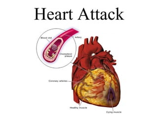
- 2. Myocardial infarction means “heart attack,” or coronary thrombosis . Myocardium is the name for the heart muscle. Infarction means the death of a muscle, tissue, or organ as a result of a blockage of the blood supply to it . Therefore a myocardial infarction is “the death of heart muscle tissue.
- 3. The heart needs oxygen to survive. The coronary arteries deliver oxygenated blood to the heart muscle. When one or more of the coronary arteries blocks, the oxygen supply to the myocardium stops, and the part of the heart supplied by that particular artery dies. This is a myocardial infarction (death of heart muscle tissue).
- 4. Thrombosis is the formation of a clot or thrombus inside an artery which occludes the flow of blood. Occlude – to cause to become closed off, to obstruct, or to block. So an occlusion is simply an obstruction or “ blockage ”. Coronary Thrombosis Thrombus Occluded (blocked) blood vessel
- 5. A thrombus , or blood clot , is the final product of the blood coagulation step in hemostasis . A thrombus is physiologic in cases of injury (begins the normal healing process), but pathologic in case of thrombosis . Occluded (blocked) blood vessel Thrombus Thrombus Occluded (blocked) blood vessel
- 6. Formation of a thrombus will cause ischemia , meaning a decrease in the blood supply to a bodily organ, tissue, or part caused by constriction or occlusion of the blood vessels . Ischemia causes hypoxia , insufficient levels of oxygen in blood or tissue . Tissues starved of oxygen will suffer necrosis , the death of cells or tissues through injury or disease . Thus, as a result of a coronary thrombosis , ischemia will lead to hypoxia , which will cause necrosis of heart tissue, which is defined as an infarct . Occluded (blocked) blood vessel Thrombus
- 7. An embolism occurs when an object, the embolus , migrates through the vascular system from one part of the body and causes an occlusion of a blood vessel in another part of the body. Embolism Formation of an Embolism embolus Occluded (blocked) blood vessel
- 8. Blood clots form the most common embolic material by far, but other possible (but not all possible) embolic materials include fat globules (a fat embolism ) , air bubbles (an air embolism ) , and septic emboli which contain pus and bacteria .
- 9. Remember, a blood clot embolus can be contrasted with a thrombus in one very important way. A thrombus is the formation of a stationary clot within a blood vessel so a thrombus is a blood clot that does not move. An embolus forms in one place and travels to another. Both a thrombus and a lodged embolus result in occlusion of a blood vessel. Embolus Thrombus
- 10. Hypoxia – insufficient levels of oxygen in blood or tissue. Ischemia – a medical term for hypoxia where there is a decrease in the blood supply to a bodily organ, tissue, or part caused by constriction or occlusion of the blood vessels. Necrosis – the death of cells or tissues through injury or disease. Occlude – to cause to become closed off, to obstruct, or to block. So an occlusion is simply an obstruction or “ blockage ”. Infarct – an area of tissue that undergoes necrosis as a result of ischemia .
- 11. Plaque
- 12. Plaque is defined as a deposit of fatty material on the inner lining of an arterial wall . Its presence represents the primary characteristic of atherosclerosis .
- 13. Atherosclerosis is the most common form of arteriosclerosis . Although the two terms are often used interchangeably, atherosclerosis refers to arteriosclerosis , or hardening of the arteries , caused by accumulation of fatty deposits, or plaques and other substances.
- 14. Plaque can build up in two different ways: Plaque can build up in the lumen of the artery, thus narrowing the blood space and restricting flow. Such plaques are often calcified, or hardened by the body. This causes increased rigidity and a thickening of the arterial wall in response to an increase in blood pressure, which furthers the arteriosclerosis .
- 15. The lumen is the interior of a vessel within the body, such as the small central space in an artery or any of their relating vessels, which blood flows through – the normal blood space . Normal Artery Normal Artery Lumen Lumen
- 16. As these plaques narrow the blood space, physical exertion may lead to ischemia . This causes a condition called angina pectoris , or chest pains with accompanying shortness of breath. Also, the narrowed artery is more susceptible to being blocked by a small embolus or thrombus due to a small plaque rupture leading to myocardial infarction or stroke.
- 17. Constricting chest pain, often radiating to the left shoulder and down the left arm, caused by an insufficient supply of blood to the heart. Coronary artery disease is a common cause of angina pectoris. Angina pectoris
- 18. As these plaques grow, they tend to occlude the artery producing a “visible” area of stenosis within the arterial lumen . Therefore, they are typically detected by cardiac stress tests or an angiogram . Stenosis - A constriction or narrowing of a duct or passage; a stricture. Cardiac stress test Angiogram Area of Stenosis Lumen Plaque Area of Stenosis
- 19. Angiography – Examination of the blood vessels using x-rays following the injection of a radiopaque substance.
- 20. Angiogram – An x-ray of one or more blood vessels produced by angiography and used in diagnosing pathology in the cardiovascular system, such as arteriosclerosis . Note the two separate areas of stenosis visible on the angiogram . These areas can be surgically “repaired” to prevent ischemia and possible major occlusion which could result in myocardial infarction .
- 21. The two most common surgical procedures used today are balloon angioplasty , often accompanied by stent placement, or coronary artery bypass . balloon angioplasty coronary artery bypass
- 22. First, angiography provides an angiogram which allows the area of stenosis to be located. Angioplasty – a surgical technique for restoring normal blood flow through an artery narrowed or blocked by atherosclerosis, either by inserting a balloon into the narrowed section and inflating it or by using a laser beam.
- 24. Before and after angioplasty
- 25. Coronary Artery Bypass Graft – open-heart surgery in which the rib cage is opened and a section of a blood vessel is grafted from the aorta to the coronary artery to bypass the blocked section of the coronary artery and improve the blood supply to the heart open-heart surgery - heart surgery in which the rib cage is spread open, the heart is stopped and blood is detoured through a heart-lung machine while a heart valve or coronary artery is surgically repaired
- 26. Coronary Artery Bypass Graft Options
- 28. Plaques can also develop within the muscular wall of an artery, where it is called an atheromatous plaque . Atheromatous plaque is defined as a buildup of fatty deposits, an atheroma , within the wall of an artery. Atheroma - A deposit or degenerative accumulation of lipid-containing plaques on the innermost layer of the wall of an artery.
- 29. Generally an atheroma becomes vulnerable if it grows more rapidly and has a thin cover separating it from the bloodstream inside the arterial lumen . Tearing of the cover is called plaque rupture . An insidiously dangerous form of plaque, called a vulnerable (unstable) plaque , is an atheromatous plaque which is particularly prone to producing sudden, major problems, such as a heart attack or stroke.
- 30. Because artery walls typically enlarge in response to enlarging vulnerable plaques, these plaques do not usually produce much stenosis of the artery lumen . Therefore, they are typically not detected by cardiac stress tests or an angiogram .
- 31. In many cases, a vulnerable plaque is one that has a thin fibrous cap and a large, soft lipid pool. These characteristics together with hemodynamic effects contribute to a high mechanical stress zone on the fibrous cap of the atheroma , making it prone to rupture.
- 34. Ventricular fibrillation (VF or V-fib) constitutes the most common electrical mechanism in cardiac arrest, and is responsible for 65 to 80% of occurrences. Another 20-30% are caused by severe bradyarrhythmias. A cardiac arrest , or circulatory arrest , is the abrupt cessation of normal circulation of the blood due to a sudden failure of the heart to contract effectively during systole .
- 36. Type III familial hyperlipoproteinemia Familial hyperlipoproteinemia marked by an abnormal tolerance of glucose and by an increase in serum concentrations of low-density lipoproteins, pre-low-density lipoproteins, cholesterol, and phospholipids as well as an endogenous increase in serum triglycerides induced by a high-carbohydrate diet. It is marked by eruptive xanthomas and atherosclerosis. Also called familial hypercholesterolemia with hyperlipemia , carbohydrate-induced hyperlipemia , familial broad-beta hyperlipoproteinemia , familial hyperbetalipoproteinemia and hyperprebetalipoproteinemia .
- 38. Blood Pressure The pressure (force) exerted by the blood on the inner walls of the arteries. Blood pressure varies with the strength of the heartbeat, the stroke volume ( volume of blood pumped with each systole – about 70 ml ), and the elasticity of the blood vessels. Arterial blood pressure is usually measured by means of a sphygmomanometer (“ blood pressure cuff ”)and reported in millimeters of mercury as a fraction, with the numerator equal to the blood pressure during systole and the denominator equal to the blood pressure during diastole .
- 39. Systole The period during the normal beating of the heart in which the chambers of the heart, especially the ventricles, contract to force blood into the aorta and pulmonary artery. Diastole The normal rhythmically occurring relaxation and dilatation of the heart chambers, especially the ventricles, during which they fill with blood. Therefore, Blood Pressure = systolic pressure diastolic pressur e
- 40. Adult blood pressure is considered normal at 120/80 (where the first number is the systolic pressure and the second is the diastolic pressure ). Hypertension Arterial disease in which chronic high blood pressure is the primary symptom. A person is considered hypertensive if one or both pressures are above the following: Systolic Pressure > 140 Diastolic Pressure > 90
- 41. Hypotension Abnormally or chronic low blood pressure . A person is considered hypotensive if one or both pressures are below the following: Systolic Pressure < 90 Diastolic Pressure < 50 Hypotension , like bradycardia, is not necessarily a “bad” thing if it is physiological (in other words “normal” for that person). It is only a problem if it is a pathological condition (in other words caused by disease).
- 42. Prehypertension A relatively new term used to refer to people who have blood pressures that constantly border on hypertensive . A person is considered pre-hypertensive if one or both pressures are chronically between the following: Systolic Pressure = 130 – 139 Diastolic Pressure = 85 – 89