SlideShare una empresa de Scribd logo
1 de 1
Descargar para leer sin conexión
-5
5
15
25
CD31 VECadherin vWF
RelativeGeneExpression
Endothelial Cell (EC) Markers
Human embryonic stem cells- a novel source for
in-vitro three dimensional vascularized oral mucosa.
Handral Harish K 1, Gopu Sriram4, Toh Wei Seong1,3 and Cao Tong1,2,3
1- Oral Sciences, Faculty of Dentistry, National University of Singapore, Singapore.
2- NUS Graduate School of Integrative Science and Engineering, National University of Singapore, Singapore.
3- Tissue Engineering Program, Life Science Institute, National University of Singapore, Singapore.
4- Experimental Dermatology, Institute of Medical Biology, A*Star, Singapore
Oral mucosa, a stratified epithelium which covers the inner lining of the mouth except
teeth part. Its main function is to act as a barrier and protecting deeper tissues from
external insults as well as preventing from the entry of microbial and other toxins into
the body. Oral mucosa has been studied since long years under both in-vivo (animal
models) and in-vitro (primary or immortalized cells) conditions. However, till date no
one has reported to establish a 3D vascularized oral mucosa from human embryonic
stem cells (hESCs). In current study, we developed a novel in-vitro 3D vascularized
oral mucosa from single source of hESCs by differentiating to keratinocytes,
endothelial cells, vascular smooth muscle cells, after which they were tri-cultured in a
PEG-Fibrin based scaffold under animal-component free medium conditions. 3D oral
mucosa carries great potential applications in high throughput screening of
pharmaceutical chemicals, dental cosmetics, drug discovery, etc.
INTRODUCTION
H1 hESC lines were purchased from WiCell Research Institute (Madison, WI) and
were propagated under feeder free conditions on Matrigel™ coated tissue culture
plates in complete mTeSR1™ culture medium. HESC cell lines were differentiated to
keratinocytes, endothelial cells and vascular smooth muscle cells by modifying
previously published protocols1,2 &3 . A PEG-Fibrin scaffold4 was considered as dermal
substitute in which hESC differentiated cells were tri-cultured. Cultured 3D
organotypic tissues were analysed by H & E staining and immuno-fluorescence
staining.
MATERIALS AND METHODS
Mesoderm
Ectoderm
A
Keratinocytes
Vascular smooth muscle cells
Endothelial cells
Blood vessel
formation
Addition of
Keratinocytes
Formation of
Stratified and
vascularized epithelia
B
Figure 1. A) Represents the hESC differentiation to ectodermal and mesodermal lineage
cells. B) Represents tri-culture of hESC differentiated cells to develop organotypic in-vitro
vascularized 3D stratified epithelia.
RESULTS-1. Differentiation
Day0 Day2 Day5Day4
Day0 Day3 Day9Day6
hESC differentiation
to Keratinocytes
hESCdifferentiation
to Vascular
progenitors
0
10
20
30
40
50
Cytokeratin-14 α6Integrin ΔNp63
RelativeGeneExpression
Keratinocyte Markers
α6Integrin Hgh/CD71Lw
2.Characterization:
2.1. mRNA level by q-PCR.
-10
-5
0
5
10
15
hESC-vSMCs hESC-EC
RelativeGeneExpression
Vascular Smooth Muscle (SMC) Markers
αSMA
SM22α
Calponin
PDGFβ
Sorted for α6H and CD 71L
Keratinocytes (α6H / CD 71L)
α6INTEGRINAPC
CD71 FITC
Vascular smooth muscle cells
Sorted for 1). CD34+&CD31+ (ECs)
2). CD34- CD31- PDGFb+ (vSMCs)
Endothelial cells
CD34-PE
CD31-APC
2.2. Flow-cytometry analysis.
ΔNP63K19
a b
2.3. Immuno-fluorescence staining.
VE-CadherinVWF α-SMA
Immuno-fluorescence staining showing the expression of K19 (a) & ΔNp63 (b) in hESC-Keratinocytes;
Von Willebrand Factor (VWF) (c) & VE-Cadherin (d) in hESC-endothelial cells; α-SMA (e) & Calponin (f) in hESC-
vascular smooth muscle cells.
Formation of network of blood vessels from GFP-
hESC-endothelial cells was confirmed by Matrigel
tube formation (g&h).
4X
Contraction of hESC-vascular smooth muscle cells was
confirmed by culturing in presence of muscarinic antagonist for
30 minutes. Arrows represents contracted cells (i & j).
k l
Epidermis
Dermis
Blood
vessels
m
(k) Z-stack images of 3D blood vessels formed by GFP-hESC-endothelial cells has been confirmed by confocal
microscopy. (l) 4x & (m) 20x images of H & E staining of 3D tissue; Immunofluorescence staining of 3D tissues
expressing K19 (n), fibronectin(o) and Collagen-type4 (p). Arrows represents presence of blood vessels.
DISCUSSION AND CONCLUSION
Testing of drugs is always as important as formulating them. Various existing in-vitro platforms used for
testing drugs, drug discovery have their own list of drawbacks such as, lack of reliable results, poor in
reflecting human in-vivo milieu, which gave opportunities to scientists to look forward for alternative in-vitro
models. Discovery of hESCs by James Thomson, has created the demand of the hESC-derived in-vitro
models. Three dimensional architecture of tissues are more promising and more reflective to human in-vivo
conditions. In current stem cell research area, we are the first to report differentiation of hESCs into various
lineages which can establish a 3D in-vitro vascularized platform from a single cell source. These models have
great industrial, pre-clinical as well as academic research value, especially in pharmaceuticals and drug
discovery.
* NOTE- Scale bar of all images 200µm.
1.Kidwai, F.K et al. 2013. Differentiation of human embryonic stem cells to clinically amenable
keratinocytes in an autogenic environment.
2.Tan, J.Y et al. 2013. Efficient differentiation of lateral plate and paraxial mesoderm subtypes from human
embryonic stem cells through GSKi-mediated differentiation.
3.Sriram, G. 2014. In-vitro vascularised tissue equivalents from human embryonic stem cell derived
endothelial and smooth muscle cells.
4.Natesan, S. et al. 2012. Engineering a bi-layered hydrogel to control ASC differentiation.
5..B.Calenic. et al. 2010. Characterization of oral keratinocyte stem cells and prospects of its differentiation
to oral epithelial equivalents.
REFERENCES
hESCs
3D Culture media
n l
c
c e
g
Calponin
Cytokeratin-19 Alpha 6 Integrin ΔNP63
3. Functional studies
C d e f
10Xh
o p
AfterBefore i j

Más contenido relacionado

La actualidad más candente

Organoid culture in cancer
Organoid culture in cancerOrganoid culture in cancer
Organoid culture in cancerSamieh Asadian
 
Gene & Tissue Culture: Presentation (Group 4)
Gene & Tissue Culture: Presentation (Group 4)Gene & Tissue Culture: Presentation (Group 4)
Gene & Tissue Culture: Presentation (Group 4)Su Shen Lim
 
William bioreactor 2014
William bioreactor 2014William bioreactor 2014
William bioreactor 2014William
 
Tissue Engineering Poster
Tissue Engineering PosterTissue Engineering Poster
Tissue Engineering PosterShasta Rizzi
 
Cimetta et al., 2013
Cimetta et al., 2013Cimetta et al., 2013
Cimetta et al., 2013Fran Flores
 
Characterization of embryoid bodies formed with different protocols 使用不同培養方式形...
Characterization of embryoid bodies formed with different protocols 使用不同培養方式形...Characterization of embryoid bodies formed with different protocols 使用不同培養方式形...
Characterization of embryoid bodies formed with different protocols 使用不同培養方式形...Honey Cheng
 
Characterization_of_embryoid_bodies_formed_with_different_protocols 2011
Characterization_of_embryoid_bodies_formed_with_different_protocols 2011Characterization_of_embryoid_bodies_formed_with_different_protocols 2011
Characterization_of_embryoid_bodies_formed_with_different_protocols 2011Cheng Han-Ni
 
The issues with prosthetic biomaterials
The issues with prosthetic biomaterialsThe issues with prosthetic biomaterials
The issues with prosthetic biomaterialsMunira Shahbuddin
 
Ch 19 _perinatal_stem_cells_isolated_from_complete_umbilical_cord_tissue_for_...
Ch 19 _perinatal_stem_cells_isolated_from_complete_umbilical_cord_tissue_for_...Ch 19 _perinatal_stem_cells_isolated_from_complete_umbilical_cord_tissue_for_...
Ch 19 _perinatal_stem_cells_isolated_from_complete_umbilical_cord_tissue_for_...ComprehensiveBiologi
 
Mammalian MSC from Selected Species: Features and Applications Christiane Ude...
Mammalian MSC from Selected Species: Features and Applications Christiane Ude...Mammalian MSC from Selected Species: Features and Applications Christiane Ude...
Mammalian MSC from Selected Species: Features and Applications Christiane Ude...Lipogems Equine & Lipogems Canine
 
GROUP 7- KNOCK IN MOUSE MODEL USED FOR DISEASE MODELLING
GROUP 7- KNOCK IN MOUSE MODEL USED FOR DISEASE MODELLINGGROUP 7- KNOCK IN MOUSE MODEL USED FOR DISEASE MODELLING
GROUP 7- KNOCK IN MOUSE MODEL USED FOR DISEASE MODELLINGVinitha Govindan Rajan
 
Excellent extracellular matrix_inspired_biomaterials
Excellent extracellular matrix_inspired_biomaterialsExcellent extracellular matrix_inspired_biomaterials
Excellent extracellular matrix_inspired_biomaterialsComprehensiveBiologi
 

La actualidad más candente (20)

International Journal of Stem Cells & Research
International Journal of Stem Cells & ResearchInternational Journal of Stem Cells & Research
International Journal of Stem Cells & Research
 
Organoid culture in cancer
Organoid culture in cancerOrganoid culture in cancer
Organoid culture in cancer
 
Gene & Tissue Culture: Presentation (Group 4)
Gene & Tissue Culture: Presentation (Group 4)Gene & Tissue Culture: Presentation (Group 4)
Gene & Tissue Culture: Presentation (Group 4)
 
A03420104
A03420104A03420104
A03420104
 
William bioreactor 2014
William bioreactor 2014William bioreactor 2014
William bioreactor 2014
 
Tissue Engineering Poster
Tissue Engineering PosterTissue Engineering Poster
Tissue Engineering Poster
 
Cimetta et al., 2013
Cimetta et al., 2013Cimetta et al., 2013
Cimetta et al., 2013
 
Whartons jelly review
Whartons jelly reviewWhartons jelly review
Whartons jelly review
 
Characterization of embryoid bodies formed with different protocols 使用不同培養方式形...
Characterization of embryoid bodies formed with different protocols 使用不同培養方式形...Characterization of embryoid bodies formed with different protocols 使用不同培養方式形...
Characterization of embryoid bodies formed with different protocols 使用不同培養方式形...
 
Characterization_of_embryoid_bodies_formed_with_different_protocols 2011
Characterization_of_embryoid_bodies_formed_with_different_protocols 2011Characterization_of_embryoid_bodies_formed_with_different_protocols 2011
Characterization_of_embryoid_bodies_formed_with_different_protocols 2011
 
The issues with prosthetic biomaterials
The issues with prosthetic biomaterialsThe issues with prosthetic biomaterials
The issues with prosthetic biomaterials
 
CH1976
CH1976CH1976
CH1976
 
Ch 19 _perinatal_stem_cells_isolated_from_complete_umbilical_cord_tissue_for_...
Ch 19 _perinatal_stem_cells_isolated_from_complete_umbilical_cord_tissue_for_...Ch 19 _perinatal_stem_cells_isolated_from_complete_umbilical_cord_tissue_for_...
Ch 19 _perinatal_stem_cells_isolated_from_complete_umbilical_cord_tissue_for_...
 
Mammalian MSC from Selected Species: Features and Applications Christiane Ude...
Mammalian MSC from Selected Species: Features and Applications Christiane Ude...Mammalian MSC from Selected Species: Features and Applications Christiane Ude...
Mammalian MSC from Selected Species: Features and Applications Christiane Ude...
 
Regenerative medicine
Regenerative medicineRegenerative medicine
Regenerative medicine
 
GROUP 7- KNOCK IN MOUSE MODEL USED FOR DISEASE MODELLING
GROUP 7- KNOCK IN MOUSE MODEL USED FOR DISEASE MODELLINGGROUP 7- KNOCK IN MOUSE MODEL USED FOR DISEASE MODELLING
GROUP 7- KNOCK IN MOUSE MODEL USED FOR DISEASE MODELLING
 
Reza Khorramirouz
Reza KhorramirouzReza Khorramirouz
Reza Khorramirouz
 
Excellent extracellular matrix_inspired_biomaterials
Excellent extracellular matrix_inspired_biomaterialsExcellent extracellular matrix_inspired_biomaterials
Excellent extracellular matrix_inspired_biomaterials
 
International Journal of Ophthalmology & Vision Research
International Journal of Ophthalmology & Vision ResearchInternational Journal of Ophthalmology & Vision Research
International Journal of Ophthalmology & Vision Research
 
Tissue Engineering slides
Tissue Engineering slidesTissue Engineering slides
Tissue Engineering slides
 

Similar a Human embryonic stem cells-a novel source for in vitro three dimensional oral mucosa

Deriving Mesenchymal Stem Cells from Human Amniotic Fluid – Potential for an ...
Deriving Mesenchymal Stem Cells from Human Amniotic Fluid – Potential for an ...Deriving Mesenchymal Stem Cells from Human Amniotic Fluid – Potential for an ...
Deriving Mesenchymal Stem Cells from Human Amniotic Fluid – Potential for an ...cordbloodsymposium
 
Menstrual Blood stem cells
Menstrual Blood stem cellsMenstrual Blood stem cells
Menstrual Blood stem cellsjayaganesh13
 
Cuckoo Search Optimization of Blebs in Human Embryonic Stem Cells
Cuckoo Search Optimization of Blebs in Human Embryonic Stem CellsCuckoo Search Optimization of Blebs in Human Embryonic Stem Cells
Cuckoo Search Optimization of Blebs in Human Embryonic Stem CellsIJMERJOURNAL
 
Umbilical cord wj_greatest___msc.vangsness2015_(1)
Umbilical cord wj_greatest___msc.vangsness2015_(1)Umbilical cord wj_greatest___msc.vangsness2015_(1)
Umbilical cord wj_greatest___msc.vangsness2015_(1)ComprehensiveBiologi
 
Trabalho 2012 teo2010 regenerative therapy
Trabalho 2012 teo2010 regenerative therapyTrabalho 2012 teo2010 regenerative therapy
Trabalho 2012 teo2010 regenerative therapyFelemon Porto
 
Martin Pera stem cells and the future of medicine
Martin Pera stem cells and the future of medicineMartin Pera stem cells and the future of medicine
Martin Pera stem cells and the future of medicineigorod
 
Fundamentals of Stem Cells
Fundamentals of Stem CellsFundamentals of Stem Cells
Fundamentals of Stem CellsDitoAnurogo1
 
Stem-cell therapy in medicine–how far we came and what we can expect?
Stem-cell therapy in medicine–how far we came and what we can expect?Stem-cell therapy in medicine–how far we came and what we can expect?
Stem-cell therapy in medicine–how far we came and what we can expect?Apollo Hospitals
 
Advance Access publication 29 January, 2004Establishment o.docx
Advance Access publication 29 January, 2004Establishment o.docxAdvance Access publication 29 January, 2004Establishment o.docx
Advance Access publication 29 January, 2004Establishment o.docxgalerussel59292
 
Organoids: A Foremost Hope For The Future
Organoids: A Foremost Hope For The FutureOrganoids: A Foremost Hope For The Future
Organoids: A Foremost Hope For The Futureshahin_ahmadian
 
Neuromics base presentation 2020
Neuromics base presentation 2020Neuromics base presentation 2020
Neuromics base presentation 2020Pete Shuster
 

Similar a Human embryonic stem cells-a novel source for in vitro three dimensional oral mucosa (20)

Virador fast 102913
Virador fast 102913Virador fast 102913
Virador fast 102913
 
Deriving Mesenchymal Stem Cells from Human Amniotic Fluid – Potential for an ...
Deriving Mesenchymal Stem Cells from Human Amniotic Fluid – Potential for an ...Deriving Mesenchymal Stem Cells from Human Amniotic Fluid – Potential for an ...
Deriving Mesenchymal Stem Cells from Human Amniotic Fluid – Potential for an ...
 
Menstrual Blood stem cells
Menstrual Blood stem cellsMenstrual Blood stem cells
Menstrual Blood stem cells
 
Cuckoo Search Optimization of Blebs in Human Embryonic Stem Cells
Cuckoo Search Optimization of Blebs in Human Embryonic Stem CellsCuckoo Search Optimization of Blebs in Human Embryonic Stem Cells
Cuckoo Search Optimization of Blebs in Human Embryonic Stem Cells
 
International Journal of Stem Cells & Research
International Journal of Stem Cells & ResearchInternational Journal of Stem Cells & Research
International Journal of Stem Cells & Research
 
Stem cells
Stem cellsStem cells
Stem cells
 
Umbilical cord wj_greatest___msc.vangsness2015_(1)
Umbilical cord wj_greatest___msc.vangsness2015_(1)Umbilical cord wj_greatest___msc.vangsness2015_(1)
Umbilical cord wj_greatest___msc.vangsness2015_(1)
 
8
88
8
 
Trabalho 2012 teo2010 regenerative therapy
Trabalho 2012 teo2010 regenerative therapyTrabalho 2012 teo2010 regenerative therapy
Trabalho 2012 teo2010 regenerative therapy
 
Name .docx
Name                                                             .docxName                                                             .docx
Name .docx
 
Martin Pera stem cells and the future of medicine
Martin Pera stem cells and the future of medicineMartin Pera stem cells and the future of medicine
Martin Pera stem cells and the future of medicine
 
Human Organoids
Human OrganoidsHuman Organoids
Human Organoids
 
Fundamentals of Stem Cells
Fundamentals of Stem CellsFundamentals of Stem Cells
Fundamentals of Stem Cells
 
Dental stem cells
Dental stem cellsDental stem cells
Dental stem cells
 
Stem-cell therapy in medicine–how far we came and what we can expect?
Stem-cell therapy in medicine–how far we came and what we can expect?Stem-cell therapy in medicine–how far we came and what we can expect?
Stem-cell therapy in medicine–how far we came and what we can expect?
 
Advance Access publication 29 January, 2004Establishment o.docx
Advance Access publication 29 January, 2004Establishment o.docxAdvance Access publication 29 January, 2004Establishment o.docx
Advance Access publication 29 January, 2004Establishment o.docx
 
Organoids: A Foremost Hope For The Future
Organoids: A Foremost Hope For The FutureOrganoids: A Foremost Hope For The Future
Organoids: A Foremost Hope For The Future
 
Neuromics base presentation 2020
Neuromics base presentation 2020Neuromics base presentation 2020
Neuromics base presentation 2020
 
Cord tissuegoldstandardarticle
Cord tissuegoldstandardarticleCord tissuegoldstandardarticle
Cord tissuegoldstandardarticle
 
Poster SVF
Poster SVFPoster SVF
Poster SVF
 

Human embryonic stem cells-a novel source for in vitro three dimensional oral mucosa

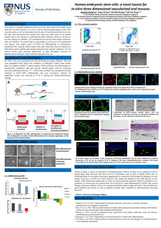
  • 1. -5 5 15 25 CD31 VECadherin vWF RelativeGeneExpression Endothelial Cell (EC) Markers Human embryonic stem cells- a novel source for in-vitro three dimensional vascularized oral mucosa. Handral Harish K 1, Gopu Sriram4, Toh Wei Seong1,3 and Cao Tong1,2,3 1- Oral Sciences, Faculty of Dentistry, National University of Singapore, Singapore. 2- NUS Graduate School of Integrative Science and Engineering, National University of Singapore, Singapore. 3- Tissue Engineering Program, Life Science Institute, National University of Singapore, Singapore. 4- Experimental Dermatology, Institute of Medical Biology, A*Star, Singapore Oral mucosa, a stratified epithelium which covers the inner lining of the mouth except teeth part. Its main function is to act as a barrier and protecting deeper tissues from external insults as well as preventing from the entry of microbial and other toxins into the body. Oral mucosa has been studied since long years under both in-vivo (animal models) and in-vitro (primary or immortalized cells) conditions. However, till date no one has reported to establish a 3D vascularized oral mucosa from human embryonic stem cells (hESCs). In current study, we developed a novel in-vitro 3D vascularized oral mucosa from single source of hESCs by differentiating to keratinocytes, endothelial cells, vascular smooth muscle cells, after which they were tri-cultured in a PEG-Fibrin based scaffold under animal-component free medium conditions. 3D oral mucosa carries great potential applications in high throughput screening of pharmaceutical chemicals, dental cosmetics, drug discovery, etc. INTRODUCTION H1 hESC lines were purchased from WiCell Research Institute (Madison, WI) and were propagated under feeder free conditions on Matrigel™ coated tissue culture plates in complete mTeSR1™ culture medium. HESC cell lines were differentiated to keratinocytes, endothelial cells and vascular smooth muscle cells by modifying previously published protocols1,2 &3 . A PEG-Fibrin scaffold4 was considered as dermal substitute in which hESC differentiated cells were tri-cultured. Cultured 3D organotypic tissues were analysed by H & E staining and immuno-fluorescence staining. MATERIALS AND METHODS Mesoderm Ectoderm A Keratinocytes Vascular smooth muscle cells Endothelial cells Blood vessel formation Addition of Keratinocytes Formation of Stratified and vascularized epithelia B Figure 1. A) Represents the hESC differentiation to ectodermal and mesodermal lineage cells. B) Represents tri-culture of hESC differentiated cells to develop organotypic in-vitro vascularized 3D stratified epithelia. RESULTS-1. Differentiation Day0 Day2 Day5Day4 Day0 Day3 Day9Day6 hESC differentiation to Keratinocytes hESCdifferentiation to Vascular progenitors 0 10 20 30 40 50 Cytokeratin-14 α6Integrin ΔNp63 RelativeGeneExpression Keratinocyte Markers α6Integrin Hgh/CD71Lw 2.Characterization: 2.1. mRNA level by q-PCR. -10 -5 0 5 10 15 hESC-vSMCs hESC-EC RelativeGeneExpression Vascular Smooth Muscle (SMC) Markers αSMA SM22α Calponin PDGFβ Sorted for α6H and CD 71L Keratinocytes (α6H / CD 71L) α6INTEGRINAPC CD71 FITC Vascular smooth muscle cells Sorted for 1). CD34+&CD31+ (ECs) 2). CD34- CD31- PDGFb+ (vSMCs) Endothelial cells CD34-PE CD31-APC 2.2. Flow-cytometry analysis. ΔNP63K19 a b 2.3. Immuno-fluorescence staining. VE-CadherinVWF α-SMA Immuno-fluorescence staining showing the expression of K19 (a) & ΔNp63 (b) in hESC-Keratinocytes; Von Willebrand Factor (VWF) (c) & VE-Cadherin (d) in hESC-endothelial cells; α-SMA (e) & Calponin (f) in hESC- vascular smooth muscle cells. Formation of network of blood vessels from GFP- hESC-endothelial cells was confirmed by Matrigel tube formation (g&h). 4X Contraction of hESC-vascular smooth muscle cells was confirmed by culturing in presence of muscarinic antagonist for 30 minutes. Arrows represents contracted cells (i & j). k l Epidermis Dermis Blood vessels m (k) Z-stack images of 3D blood vessels formed by GFP-hESC-endothelial cells has been confirmed by confocal microscopy. (l) 4x & (m) 20x images of H & E staining of 3D tissue; Immunofluorescence staining of 3D tissues expressing K19 (n), fibronectin(o) and Collagen-type4 (p). Arrows represents presence of blood vessels. DISCUSSION AND CONCLUSION Testing of drugs is always as important as formulating them. Various existing in-vitro platforms used for testing drugs, drug discovery have their own list of drawbacks such as, lack of reliable results, poor in reflecting human in-vivo milieu, which gave opportunities to scientists to look forward for alternative in-vitro models. Discovery of hESCs by James Thomson, has created the demand of the hESC-derived in-vitro models. Three dimensional architecture of tissues are more promising and more reflective to human in-vivo conditions. In current stem cell research area, we are the first to report differentiation of hESCs into various lineages which can establish a 3D in-vitro vascularized platform from a single cell source. These models have great industrial, pre-clinical as well as academic research value, especially in pharmaceuticals and drug discovery. * NOTE- Scale bar of all images 200µm. 1.Kidwai, F.K et al. 2013. Differentiation of human embryonic stem cells to clinically amenable keratinocytes in an autogenic environment. 2.Tan, J.Y et al. 2013. Efficient differentiation of lateral plate and paraxial mesoderm subtypes from human embryonic stem cells through GSKi-mediated differentiation. 3.Sriram, G. 2014. In-vitro vascularised tissue equivalents from human embryonic stem cell derived endothelial and smooth muscle cells. 4.Natesan, S. et al. 2012. Engineering a bi-layered hydrogel to control ASC differentiation. 5..B.Calenic. et al. 2010. Characterization of oral keratinocyte stem cells and prospects of its differentiation to oral epithelial equivalents. REFERENCES hESCs 3D Culture media n l c c e g Calponin Cytokeratin-19 Alpha 6 Integrin ΔNP63 3. Functional studies C d e f 10Xh o p AfterBefore i j