1. Intensive Tuberculosis screening in HIV infected persons
not yet on Antiretroviral Treatment in Macha,
Rural Zambia
J.F.M. Sikalima1
, J.H. van Dijk2,3
, G.M. Hagoort2
, L.M. Chikobolo1
, M. Musonda1
, J.L. Nouwen2
1
Macha Research Trust, Clinical Research Department, Choma, Zambia; 2
Erasmus University Rotterdam, Internal Medicine - Infectious Diseases, Rotterdam, The Netherlands;
3
Macha Research Trust / Macha Hospital, Clinical Research Department, Choma, Zambia
.
Erasmus MC
University Medical Center Rotterdam
Medical Microbiology & Infectious Diseases
‘s Gravendijkwal 230
3015 CE Rotterdam
The Netherlands
Phone +31 10 463 3511
Fax +31 10 463 3875
E-mail j.l.nouwen@erasmusmc.nl
Introduction
Tuberculosis (TB) is the most common cause of morbidity and mortality in individuals
with HIV-1 infection in sub-Saharan Africa. World Health Organization (WHO) estimates
that only 50% of TB cases are detected, creating a need to intensify active case finding
via HIV patient screening using more sensitive laboratory diagnostics.
In this study, we aimed to determine the additional value of screening Antiretroviral
Treatment (ART)-naïve HIV patients presenting with a cough, comparing Fluorescent
Light Emitting Diode (LED) microscopy (FM) with the traditional Ziehl-Neelsen Staining
light microscopy (ZN), using TB culture as the Gold Standard.
Methods
All new HIV-infected pre-ART adult patients
seeking care at Macha Hospital in rural southern
Zambia between April 2010 and March 2012 were
screened.
Those presenting with a cough, were invited to
enroll in the study and asked to produce 3 sputum
samples.
Information on TB history and exposure was
collected from medical records.
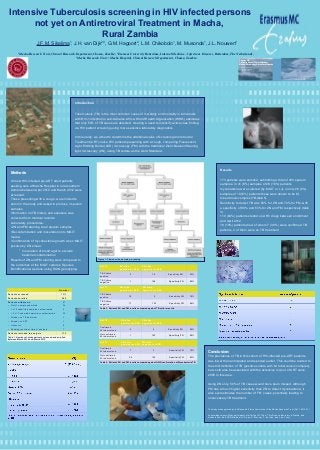
Laboratory procedures:
•ZN and FM staining to all sputum samples
•Decontamination and inoculation onto MGIT
media.
•Confirmation of mycobacterial growth when MGIT
positive by ZN smear.
• Inoculation of blood agar to exclude
bacterial contamination
Results of ZN and FM staining were compared to
the outcomes of the MGIT cultures. Species
identification was done using HAIN genotyping.
Results
173 patients were enrolled, submitting a total of 493 sputum
samples. In 41 (8%) samples of 28 (16%) patients
mycobacteria were cultured (by MGIT or LJ); in only 28 (6%)
samples of 10 (6%) patients these were shown to be M.
tuberculosis complex (TB, table 5).
Sensitivity to detect TB was 50% for ZN and 70% for FM, with
a specificity of 99% and 83% for ZN and FM respectively (table
3).
110 (64%) patients started on ARV drugs between enrollment
and April 2012.
18 (10%) patients died, of whom 7 (39%) were confirmed TB
patients, 3 of them were on TB treatment.
Conclusion:
The prevalence of TB in this cohort of HIV-infected pre-ART patients
was lower than anticipated and reported earlier. This could be related to
the strict definition of TB (positive culture with M. tuberculosis complex),
but could also be associated with the extensive roll-out of ART since
2005 in this area.
Using ZN only, 50% of TB cases would have been missed. Although
FM has a much higher sensitivity than ZN to detect mycobacteria, it
also overestimates the number of ‘TB’ cases, potentially leading to
unnecessary TB treatment.
The study was approved by the Research Ethics Committee of the Macha Research Trust (Ref: 1.2010.01).
Acknowledgements: Study participants, staff at the HIV Clinic, TB office and laboratory in Macha, and
students from the EUR Rotterdam (V. Kraak, G. Brouwer, F. de Vries, and L. de Vries).
N=173 ZN slide
positive for AFB
ZN slide
negative for AFB
TB Culture
positive 6 18 Sensitivity ZN 33%
TB Culture
negative 1 148 Specificity ZN 99%
N=173 FM slide
positive for AFB
FM slide
negative for AFB
TB Culture
positive 19 5 Sensitivity FM 79%
TB Culture
negative 17 132 Specificity FM 89%
Table 3. Patient’s ZN and FM results compared against TB culture results
N=173 ZN slide
positive for AFB
ZN slide
negative for AFB
Confirmed
M. tuberculosis 5 5 Sensitivity ZN 50%
Not confirmed
M. tuberculosis 1 162 Specificity ZN 99%
N=173 FM slide
positive for AFB
FM slide
negative for AFB
Confirmed
M. tuberculosis 7 3 Sensitivity FM 70%
Not confirmed
M. tuberculosis 28 135 Specificity FM 83%
Table 4. Patient’s ZN and FM results compared against HAIN confirmation of Mycobacteria TB
Number
Patients screened 1012
Patients enrolled 240
Patients withdrawn 67
• Only 1 sample submitted 24
• 1 of 2 submitted samples contaminated 4
• > 2 of 3 submitted samples contaminated 22
• Already on TB treatment 1
• Already on ART 1
• Unknown 8
• Pending results at time of analysis 7
Patients included in analysis 173
Table 1. Number of study patients screened and enrolled
between March 2010 and March 2012
Figure 1. Flowchart on sample processing