SlideShare una empresa de Scribd logo
1 de 40
BACHELOR OF SECONDARY EDUCATION
CENTRAL BICOL STATE UNIVERSITY OF
          AGRICULTURE
INTRODUCTION
Electron Microscopy involves the study of different
 specimens by using an electron microscope.
  Electron microscopes are scientific instruments
 that use a beam of energetic electrons to examine
 objects on a very fine scale.
Through the use of an electron microscope we can see
 things that we would not normally be able to see with
 our naked eyes and has greater magnification than
 light microscope.
  This required 10,000x plus magnification which was
 not possible using current optical microscopes
TYPES OF ELECTRON
            MICROSCOPE
1. TRANSMISSION ELECTRON MICROSCOPE-
   form images using electrons that are transmitted
   through a specimen.
2. SCANNING ELECTRON MICROSCOPE-
   utilize electron that have bounced off the surface
   of the specimen.
The transmission electron microscope (TEM) was the first type
of Electron Microscope to be developed and is patterned exactly on
the light transmission microscope except that a focused beam of
electrons is used instead of light to "see through" the specimen. It
was developed by Max Knoll and Ernst Ruska in Germany in 1931.
TRANSMISSION ELECTRON
         MICROSCOPY (TEM)
 Transmission electron microscopy (TEM) is a
 microscopy technique whereby a beam of electrons is
 transmitted through an ultra thin specimen, interacting
 with the specimen as it passes through. An image is formed
 from the interaction of the electrons transmitted through
 the specimen; the image is magnified and focused onto an
 imaging device, such as a fluorescent screen, on a layer of
 photographic film, or to be detected by a sensor such as a
 CCD camera.
DIFFERENCES BETWEEN OM AND EM
    OPTICAL MICROSCOPE                   ELECTRON MICROSCOPE


1. The source of light.         1. The light source is replaced by a beam of
2. The specimen.                   very fast moving electrons.
3. The lenses that makes the    2. The specimen usually has to be specially
   specimen seem bigger.           prepared and held inside a vacuum
4. The magnified image of the      chamber from which the air has been
   specimen that you see.          pumped out (because electrons do not
                                   travel very far in air).
                                3. The lenses are replaced by a series of coil-
                                   shaped electromagnets through which
                                   the electron beam travels.
                                4. The image is formed as a photograph
                                   (called an electron micrograph) or as an
                                   image on a TV screen.
MICROSCOPE   RESOLUTION MAGNIFICATION
OPTICAL      200 nm     1000x

TEM          0.2 nm     500000x
DIAGRAM TO REPRESENT TEM’S WORKING
                                  Virtual Source


                                                   First Condenser Lens


                                                   Second Condenser Lens
   Condenser Aperture

                         Sample

                                                   Objective Lens
   Objective Aperture

Selected Area Aperture


                                                   First Intermediate Lens

                                                   Second Intermediate Lens

                                                   Projector Lens

Main Screen (Phosphor)
DIFFERENT COMPONENTS OF TEM
                               1.       HIGH TENSION CABLE


2.     ELECTRON EMITTER
                               3.   STEPPER MOTORS FOR
                               CENTERING THE ELECTRON BEAM


4.     CONDENSER               11.     VACUUM PUMP LEADS


5.     APERTURE CONTROLS

                                12.         GONIOMETER
 6.    SPECIMEN HOLDER
                                      13. VACUUM AND
 7.    OBJECTIVE LENS
                                      MAGNIFICATION CONTROL
 8.     PROJECTOR LENS
                                      14.     FOCUSING CONTROL
 9.     OPTICAL LENS


 10.   FLUORESCENT SCREEN
SOME TYPICAL TEM MODELS
TEM SAMPLE PREPARATION
 Cleaning the surface of the specimen
 The proper cleaning of the surface of the sample is
 important because the surface can contain a variety of
 unwanted deposits, such as dust, silt, and detritus,
 media components, or other contaminants.
 The best way to clean the surface of specimen from
 contaminants is to carefully rinse them three times for
 10 min in 0.1 M cacodylic acid buffer (pH 7.3) at room
 temperature.
TEM SAMPLE PREPARATION
 Primary fixation of the specimen
 Fixation can be achieved by perfusion and
 microinjection, immersions, or with vapours using
 various fixatives including aldehydes(glutaraldehyde),
 osmium tetroxide,, tannic acid, or thiocarbohydrazide.

 FIXATIVES- are chemicals that denature and
 precipitate cellular macromolecules.
Some common fixatives:
1. GLUTARALDEHYDE- is a 5-carbon with an aldehyde
   group at each end of the molecule. An aldehyde
   groups reacts with amino groups and cross link with
   the proteins into an insoluble network.
2. OSMIUM- heavy metal that reacts primarily with
   fatty acids leading to the preservation of cellular
   membranes.
TEM SAMPLE PREPARATION
 Rinsing of the specimen
 After the fixation step, samples must be rinsed in
 order to remove the excess fixative. To remove excess
 glutaraldehyde from the samples, the specimen should
 be subjected to a thorough but carefully conducted
 rinsing procedure. Specimens can be washed in 0.1 M
 cacodylic acid buffer (pH 7.3), starting with one time
 for 10 min, and then three times for 20 min at 4 oC.
TEM SAMPLE PREPARATION
 Secondary fixation of the specimen
 Specimen can be successfully stabilized for TEM
 investigation by post fixation with 1% osmium
 tetroxide prepared in 0.1 M cacodylic acid buffer (pH
 7.3) for 1.5 hrs at room temperature (immersion
 fixation).
TEM SAMPLE PREPARATION
 Dehydrating the specimen
 For TEM investigation, specimen can be dehydrated in
 a graded series of ethanol. More specifically, the
 following protocol is useful: Dehydration of specimen
 in 50% ethanol for 5 min, 70% ethanol for 10 min, 80%
 ethanol for 10 min, 90% ethanol for 15 min, and 99.9%
 ethanol (dried with a 4-mesh molecular sieve) twice
 for 20 min at room temperature. This process allows
 the water in the samples to be slowly exchanged
 through liquids with lower surface tensions.
TEM SAMPLE PREPARATION
 Infiltration of the specimen with a transitional
 solvent
 The reason why this step is required is that the ethanol is
 not miscible with the plastic embedding medium I found
 most suitable for TEM investigation of the specimen
 (mollicutes). The replacement of the dehydration solution
 by another intermediary solvent (i.e., propylene oxide) is
 thus necessary [1, 23, 26]. This process is essentially an
 alcohol substitution. The immersion of mollicutes in
 propylene oxide twice for 20 min at room temperature is
 sufficient before attempting to embed the specimens in a
 resin.
TEM SAMPLE PREPARATION
 Infiltration with resin and embedding the specimen
 Mollicutes can be embedded in a variety of different media depending on the use (e.g.,
 conventional TEM or immuno TEM). For conventional TEM of mollicutes, the epoxy
 resin Durcupan ACM is quite suitable. The following protocol can be used: Immersion of
 mollicutes in propylene-oxide/Durcupan-ACM (1:1; v/v) at room temperature overnight
 (use gloves and a fume hood, and leave the specimen container open for the propylene
 oxide to evaporate). The next day, the specimens should be immersed in a freshly
 prepared Durcupan ACM mixture (pure) and left for 2 hrs at room temperature. A
 second Durcupan ACM mixture (pure) is then prepared and used as the embedding
 medium (free of air bubbles!).ible with the plastic embedding medium I found most
 suitable for TEM investigation of mollicutes. The replacement of the dehydration
 solution by another intermediary solvent (i.e., propylene oxide) is thus necessary [1, 23,
 26]. This process is essentially an alcohol substitution. The immersion of mollicutes in
 propylene oxide twice for 20 min at room temperature is sufficient before attempting to
 embed the specimens in a resin. Polymerization of the epoxy mixture can be achieved by
 placing the specimens in a drying cabinet for 2 days at 40 oC and for an additional 2 days
 at 60 oC. Leaving the samples after heat polymerization for an additional 1-2 weeks at
 room temperature can improve the subsequent cutting experience as the resin blocks
 continue to harden during this time.
TEM SAMPLE PREPARATION
 Sectioning and staining of the specimen
 The procedure for cutting specimens into semithin
 and ultrathin slices (sections) is known as microtomy
 and ultramicrotomy, respectively. Semithin sections
 (0.5 μm to 2 μm) were typically stained with toluidine
 blue for 1 min on a hot plate (70 oC to 90 oC),
 examined by LM, and used for identifying the
 specimen within the resin block before proceeding
 with ultramicrotomy. Ultrathin sections (about 70 nm
 to 90 nm) were typically stained with uranyl acetate
 followed by lead citrate.
OTHER METHODS OF
SAMPLE PREPARATION
       (TEM)
Cryofixation and the use of Frozen specimen
 CRYOFIXATION-like chemical fixatives it stops
 metabolic processes and preserves biological
 structure.
 The method involves ultra-rapid cooling of small
 samples to liquid nitrogen temperature (-196 oC) or
 below, thus stopping all motion and metabolic activity,
 and preserving the internal structure by freezing all
 fluid phases solid.
 The ultimate objective is to freeze the specimen so
 rapidly (at 104 to 106 K per second) that ice crystals are
 unable to form, or are prevented from growing
 sufficiently large to cause damage to the specimen's
 ultrastructure.
NEGATIVE STAINING
 The main purpose of negative-staining is to surround
 or embed the biological object in a suitable electron
 dense material which provides high contrast and good
 preservation. This method is capable of providing
 information about structural details often finer than
 those visible in thin sections, replicas, or shadowed
 specimens. In addition to the possibility of obtaining a
 spectacular enhancement of contrast, negative-
 staining has the advantage of speed and simplicity.
 The technique has mainly been used to examine
 particulate (purified) specimens - e.g.. ribosomes,
 enzyme molecules, viruses, bacteriophages,
 microtubules, actin filaments, etc. at a resolution of
 1.5-2.5 nm. This technique generally allows the shape,
 size, and the surface structure of the object to be
 studied as well as provide information about subunit
 stoichiometries and symmetry in oligomeric
 complexes. Any surface of the specimen accessible to
 water can potentially be stained, and thus, that part of
 the specimen will be imaged at high contrast.
SHADOW CASTING
 The grid containing the specimen are placed in sealed
  chamber which is the evacuated by vacuum pump.
 The chamber contains a filament composed of a heavy
  metal together with carbon.
The shadow cast by the nanoparticles (NPs) on metal deposition is
visible in the high-magnification images.
FREEZE FRACTURE REPLICATION
AND FREEZE ETCHING
 Small pieces of tissue are placed on a small metal disk
  and rapidly frozen.
 The disk is then mounted on a cooled stage within a
  vacuum chamber and a frozen tissue block is struck by
  a knife edge.
 The resulting fracture plane spreads out from the point
  of contact, splitting the tissue into two pieces.
Freeze fracturing can separate the two phospholipid leaflets that form
every cellular membrane
(a) A preparation of cells or tissues is
quickly frozen in liquid nitrogen at −196
°C, which instantly immobilizes cell
components. (b) The block of frozen cells
is fractured with a sharp blow from a cold
knife. The fracture plane is irregular, often
between the leaflets of the plasma or an
organelle membrane. (c) Membrane
proteins and particles remain bound to one
leaflet or the other, as illustrated in the
expanded view of a fractured membrane.
 shadowing
    coating specimen with a thin film of a heavy metal
 freeze-etching
    freeze specimen then fracture along lines of greatest
     weakness (e.g., membranes)
GRID IS A SIEVE WOVEN FROM A THIN METAL WIRE,USUALLY
NICKEL OR COPPER GRIDS OF 3 mm DIAMETER ARE
COMMERCIALLY AVAILABLE WITH DIFFERENT MESH
SIZES(GENERALLY OF 100-200 µm SIZE)
Staphylococcus aureus platinum replica image shot on a TEM at
                   50,000X magnification
A TEM image of the polio virus.
CONCLUSION and
            RECOMMENDATION
 Sample preparation for TEM involves more and some
 different steps than those for SEM. Like in any multi-step
 preparation procedure, virtually every step can affect the
 quality of the final electron micrograph. A single mistake
 in one of these steps will affect all remaining steps, and
 thus the outcome of the entire study. It is therefore
 important that the investigator plans and executes every
 step in great detail. These procedures involve a significant
 time commitment and require patience and skills that
 come only with practice. It is important to mention that
 most of the chemicals used in EM are dangerous.
 Investigator must be aware of potential hazards such as
 fire, chemical, electrical, and physical associated with these
 items.
REFERENCES
 http://cms.mse.berkeley.edu/publications/MRS_Spring_20
    04_Xu.pdf
   http://www.sciencepub.org/nature/0403/03-0171-
    mahongbao-ns.pdf
   http://www.arl.army.mil/arlreports/2003/ARL-TR-3128.pdf
   http://stem.ornl.gov/papers/reviews/pdfs/NIST.pdf
   http://cimewww.epfl.ch/people/cayron/Fichiers/theseboo
    k-chap3.pdf
   http://em-outreach.ucsd.edu/web-course/Sec-II.A.3/Sec-
    II.A.3.html
   http://en.wikipedia.org/wiki/Cryofixation
BSEU Secondary Education Degree

Más contenido relacionado

La actualidad más candente

Transmission electron microscope
Transmission electron microscopeTransmission electron microscope
Transmission electron microscopeSenthil Arasan
 
Atomic force microscopy
Atomic force microscopyAtomic force microscopy
Atomic force microscopySonu Bishnoi
 
Scanning Electron Microscope (SEM)
Scanning Electron Microscope (SEM)Scanning Electron Microscope (SEM)
Scanning Electron Microscope (SEM)Amna Jalil
 
Electron microscope ppt
Electron microscope pptElectron microscope ppt
Electron microscope pptNithyaNandapal
 
Scanning electron microscope
Scanning electron microscopeScanning electron microscope
Scanning electron microscopeAkhtar Kamal
 
SCANNING PROBE MICROSCOPY .
SCANNING PROBE MICROSCOPY .SCANNING PROBE MICROSCOPY .
SCANNING PROBE MICROSCOPY .sana shaikh
 
Principle of transmission electron microscope.
Principle of transmission electron microscope.Principle of transmission electron microscope.
Principle of transmission electron microscope.naren
 
Difference between SEM and TEM
Difference between SEM and TEMDifference between SEM and TEM
Difference between SEM and TEMVrushali Tambe
 
ELECTRON MICROSCOPY (TEM & SEM)
ELECTRON MICROSCOPY (TEM & SEM)ELECTRON MICROSCOPY (TEM & SEM)
ELECTRON MICROSCOPY (TEM & SEM)ADITYA ARYA
 
AFM (Atomic Force Microscopy)
AFM (Atomic Force Microscopy)AFM (Atomic Force Microscopy)
AFM (Atomic Force Microscopy)Chemist Sohaib
 
Scanning Electron Microscopy (SEM) lecture
Scanning Electron Microscopy (SEM) lectureScanning Electron Microscopy (SEM) lecture
Scanning Electron Microscopy (SEM) lectureSaurabh Bhargava
 
Atomic force microscopy
Atomic force microscopy Atomic force microscopy
Atomic force microscopy tabirsir
 
Electron microscope (TEM & SEM)
Electron microscope (TEM & SEM)Electron microscope (TEM & SEM)
Electron microscope (TEM & SEM)Subhankar Das
 
Scanning Tunneling Microscope
Scanning Tunneling MicroscopeScanning Tunneling Microscope
Scanning Tunneling MicroscopeHilal Aybike Can
 
Energy Dispersive Spectroscopy
Energy Dispersive SpectroscopyEnergy Dispersive Spectroscopy
Energy Dispersive SpectroscopySaad Shaukat
 

La actualidad más candente (20)

Transmission electron microscope
Transmission electron microscopeTransmission electron microscope
Transmission electron microscope
 
Atomic force microscopy
Atomic force microscopyAtomic force microscopy
Atomic force microscopy
 
Scanning Electron Microscope (SEM)
Scanning Electron Microscope (SEM)Scanning Electron Microscope (SEM)
Scanning Electron Microscope (SEM)
 
Electron microscope ppt
Electron microscope pptElectron microscope ppt
Electron microscope ppt
 
Scanning electron microscope
Scanning electron microscopeScanning electron microscope
Scanning electron microscope
 
SCANNING PROBE MICROSCOPY .
SCANNING PROBE MICROSCOPY .SCANNING PROBE MICROSCOPY .
SCANNING PROBE MICROSCOPY .
 
Raman spectroscopy
Raman spectroscopyRaman spectroscopy
Raman spectroscopy
 
Principle of transmission electron microscope.
Principle of transmission electron microscope.Principle of transmission electron microscope.
Principle of transmission electron microscope.
 
Difference between SEM and TEM
Difference between SEM and TEMDifference between SEM and TEM
Difference between SEM and TEM
 
Scanning electron microscopy (sem)
Scanning electron microscopy (sem)Scanning electron microscopy (sem)
Scanning electron microscopy (sem)
 
ELECTRON MICROSCOPY (TEM & SEM)
ELECTRON MICROSCOPY (TEM & SEM)ELECTRON MICROSCOPY (TEM & SEM)
ELECTRON MICROSCOPY (TEM & SEM)
 
AFM (Atomic Force Microscopy)
AFM (Atomic Force Microscopy)AFM (Atomic Force Microscopy)
AFM (Atomic Force Microscopy)
 
STM ppt
STM pptSTM ppt
STM ppt
 
Spectroscopy
Spectroscopy   Spectroscopy
Spectroscopy
 
Scanning Electron Microscopy (SEM) lecture
Scanning Electron Microscopy (SEM) lectureScanning Electron Microscopy (SEM) lecture
Scanning Electron Microscopy (SEM) lecture
 
Atomic force microscopy
Atomic force microscopy Atomic force microscopy
Atomic force microscopy
 
Electron microscope (TEM & SEM)
Electron microscope (TEM & SEM)Electron microscope (TEM & SEM)
Electron microscope (TEM & SEM)
 
Scanning Tunneling Microscope
Scanning Tunneling MicroscopeScanning Tunneling Microscope
Scanning Tunneling Microscope
 
Scanning electron microscopy
Scanning electron microscopyScanning electron microscopy
Scanning electron microscopy
 
Energy Dispersive Spectroscopy
Energy Dispersive SpectroscopyEnergy Dispersive Spectroscopy
Energy Dispersive Spectroscopy
 

Destacado

Transmission Electron Microscope
Transmission Electron MicroscopeTransmission Electron Microscope
Transmission Electron MicroscopeManoranjan Ghosh
 
Electron microscopy
Electron microscopyElectron microscopy
Electron microscopysuniu
 
Electron microscope
Electron microscopeElectron microscope
Electron microscopeshabeel pn
 
Electron microscope
Electron microscopeElectron microscope
Electron microscopejakkaas
 
Principle of transmission electron microscope.
Principle of transmission electron microscope.Principle of transmission electron microscope.
Principle of transmission electron microscope.naren
 
Electron microscopy
Electron microscopyElectron microscopy
Electron microscopyGanga Huvin
 
Atomic force microscopy(afm)
Atomic force microscopy(afm) Atomic force microscopy(afm)
Atomic force microscopy(afm) vishal gupta
 
atomic force microscopy AFM
atomic force microscopy AFMatomic force microscopy AFM
atomic force microscopy AFMAmare Worku
 
Principle & Applications of Transmission Electron Microscopy (TEM) & High Res...
Principle & Applications of Transmission Electron Microscopy (TEM) & High Res...Principle & Applications of Transmission Electron Microscopy (TEM) & High Res...
Principle & Applications of Transmission Electron Microscopy (TEM) & High Res...Gulfam Raza
 

Destacado (11)

Transmission Electron Microscope
Transmission Electron MicroscopeTransmission Electron Microscope
Transmission Electron Microscope
 
Sem and tem
Sem and temSem and tem
Sem and tem
 
Electron microscopy
Electron microscopyElectron microscopy
Electron microscopy
 
Electron microscope
Electron microscopeElectron microscope
Electron microscope
 
Electron microscope
Electron microscopeElectron microscope
Electron microscope
 
Principle of transmission electron microscope.
Principle of transmission electron microscope.Principle of transmission electron microscope.
Principle of transmission electron microscope.
 
Electron microscopy
Electron microscopyElectron microscopy
Electron microscopy
 
Atomic force microscopy(afm)
Atomic force microscopy(afm) Atomic force microscopy(afm)
Atomic force microscopy(afm)
 
SEM,TEM & AFM
SEM,TEM & AFMSEM,TEM & AFM
SEM,TEM & AFM
 
atomic force microscopy AFM
atomic force microscopy AFMatomic force microscopy AFM
atomic force microscopy AFM
 
Principle & Applications of Transmission Electron Microscopy (TEM) & High Res...
Principle & Applications of Transmission Electron Microscopy (TEM) & High Res...Principle & Applications of Transmission Electron Microscopy (TEM) & High Res...
Principle & Applications of Transmission Electron Microscopy (TEM) & High Res...
 

Similar a BSEU Secondary Education Degree

Electron microscope
Electron microscopeElectron microscope
Electron microscopeReena Rai
 
Basic overview of nanotechnology
Basic overview of nanotechnologyBasic overview of nanotechnology
Basic overview of nanotechnologySAMEER PAL
 
Light Microscope and Electron Microscope [Best one]
Light Microscope and Electron Microscope [Best one]Light Microscope and Electron Microscope [Best one]
Light Microscope and Electron Microscope [Best one]abdelrahman_asar
 
5. Microsocope ELECTRON MICROSCOPE (TEM & SEM ) - Basics
5. Microsocope   ELECTRON MICROSCOPE (TEM & SEM ) - Basics5. Microsocope   ELECTRON MICROSCOPE (TEM & SEM ) - Basics
5. Microsocope ELECTRON MICROSCOPE (TEM & SEM ) - BasicsNethravathi Siri
 
Tem or transmitted electron microscopy
Tem or transmitted electron microscopyTem or transmitted electron microscopy
Tem or transmitted electron microscopyvijayakumars66
 
chapter 2- Microscopy.pptx Microscopy related with medicine
chapter 2- Microscopy.pptx Microscopy related with medicinechapter 2- Microscopy.pptx Microscopy related with medicine
chapter 2- Microscopy.pptx Microscopy related with medicineHarikantSingh4
 
Presentationon optical and electron microscopy by deepak kumar
Presentationon optical and electron  microscopy by deepak kumar Presentationon optical and electron  microscopy by deepak kumar
Presentationon optical and electron microscopy by deepak kumar Drx Kumar
 
ELECTRON MICROSCOPE AND ITS APPLICATION.pptx
ELECTRON MICROSCOPE AND ITS APPLICATION.pptxELECTRON MICROSCOPE AND ITS APPLICATION.pptx
ELECTRON MICROSCOPE AND ITS APPLICATION.pptxPallaviKumari112
 
Recent advancement in microscopic techniques cryoem and super
Recent advancement in microscopic techniques  cryoem and superRecent advancement in microscopic techniques  cryoem and super
Recent advancement in microscopic techniques cryoem and superDanishNigar
 
Microscope anwesha.pptx
Microscope anwesha.pptxMicroscope anwesha.pptx
Microscope anwesha.pptxAnwesha Mandal
 
Transmission electron microscope
Transmission electron microscopeTransmission electron microscope
Transmission electron microscopesoniaangeline
 
Electron microscope - SEM and TEM
Electron microscope - SEM and TEMElectron microscope - SEM and TEM
Electron microscope - SEM and TEMgaurav raja
 
MICROSCOPY.pdf..........................
MICROSCOPY.pdf..........................MICROSCOPY.pdf..........................
MICROSCOPY.pdf..........................YolywinSanico
 
Fluorescence and electron Microscope.pptx
Fluorescence  and electron Microscope.pptxFluorescence  and electron Microscope.pptx
Fluorescence and electron Microscope.pptxsaraso888
 
2 Microscopy and staining.pptx
2 Microscopy and staining.pptx2 Microscopy and staining.pptx
2 Microscopy and staining.pptxNeelGamers
 
Microscopy
MicroscopyMicroscopy
Microscopytrixie19
 
2. Microscopy (Biochemistry)
2. Microscopy (Biochemistry)2. Microscopy (Biochemistry)
2. Microscopy (Biochemistry)Jay Khaniya
 

Similar a BSEU Secondary Education Degree (20)

Electron microscope
Electron microscopeElectron microscope
Electron microscope
 
Transmission electron microscope
Transmission electron microscope   Transmission electron microscope
Transmission electron microscope
 
Basic overview of nanotechnology
Basic overview of nanotechnologyBasic overview of nanotechnology
Basic overview of nanotechnology
 
Light Microscope and Electron Microscope [Best one]
Light Microscope and Electron Microscope [Best one]Light Microscope and Electron Microscope [Best one]
Light Microscope and Electron Microscope [Best one]
 
5. Microsocope ELECTRON MICROSCOPE (TEM & SEM ) - Basics
5. Microsocope   ELECTRON MICROSCOPE (TEM & SEM ) - Basics5. Microsocope   ELECTRON MICROSCOPE (TEM & SEM ) - Basics
5. Microsocope ELECTRON MICROSCOPE (TEM & SEM ) - Basics
 
microbiology
microbiologymicrobiology
microbiology
 
Tem or transmitted electron microscopy
Tem or transmitted electron microscopyTem or transmitted electron microscopy
Tem or transmitted electron microscopy
 
chapter 2- Microscopy.pptx Microscopy related with medicine
chapter 2- Microscopy.pptx Microscopy related with medicinechapter 2- Microscopy.pptx Microscopy related with medicine
chapter 2- Microscopy.pptx Microscopy related with medicine
 
Presentationon optical and electron microscopy by deepak kumar
Presentationon optical and electron  microscopy by deepak kumar Presentationon optical and electron  microscopy by deepak kumar
Presentationon optical and electron microscopy by deepak kumar
 
ELECTRON MICROSCOPE AND ITS APPLICATION.pptx
ELECTRON MICROSCOPE AND ITS APPLICATION.pptxELECTRON MICROSCOPE AND ITS APPLICATION.pptx
ELECTRON MICROSCOPE AND ITS APPLICATION.pptx
 
Recent advancement in microscopic techniques cryoem and super
Recent advancement in microscopic techniques  cryoem and superRecent advancement in microscopic techniques  cryoem and super
Recent advancement in microscopic techniques cryoem and super
 
Microscope anwesha.pptx
Microscope anwesha.pptxMicroscope anwesha.pptx
Microscope anwesha.pptx
 
Transmission electron microscope
Transmission electron microscopeTransmission electron microscope
Transmission electron microscope
 
Electron microscope - SEM and TEM
Electron microscope - SEM and TEMElectron microscope - SEM and TEM
Electron microscope - SEM and TEM
 
MICROSCOPY.pdf..........................
MICROSCOPY.pdf..........................MICROSCOPY.pdf..........................
MICROSCOPY.pdf..........................
 
Microscopy
MicroscopyMicroscopy
Microscopy
 
Fluorescence and electron Microscope.pptx
Fluorescence  and electron Microscope.pptxFluorescence  and electron Microscope.pptx
Fluorescence and electron Microscope.pptx
 
2 Microscopy and staining.pptx
2 Microscopy and staining.pptx2 Microscopy and staining.pptx
2 Microscopy and staining.pptx
 
Microscopy
MicroscopyMicroscopy
Microscopy
 
2. Microscopy (Biochemistry)
2. Microscopy (Biochemistry)2. Microscopy (Biochemistry)
2. Microscopy (Biochemistry)
 

Más de Jessa Ariño

Internet and telecommunications
Internet and telecommunicationsInternet and telecommunications
Internet and telecommunicationsJessa Ariño
 
Education systems of australia, china, japan
Education systems of australia, china, japanEducation systems of australia, china, japan
Education systems of australia, china, japanJessa Ariño
 
Qualitative analysis of group 4 cations
Qualitative analysis of group 4 cationsQualitative analysis of group 4 cations
Qualitative analysis of group 4 cationsJessa Ariño
 
It enters a new learning environment
It enters a new learning environmentIt enters a new learning environment
It enters a new learning environmentJessa Ariño
 
Namingandclassification
NamingandclassificationNamingandclassification
NamingandclassificationJessa Ariño
 

Más de Jessa Ariño (6)

Internet and telecommunications
Internet and telecommunicationsInternet and telecommunications
Internet and telecommunications
 
Education systems of australia, china, japan
Education systems of australia, china, japanEducation systems of australia, china, japan
Education systems of australia, china, japan
 
Qualitative analysis of group 4 cations
Qualitative analysis of group 4 cationsQualitative analysis of group 4 cations
Qualitative analysis of group 4 cations
 
Completiontestppt
CompletiontestpptCompletiontestppt
Completiontestppt
 
It enters a new learning environment
It enters a new learning environmentIt enters a new learning environment
It enters a new learning environment
 
Namingandclassification
NamingandclassificationNamingandclassification
Namingandclassification
 

Último

Earth Day Presentation wow hello nice great
Earth Day Presentation wow hello nice greatEarth Day Presentation wow hello nice great
Earth Day Presentation wow hello nice greatYousafMalik24
 
What is Model Inheritance in Odoo 17 ERP
What is Model Inheritance in Odoo 17 ERPWhat is Model Inheritance in Odoo 17 ERP
What is Model Inheritance in Odoo 17 ERPCeline George
 
4.18.24 Movement Legacies, Reflection, and Review.pptx
4.18.24 Movement Legacies, Reflection, and Review.pptx4.18.24 Movement Legacies, Reflection, and Review.pptx
4.18.24 Movement Legacies, Reflection, and Review.pptxmary850239
 
Visit to a blind student's school🧑‍🦯🧑‍🦯(community medicine)
Visit to a blind student's school🧑‍🦯🧑‍🦯(community medicine)Visit to a blind student's school🧑‍🦯🧑‍🦯(community medicine)
Visit to a blind student's school🧑‍🦯🧑‍🦯(community medicine)lakshayb543
 
HỌC TỐT TIẾNG ANH 11 THEO CHƯƠNG TRÌNH GLOBAL SUCCESS ĐÁP ÁN CHI TIẾT - CẢ NĂ...
HỌC TỐT TIẾNG ANH 11 THEO CHƯƠNG TRÌNH GLOBAL SUCCESS ĐÁP ÁN CHI TIẾT - CẢ NĂ...HỌC TỐT TIẾNG ANH 11 THEO CHƯƠNG TRÌNH GLOBAL SUCCESS ĐÁP ÁN CHI TIẾT - CẢ NĂ...
HỌC TỐT TIẾNG ANH 11 THEO CHƯƠNG TRÌNH GLOBAL SUCCESS ĐÁP ÁN CHI TIẾT - CẢ NĂ...Nguyen Thanh Tu Collection
 
DATA STRUCTURE AND ALGORITHM for beginners
DATA STRUCTURE AND ALGORITHM for beginnersDATA STRUCTURE AND ALGORITHM for beginners
DATA STRUCTURE AND ALGORITHM for beginnersSabitha Banu
 
Choosing the Right CBSE School A Comprehensive Guide for Parents
Choosing the Right CBSE School A Comprehensive Guide for ParentsChoosing the Right CBSE School A Comprehensive Guide for Parents
Choosing the Right CBSE School A Comprehensive Guide for Parentsnavabharathschool99
 
ENGLISH6-Q4-W3.pptxqurter our high choom
ENGLISH6-Q4-W3.pptxqurter our high choomENGLISH6-Q4-W3.pptxqurter our high choom
ENGLISH6-Q4-W3.pptxqurter our high choomnelietumpap1
 
Incoming and Outgoing Shipments in 3 STEPS Using Odoo 17
Incoming and Outgoing Shipments in 3 STEPS Using Odoo 17Incoming and Outgoing Shipments in 3 STEPS Using Odoo 17
Incoming and Outgoing Shipments in 3 STEPS Using Odoo 17Celine George
 
Full Stack Web Development Course for Beginners
Full Stack Web Development Course  for BeginnersFull Stack Web Development Course  for Beginners
Full Stack Web Development Course for BeginnersSabitha Banu
 
ANG SEKTOR NG agrikultura.pptx QUARTER 4
ANG SEKTOR NG agrikultura.pptx QUARTER 4ANG SEKTOR NG agrikultura.pptx QUARTER 4
ANG SEKTOR NG agrikultura.pptx QUARTER 4MiaBumagat1
 
Proudly South Africa powerpoint Thorisha.pptx
Proudly South Africa powerpoint Thorisha.pptxProudly South Africa powerpoint Thorisha.pptx
Proudly South Africa powerpoint Thorisha.pptxthorishapillay1
 
Karra SKD Conference Presentation Revised.pptx
Karra SKD Conference Presentation Revised.pptxKarra SKD Conference Presentation Revised.pptx
Karra SKD Conference Presentation Revised.pptxAshokKarra1
 
Judging the Relevance and worth of ideas part 2.pptx
Judging the Relevance  and worth of ideas part 2.pptxJudging the Relevance  and worth of ideas part 2.pptx
Judging the Relevance and worth of ideas part 2.pptxSherlyMaeNeri
 
Gas measurement O2,Co2,& ph) 04/2024.pptx
Gas measurement O2,Co2,& ph) 04/2024.pptxGas measurement O2,Co2,& ph) 04/2024.pptx
Gas measurement O2,Co2,& ph) 04/2024.pptxDr.Ibrahim Hassaan
 

Último (20)

Earth Day Presentation wow hello nice great
Earth Day Presentation wow hello nice greatEarth Day Presentation wow hello nice great
Earth Day Presentation wow hello nice great
 
What is Model Inheritance in Odoo 17 ERP
What is Model Inheritance in Odoo 17 ERPWhat is Model Inheritance in Odoo 17 ERP
What is Model Inheritance in Odoo 17 ERP
 
4.18.24 Movement Legacies, Reflection, and Review.pptx
4.18.24 Movement Legacies, Reflection, and Review.pptx4.18.24 Movement Legacies, Reflection, and Review.pptx
4.18.24 Movement Legacies, Reflection, and Review.pptx
 
Visit to a blind student's school🧑‍🦯🧑‍🦯(community medicine)
Visit to a blind student's school🧑‍🦯🧑‍🦯(community medicine)Visit to a blind student's school🧑‍🦯🧑‍🦯(community medicine)
Visit to a blind student's school🧑‍🦯🧑‍🦯(community medicine)
 
HỌC TỐT TIẾNG ANH 11 THEO CHƯƠNG TRÌNH GLOBAL SUCCESS ĐÁP ÁN CHI TIẾT - CẢ NĂ...
HỌC TỐT TIẾNG ANH 11 THEO CHƯƠNG TRÌNH GLOBAL SUCCESS ĐÁP ÁN CHI TIẾT - CẢ NĂ...HỌC TỐT TIẾNG ANH 11 THEO CHƯƠNG TRÌNH GLOBAL SUCCESS ĐÁP ÁN CHI TIẾT - CẢ NĂ...
HỌC TỐT TIẾNG ANH 11 THEO CHƯƠNG TRÌNH GLOBAL SUCCESS ĐÁP ÁN CHI TIẾT - CẢ NĂ...
 
DATA STRUCTURE AND ALGORITHM for beginners
DATA STRUCTURE AND ALGORITHM for beginnersDATA STRUCTURE AND ALGORITHM for beginners
DATA STRUCTURE AND ALGORITHM for beginners
 
FINALS_OF_LEFT_ON_C'N_EL_DORADO_2024.pptx
FINALS_OF_LEFT_ON_C'N_EL_DORADO_2024.pptxFINALS_OF_LEFT_ON_C'N_EL_DORADO_2024.pptx
FINALS_OF_LEFT_ON_C'N_EL_DORADO_2024.pptx
 
Choosing the Right CBSE School A Comprehensive Guide for Parents
Choosing the Right CBSE School A Comprehensive Guide for ParentsChoosing the Right CBSE School A Comprehensive Guide for Parents
Choosing the Right CBSE School A Comprehensive Guide for Parents
 
Raw materials used in Herbal Cosmetics.pptx
Raw materials used in Herbal Cosmetics.pptxRaw materials used in Herbal Cosmetics.pptx
Raw materials used in Herbal Cosmetics.pptx
 
ENGLISH6-Q4-W3.pptxqurter our high choom
ENGLISH6-Q4-W3.pptxqurter our high choomENGLISH6-Q4-W3.pptxqurter our high choom
ENGLISH6-Q4-W3.pptxqurter our high choom
 
TataKelola dan KamSiber Kecerdasan Buatan v022.pdf
TataKelola dan KamSiber Kecerdasan Buatan v022.pdfTataKelola dan KamSiber Kecerdasan Buatan v022.pdf
TataKelola dan KamSiber Kecerdasan Buatan v022.pdf
 
Incoming and Outgoing Shipments in 3 STEPS Using Odoo 17
Incoming and Outgoing Shipments in 3 STEPS Using Odoo 17Incoming and Outgoing Shipments in 3 STEPS Using Odoo 17
Incoming and Outgoing Shipments in 3 STEPS Using Odoo 17
 
Model Call Girl in Tilak Nagar Delhi reach out to us at 🔝9953056974🔝
Model Call Girl in Tilak Nagar Delhi reach out to us at 🔝9953056974🔝Model Call Girl in Tilak Nagar Delhi reach out to us at 🔝9953056974🔝
Model Call Girl in Tilak Nagar Delhi reach out to us at 🔝9953056974🔝
 
Full Stack Web Development Course for Beginners
Full Stack Web Development Course  for BeginnersFull Stack Web Development Course  for Beginners
Full Stack Web Development Course for Beginners
 
ANG SEKTOR NG agrikultura.pptx QUARTER 4
ANG SEKTOR NG agrikultura.pptx QUARTER 4ANG SEKTOR NG agrikultura.pptx QUARTER 4
ANG SEKTOR NG agrikultura.pptx QUARTER 4
 
Proudly South Africa powerpoint Thorisha.pptx
Proudly South Africa powerpoint Thorisha.pptxProudly South Africa powerpoint Thorisha.pptx
Proudly South Africa powerpoint Thorisha.pptx
 
YOUVE GOT EMAIL_FINALS_EL_DORADO_2024.pptx
YOUVE GOT EMAIL_FINALS_EL_DORADO_2024.pptxYOUVE GOT EMAIL_FINALS_EL_DORADO_2024.pptx
YOUVE GOT EMAIL_FINALS_EL_DORADO_2024.pptx
 
Karra SKD Conference Presentation Revised.pptx
Karra SKD Conference Presentation Revised.pptxKarra SKD Conference Presentation Revised.pptx
Karra SKD Conference Presentation Revised.pptx
 
Judging the Relevance and worth of ideas part 2.pptx
Judging the Relevance  and worth of ideas part 2.pptxJudging the Relevance  and worth of ideas part 2.pptx
Judging the Relevance and worth of ideas part 2.pptx
 
Gas measurement O2,Co2,& ph) 04/2024.pptx
Gas measurement O2,Co2,& ph) 04/2024.pptxGas measurement O2,Co2,& ph) 04/2024.pptx
Gas measurement O2,Co2,& ph) 04/2024.pptx
 

BSEU Secondary Education Degree

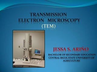
  • 1. BACHELOR OF SECONDARY EDUCATION CENTRAL BICOL STATE UNIVERSITY OF AGRICULTURE
  • 2. INTRODUCTION Electron Microscopy involves the study of different specimens by using an electron microscope.  Electron microscopes are scientific instruments that use a beam of energetic electrons to examine objects on a very fine scale. Through the use of an electron microscope we can see things that we would not normally be able to see with our naked eyes and has greater magnification than light microscope.  This required 10,000x plus magnification which was not possible using current optical microscopes
  • 3.
  • 4. TYPES OF ELECTRON MICROSCOPE 1. TRANSMISSION ELECTRON MICROSCOPE- form images using electrons that are transmitted through a specimen. 2. SCANNING ELECTRON MICROSCOPE- utilize electron that have bounced off the surface of the specimen.
  • 5. The transmission electron microscope (TEM) was the first type of Electron Microscope to be developed and is patterned exactly on the light transmission microscope except that a focused beam of electrons is used instead of light to "see through" the specimen. It was developed by Max Knoll and Ernst Ruska in Germany in 1931.
  • 6. TRANSMISSION ELECTRON MICROSCOPY (TEM)  Transmission electron microscopy (TEM) is a microscopy technique whereby a beam of electrons is transmitted through an ultra thin specimen, interacting with the specimen as it passes through. An image is formed from the interaction of the electrons transmitted through the specimen; the image is magnified and focused onto an imaging device, such as a fluorescent screen, on a layer of photographic film, or to be detected by a sensor such as a CCD camera.
  • 7. DIFFERENCES BETWEEN OM AND EM OPTICAL MICROSCOPE ELECTRON MICROSCOPE 1. The source of light. 1. The light source is replaced by a beam of 2. The specimen. very fast moving electrons. 3. The lenses that makes the 2. The specimen usually has to be specially specimen seem bigger. prepared and held inside a vacuum 4. The magnified image of the chamber from which the air has been specimen that you see. pumped out (because electrons do not travel very far in air). 3. The lenses are replaced by a series of coil- shaped electromagnets through which the electron beam travels. 4. The image is formed as a photograph (called an electron micrograph) or as an image on a TV screen.
  • 8. MICROSCOPE RESOLUTION MAGNIFICATION OPTICAL 200 nm 1000x TEM 0.2 nm 500000x
  • 9.
  • 10. DIAGRAM TO REPRESENT TEM’S WORKING Virtual Source First Condenser Lens Second Condenser Lens Condenser Aperture Sample Objective Lens Objective Aperture Selected Area Aperture First Intermediate Lens Second Intermediate Lens Projector Lens Main Screen (Phosphor)
  • 11. DIFFERENT COMPONENTS OF TEM 1. HIGH TENSION CABLE 2. ELECTRON EMITTER 3. STEPPER MOTORS FOR CENTERING THE ELECTRON BEAM 4. CONDENSER 11. VACUUM PUMP LEADS 5. APERTURE CONTROLS 12. GONIOMETER 6. SPECIMEN HOLDER 13. VACUUM AND 7. OBJECTIVE LENS MAGNIFICATION CONTROL 8. PROJECTOR LENS 14. FOCUSING CONTROL 9. OPTICAL LENS 10. FLUORESCENT SCREEN
  • 13. TEM SAMPLE PREPARATION  Cleaning the surface of the specimen The proper cleaning of the surface of the sample is important because the surface can contain a variety of unwanted deposits, such as dust, silt, and detritus, media components, or other contaminants. The best way to clean the surface of specimen from contaminants is to carefully rinse them three times for 10 min in 0.1 M cacodylic acid buffer (pH 7.3) at room temperature.
  • 14. TEM SAMPLE PREPARATION  Primary fixation of the specimen Fixation can be achieved by perfusion and microinjection, immersions, or with vapours using various fixatives including aldehydes(glutaraldehyde), osmium tetroxide,, tannic acid, or thiocarbohydrazide. FIXATIVES- are chemicals that denature and precipitate cellular macromolecules.
  • 15. Some common fixatives: 1. GLUTARALDEHYDE- is a 5-carbon with an aldehyde group at each end of the molecule. An aldehyde groups reacts with amino groups and cross link with the proteins into an insoluble network. 2. OSMIUM- heavy metal that reacts primarily with fatty acids leading to the preservation of cellular membranes.
  • 16. TEM SAMPLE PREPARATION  Rinsing of the specimen After the fixation step, samples must be rinsed in order to remove the excess fixative. To remove excess glutaraldehyde from the samples, the specimen should be subjected to a thorough but carefully conducted rinsing procedure. Specimens can be washed in 0.1 M cacodylic acid buffer (pH 7.3), starting with one time for 10 min, and then three times for 20 min at 4 oC.
  • 17. TEM SAMPLE PREPARATION  Secondary fixation of the specimen Specimen can be successfully stabilized for TEM investigation by post fixation with 1% osmium tetroxide prepared in 0.1 M cacodylic acid buffer (pH 7.3) for 1.5 hrs at room temperature (immersion fixation).
  • 18. TEM SAMPLE PREPARATION  Dehydrating the specimen For TEM investigation, specimen can be dehydrated in a graded series of ethanol. More specifically, the following protocol is useful: Dehydration of specimen in 50% ethanol for 5 min, 70% ethanol for 10 min, 80% ethanol for 10 min, 90% ethanol for 15 min, and 99.9% ethanol (dried with a 4-mesh molecular sieve) twice for 20 min at room temperature. This process allows the water in the samples to be slowly exchanged through liquids with lower surface tensions.
  • 19. TEM SAMPLE PREPARATION  Infiltration of the specimen with a transitional solvent The reason why this step is required is that the ethanol is not miscible with the plastic embedding medium I found most suitable for TEM investigation of the specimen (mollicutes). The replacement of the dehydration solution by another intermediary solvent (i.e., propylene oxide) is thus necessary [1, 23, 26]. This process is essentially an alcohol substitution. The immersion of mollicutes in propylene oxide twice for 20 min at room temperature is sufficient before attempting to embed the specimens in a resin.
  • 20. TEM SAMPLE PREPARATION  Infiltration with resin and embedding the specimen Mollicutes can be embedded in a variety of different media depending on the use (e.g., conventional TEM or immuno TEM). For conventional TEM of mollicutes, the epoxy resin Durcupan ACM is quite suitable. The following protocol can be used: Immersion of mollicutes in propylene-oxide/Durcupan-ACM (1:1; v/v) at room temperature overnight (use gloves and a fume hood, and leave the specimen container open for the propylene oxide to evaporate). The next day, the specimens should be immersed in a freshly prepared Durcupan ACM mixture (pure) and left for 2 hrs at room temperature. A second Durcupan ACM mixture (pure) is then prepared and used as the embedding medium (free of air bubbles!).ible with the plastic embedding medium I found most suitable for TEM investigation of mollicutes. The replacement of the dehydration solution by another intermediary solvent (i.e., propylene oxide) is thus necessary [1, 23, 26]. This process is essentially an alcohol substitution. The immersion of mollicutes in propylene oxide twice for 20 min at room temperature is sufficient before attempting to embed the specimens in a resin. Polymerization of the epoxy mixture can be achieved by placing the specimens in a drying cabinet for 2 days at 40 oC and for an additional 2 days at 60 oC. Leaving the samples after heat polymerization for an additional 1-2 weeks at room temperature can improve the subsequent cutting experience as the resin blocks continue to harden during this time.
  • 21. TEM SAMPLE PREPARATION  Sectioning and staining of the specimen The procedure for cutting specimens into semithin and ultrathin slices (sections) is known as microtomy and ultramicrotomy, respectively. Semithin sections (0.5 μm to 2 μm) were typically stained with toluidine blue for 1 min on a hot plate (70 oC to 90 oC), examined by LM, and used for identifying the specimen within the resin block before proceeding with ultramicrotomy. Ultrathin sections (about 70 nm to 90 nm) were typically stained with uranyl acetate followed by lead citrate.
  • 22. OTHER METHODS OF SAMPLE PREPARATION (TEM)
  • 23. Cryofixation and the use of Frozen specimen  CRYOFIXATION-like chemical fixatives it stops metabolic processes and preserves biological structure.  The method involves ultra-rapid cooling of small samples to liquid nitrogen temperature (-196 oC) or below, thus stopping all motion and metabolic activity, and preserving the internal structure by freezing all fluid phases solid.
  • 24.  The ultimate objective is to freeze the specimen so rapidly (at 104 to 106 K per second) that ice crystals are unable to form, or are prevented from growing sufficiently large to cause damage to the specimen's ultrastructure.
  • 25. NEGATIVE STAINING  The main purpose of negative-staining is to surround or embed the biological object in a suitable electron dense material which provides high contrast and good preservation. This method is capable of providing information about structural details often finer than those visible in thin sections, replicas, or shadowed specimens. In addition to the possibility of obtaining a spectacular enhancement of contrast, negative- staining has the advantage of speed and simplicity.
  • 26.  The technique has mainly been used to examine particulate (purified) specimens - e.g.. ribosomes, enzyme molecules, viruses, bacteriophages, microtubules, actin filaments, etc. at a resolution of 1.5-2.5 nm. This technique generally allows the shape, size, and the surface structure of the object to be studied as well as provide information about subunit stoichiometries and symmetry in oligomeric complexes. Any surface of the specimen accessible to water can potentially be stained, and thus, that part of the specimen will be imaged at high contrast.
  • 27. SHADOW CASTING  The grid containing the specimen are placed in sealed chamber which is the evacuated by vacuum pump.  The chamber contains a filament composed of a heavy metal together with carbon.
  • 28.
  • 29. The shadow cast by the nanoparticles (NPs) on metal deposition is visible in the high-magnification images.
  • 30. FREEZE FRACTURE REPLICATION AND FREEZE ETCHING  Small pieces of tissue are placed on a small metal disk and rapidly frozen.  The disk is then mounted on a cooled stage within a vacuum chamber and a frozen tissue block is struck by a knife edge.  The resulting fracture plane spreads out from the point of contact, splitting the tissue into two pieces.
  • 31. Freeze fracturing can separate the two phospholipid leaflets that form every cellular membrane (a) A preparation of cells or tissues is quickly frozen in liquid nitrogen at −196 °C, which instantly immobilizes cell components. (b) The block of frozen cells is fractured with a sharp blow from a cold knife. The fracture plane is irregular, often between the leaflets of the plasma or an organelle membrane. (c) Membrane proteins and particles remain bound to one leaflet or the other, as illustrated in the expanded view of a fractured membrane.
  • 32.  shadowing  coating specimen with a thin film of a heavy metal  freeze-etching  freeze specimen then fracture along lines of greatest weakness (e.g., membranes)
  • 33.
  • 34. GRID IS A SIEVE WOVEN FROM A THIN METAL WIRE,USUALLY NICKEL OR COPPER GRIDS OF 3 mm DIAMETER ARE COMMERCIALLY AVAILABLE WITH DIFFERENT MESH SIZES(GENERALLY OF 100-200 µm SIZE)
  • 35. Staphylococcus aureus platinum replica image shot on a TEM at 50,000X magnification
  • 36. A TEM image of the polio virus.
  • 37. CONCLUSION and RECOMMENDATION  Sample preparation for TEM involves more and some different steps than those for SEM. Like in any multi-step preparation procedure, virtually every step can affect the quality of the final electron micrograph. A single mistake in one of these steps will affect all remaining steps, and thus the outcome of the entire study. It is therefore important that the investigator plans and executes every step in great detail. These procedures involve a significant time commitment and require patience and skills that come only with practice. It is important to mention that most of the chemicals used in EM are dangerous. Investigator must be aware of potential hazards such as fire, chemical, electrical, and physical associated with these items.
  • 38. REFERENCES  http://cms.mse.berkeley.edu/publications/MRS_Spring_20 04_Xu.pdf  http://www.sciencepub.org/nature/0403/03-0171- mahongbao-ns.pdf  http://www.arl.army.mil/arlreports/2003/ARL-TR-3128.pdf  http://stem.ornl.gov/papers/reviews/pdfs/NIST.pdf  http://cimewww.epfl.ch/people/cayron/Fichiers/theseboo k-chap3.pdf  http://em-outreach.ucsd.edu/web-course/Sec-II.A.3/Sec- II.A.3.html  http://en.wikipedia.org/wiki/Cryofixation