SlideShare una empresa de Scribd logo
1 de 64
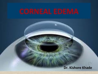
CORNEAL EDEMA


         Dr. Kishore Khade
cornea
Thickness of the cornea in
 the centre is 0.52mm
While at periphery is
 0.67mm.
Epithelium is 50-90μm
thick
BM 8-14μm
Stroma 0.5mm thick &
constitute most of cornea.
Dm is 10-12μm
Endothelium 18-20μm
with 2400-3000cells/mm2
CORNEA
• To perform its primary function of refraction
  of light the cornea must be relatively thin &
  dehydrated with smooth anterior surface.
• In normal cornea, optical transparency is
  directly related to the state of hydration of
  the tissue.
• If cornea swells, it increases its thickness , its
  surface becomes irregular both changes
  downgrades its optic properties.
CORNEAL HYDRATION
• Cornea is relatively in dehydrated state for its
  transperancy.
• Water content of cornea is 80% which is
  highest of any connective tissue in body.
• If hydration becomes above 80% its central
  thickness increases & transperancy reduces.
• Cornea swells only in the direction of its
  thickness therefore, corneal thickness &
  hydration are linearly related.
CORNEAL HYDRATION
• Hydration is maintained by:-
A}Factors draw water in to the cornea:-
1)Swelling pressure of stromal matrix ( GAGs).
2)Intraocular pressure.
B} factors which prevent flow of water into the
  cornea:-
1)Mechanical barriers.
2) Na+-K+ active pump of endothelium.
stromal                 barrier
pressure


           Hydration
                                 IOP
Na-k
pump

                   Evaporation
{A} Stromal swelling pressure
• Pressure exerted by corneal stroma mainly
  GAGs is stromal pressure (SP).
• Sp is 60 mmhg, is a keystone of corneal
  biophysics.
• Anionic charges on GAGs molecule expands
  the tissue, draws fluid with equal but negative
  pressure called imbibation pressure.
Contd.....
• In vivo imbibation pressure is reduced by IOP,
  so IP= IOP – SP
• IP= 17- 60, IP = -43mmhg.
• In vitro IP = SP
• Sp generates interfibrillar tension may be
  biophysical mechanism to maintain fibrils
  normal arrangement.
• Cornea has an swelling pressure, which is
  maintained by endothelial metabolic pump
{B} Barrier mechanism
• Both epithelium & endothelium acts as a
  barrier for excessive flow of water & diffusion
  of electrolytes into stroma.
• As compare to endothelium, epithelium offers
  twice resistance.
• Endothelium allows diffusion of small solutes
  like NaCl & urea, while epithelium produces
  hypertonicity of the solution bathing the
  cornea.
Barrier mechanism
• Endothelial cells are attached to each other by
  discontinuous tight junctions i.e maculae
  occludentes.
• Endothelial barrier function is mainly calcium
  dependent.
• A calcium free solution will reduce the barrier
  function & cause stromal edema.
{C} Na - K pump
                    +    +


• Present in endothelium, several fold more
  active than its epithelium counterparts.
• Activated ATPase mediates active extrusion of
  Na from stroma to the aqueous.
• It causes diffusion gradient for water.
• Na conc in aqueous is more compare to
  stroma, which draws water from the stroma.
Sodium activity across
    endothelium
    stroma
             Na-K pump

   Na 134
   meq/l
                     Aqueous
                     humour
• Bicarbonate dependent ATPase has also been
  reported in the endothelial cells.
• Depletion of bicarbonates induces swelling.
• Carbonic anhydrase enzyme has also been
  implicated in fluid transport, CAE inhibitors
  decreases flow of fluid from stroma to
  aqueous ( found only in endothelium).
Wounded
 cornea     142.3meq/l

                 Aqueous
NA               humour
149.8
Meq/l
           H2O
{D} EVAPORATION
• Evaporation of water from precorneal tearfilm
  increases its osmolarity relative to cornea.
• Hypertonicity of tear film could draw water
  from cornea.
• However this water loss is readily replaced by
  aqueous, it results in only a little corneal
  dehydration.
{E} Intra ocular pressure
• Most of early writers assume corneal edema is
  due to mechanical forcing of aqueous into the
  cornea.
• But experimental event found out that to
  achieve this,effect pressure required is
  200mmHg.
• More likely explanation is that the
  determining factor is endothelial damage.
EDEMA
• Word edema is derived from greek word
  oídēma 1400bc , means "swelling“.
• Formerly known as dropsy or hydropsy which
  means accumulation of excessive fluid.
• Etiology of corneal edema:
   SECONDARY CAUSES
   PRIMARY CAUSES
CORNEAL EDEMA
[I] Mechanical trauma
1) Blunt non penetrating injury causes edema
  by injury to endothelium, mostly it is
  reversible.
2) Perforating injuries cause direct damage to
  the cornea, intra ocular FB in AC can cause
  edema mainly in inferior periphery where FB
  mainly settles.
3) Forceps delivery cause pressure on globe,
  may cause edema due to DM tear.
DESCEMET’s TEAR
Mechanical trauma
4) Noxious chemicals mainly alkalies which
  penetrates cornea cause endothelium
  damage.
5)Intraocular sx can cause acute endothelial loss
  most notably in superior & central cornea.
6) BROWN McLEAN syndrome: peripheral
  edema with brown black discolouration of
  underlying endothelium seen in ICCE,
  ECCE,CCPE, pars plana vitrectomy.
Brown Mclean Syndrome
Mechanical trauma
7)Cold induced reversible corneal edema has been
  reported in trigeminal nerve dysfunction.
8) Certain systemic medications like amantadine &
  cefaclor can cause edema.
9) Lasers used for iridotomy can cause focal corneal
  edema.
10) High altitude corneal decompensation has been
  reported causing hypoxia induced corneal
  edema.
[ II ] GLAUCOMA
• Acute rise in IOP which exceeds swelling
  pressure of stroma causes epithelial edema.
• Hypoxic Endothelial decompensation occurs
  due to diminished aqueous flow.
• When corneal endothelium is compromised,
  edema occurs even @ lower level of IOP.
• Chronic elevation of IOP permanently
  damages the endothelium.
• Irreversible corneal edema may occur.
GLAUCOMA
GLAUCOMA
• Penetrating keratoplasty is the only treatment
  of choice in irreversible corneal edema, but
  IOP must be first controlled.
• In hypotony, AC is shallow or flat. Mechanical
  trauma by cornea iris or iris corneal touch
  leads to edema.
• Normal human volunteers experiment study
 can be explained by hypotony induced edema,
(corneal edema occurs in tightly patched eye).
[ III ] Contact lenses
• Most common cause of corneal edema is
  prolonged use of contact lens.
• It is mainly due to insufficient supply of
  oxygen to epithelium.
• Edema presents as microcystic epithelial
  edema near the center of resting position of
  the lens.
• It is best seen with scattered illumination of
  slit lamp referred as Sattler’s veil.
Contd....
• If allowed to continue, it‘ll cause stromal
  edema, descemet’s membrane folds.
• Edema easily clears if contact lens is removed.
• Even altering the fit of contact lens is also
  successful in reducing edema if it provides
  sufficient oxygen to the epithelium.
• The response & recovery from edema is
  independent of age.
[IV] ICE syndrome
• Iridocorneal endothelial syndrome is basically
  spectrum of disorders that includes
        A) Progressive iris atrophy.
        B) Chandler’s syndrome.
       C) Iris nevus syndrome ( Cogan Reese)
Chandler syndrome
• Corneal endothelial abnormalities (hammered
  silver).
• Presents with blurred vision & haloes due to
  corneal edema
• Corectopia may be mild to moderate
• Glaucoma may be less severe & but at
  presentation IOP may be normal.
• Chandler syndrome have more severe edema.
Cogan Resse syndrome
Characterised by diffuse
 naevus Which covers
iris or iris nodule.
Iris atrophy may be
absent in
50% of patients,
but corectopia &
Glaucoma
May be severe.
Iris atrophy
Essential iris atrophy
characterised by distortion
Of pupil, peripheral anterior
 synechie & iris atrophy
With full thickness holes.
Glaucoma commonly
 present in the involved eye.
Unilateral occurs in 4th & 5th
Decades of life in caucasians
[V] Essential corneal edema
• Idiopathic , episodic & often cyclic may be
  unilateral.
• Presents with typical features of corneal edema
  like fb sensation,diminished vision & haloes
  which persists for months then disappears.
• Recurrent erosions on cornea may be noted.
• It may progress to the formation of bullae with
  ciliary injection & urgent symptoms of pain &
  photophobia.
Essential corneal edema
• Pupils may be semidilated & sluggishly
  reacting to light.
• If secondary infection does not set up attack
  passes of & the condition eventually cleans
  up.
• Some of these cases may be early
  presentation of dystrophic changes like Fuch’s
  dystrophy.
[VI] Metabolic disorders
• Some vague concepts suggested by corneal
  edema occuring in some metabolic conditions
  like myxedema.
• It has also seen in hypercholesterolemia.
• In malaria mainly in patients taking mepacrine
  for its treatment, in this condition edema is
  limited to basal layer of epithelium &
  superficial layer of stroma.
PRIMARY CAUSES
A) Primary endothelial dystrophies: dystrophies
   involving endothelium & descemet’s
   membrane causes symmetrical marked
   stromal edema which is gradually progressive
   over a period of years.
B) Primary endothelial dystrophy which develop
   later in life are fuch’s dystrophy.
Primary Endothelial Dystrophy
• Congenital hereditary endothelial dystrophy:
  characterised by diffuse edema at birth or
  soon thereafter, without significant anterior
  segment abnormalities.
• Posterior polymorphous dystrophy: B/L
  vesicular or linear lesions at the level of
  descemet’s membrane & endothelium is
  present, it presents with congenital corneal
  edema.
FUCH’s dytrophy
AD pattern of inheritance,
Earliest changes are limited
To posterior cornea &
presents with central B/L
asymmetrical corneal
Guttata.
In fuch’s dystrophy
endothelial cells transform
into fibroblast
Like cells capable of
secreting collagen fibrils.
Contribute to BM thickening.
FUCH’s dytrophy
• Progressive endothelial decompensation leads
  to stromal & epithelial edema.
• Fluid in the stroma permeates the epitelial
  layer causes microcystic epithelial edema.
• Individual epithelial cells burst, intercellular
  edema occurs & typical blisters or bullae
  formed.
• These changes are confined to centre of
  cornea initially.
Bullous keratopathy
It represents the terminal
stage of severe or
Prolonged epithelial
edema.
In the affected area the
 epithelium is steamy
Irregular & on its surface
 one or more large bullae
Appears, raised in the
form of blebs.
Bullous keratopathy
• After 2-3 days the bullae rupture only to
  reappear, the cycle associated with considerable
  irritation & pain.
• Treatment is extremely difficult; retrobulbar
  injection of alcohol, removal of endothelium by
  scraping, cauterisation of cornea by tincture
  iodine, trichloro-acetic acid.
• Bandage contact lens to relieve pain.
• Lamellar grafting, if measures of therapy fails & if
  recurrence persists treatment is enucleation.
Manifestations of edema
 Depends upon cause & degree of the
  condition.
 Mild discomfort in conditions like fuch’s
  dystrophy.
 Severe neuralgic pain is seen in bullous
  keratopathy.
 Colour haloes.
 Severe visual loss.
Visual acuity
• Small amount of epithelial edema can result in
  substantial reduction in visual acuity.
• Although 70% stromal edema is compatible
  with normal visual acuity.
• Decreased acuity is more severe in early
  morning.
• IOP, iritis glaucoma & optic nerve changes
  may contribute to reduced acuity.
Pain & discomfort
• As edema increases epithelium is detached
  from basement membrane to form bullae.
• This rupture of bullae causes severe pain,
  photophobia, epiphora & narrowing of
  palpebral fissure.
• Photophobia is due to light scattering in the
  edematous cornea.
• Coloured haloes.
Coloured haloes
Evaluation of corneal morphology
Slit lamp examination.
Specular biomicroscopy.
Pachymetry.
Optical coherence tomography.
Scheimpflug camera.
Orbscan.
Slit lamp examination
Slit lamp examination reveals
cornea guttata, stromal
density,
Descemet’s membrane folds.
Bullae are easily observed by
slitlamp. In case of chronic
Corneal edema
neovascularization,pannus
 formation or dystrophies
are visible. In case of stromal
edema thickness exceeds 0.6mm.
Specular biomicroscopy
Measures cell density.
Normally endothelial
cell count is 3000 --
2000 cells/mm2.
Cell count less <1000
poorly tolerate ocular
Surgery.
Used in assesing cells
in Corneal grafting,
LASIK, dystrophies
 corneal edema.
Pachymetry
Pachymetry
• Corneal pachymetry;- is the process of measuring
  the thickness of the cornea. It can be done using
  contact methods, such as ultrasound and confocal
  microscopy (CONFOSCAN), or noncontact methods
  such as optical biometry with a single Scheimpflug
  camera (such as SIRIUS or PENTACAM), or Optical
  Coherence Tomography (OCT) and online Optical
  Coherence Pachymetry (OCP, such as ORBSCAN).
Optical coherence tomography
is an established
medical imaging technique.
It is widely used to obtain
high-resolution images
of the retina and the
anterior segment of the eye.
Corneal thickness can also be
measured.
Scheimpflug camera.
Pentacam trade name,
Is a diagnostic unit able
To perform five functions;

Image of AS.
3D anterior chamber
 analyzer.
pachymetry.
Corneal topography.
Cataract analyzer.
ORBSCAN II
The Orbscan II topographer
analyses the physical
shape / contours of
cornea and allows the surgeon
 to decide if it has suitable
shape, is healthy and thick
enough for treatment.
It is the only
topographer currently available
that measures the shape of both
the front and back surface of the
entire cornea (other systems
only measure the front surface)
 and can therefore provide
acomplete picture of the thickness of cornea
management
• In all cases of corneal edema if documented IOP
  is high, its control with topical anti glaucoma
  drugs or systemic CAE inhibitors are given.
• Even in moderately elevated IOP patients control
  may significantly reduces epithelial edema.
• If IOP remains elevated despite maximum
  tolerated medical therapy surgical intervention
  must be considered.
• Cyclocryotherapy/ trabeculectomy are
  considered.
Local therapy
• Early morning reduced vision can be improved
  by exposing eyes to warm air eg:hair dryer.
• Topical application of hyperosmotic agents
  like 5%NaCl solution or ointment can reduce
  edema to an little extend.
• If inflammation is a contributory cause of
  corneal edema topical application steroids
  may be very helpful.
Local therapy
• If the etiology of edema is not apperent 10
  days course of topical steroids can serve as
  diagnostic as well as therapeutic purpose.
• In patients with early corneal decompensation
  & mild edema a careful refraction may
  improve vision.
• Therapeutic hydrophilic contact lens can be
  used .
Local therapy
• Thin hydrophilic lens fitted flat on cornea
  allow maximum contact between lens &
  irregular cornea.
• Application of radiodiathermy or other forms
  of electrocautery on bowman’s membrane.
• This produces adhesion between stroma &
  basal layer of epithelium preventing formation
  of bullae.
management
• Gundersen conjunctival flap with or without
  lamellar keratectomy can be performed to
  relieve pain.
• Retrobulbar alcohol injection or tarsorrhaphy
  also may relieve pain.
• In painful blind eye or in absolute glaucoma
  enucleation may represent the optimal
  procedure.
Penetrating keratoplasty
• If visual recovery exists penetrating
  keratoplasty is performed before
  cauterisation of BM or conjuntival flap
  procedure.
• In case of multiple graft failure & corneal
  thickness exceeds 1.5mm, the use of
  keratoprosthesis has been advocated.
• Keraoprosthesis is considered as an last ditch
  effort for visual rehabilitation.
keratoplasty
Keratoprosthesis

Keratoprosthesis causes
high rate of failure &
Complications.
In case of bilateral corneal
edema
Keratoprosthesis in worst
eye & keratoplasty in
fellow is
Performed.
Keratoprosthesis
THANKYOU

Más contenido relacionado

La actualidad más candente (20)

Bullous keratopathy
Bullous keratopathyBullous keratopathy
Bullous keratopathy
 
Corneal topography
Corneal topographyCorneal topography
Corneal topography
 
Congenital ptosis
Congenital ptosisCongenital ptosis
Congenital ptosis
 
Presbyopia
PresbyopiaPresbyopia
Presbyopia
 
Keratoconus
KeratoconusKeratoconus
Keratoconus
 
Evaluation of ptosis
Evaluation of ptosis Evaluation of ptosis
Evaluation of ptosis
 
Concomitant and Incomitant, AHP and Hess chart
Concomitant and Incomitant, AHP and Hess chartConcomitant and Incomitant, AHP and Hess chart
Concomitant and Incomitant, AHP and Hess chart
 
Congenital cataract
Congenital cataractCongenital cataract
Congenital cataract
 
Refrective surgery ppt
Refrective surgery pptRefrective surgery ppt
Refrective surgery ppt
 
Slit lamp examination lecture
Slit lamp examination lectureSlit lamp examination lecture
Slit lamp examination lecture
 
Types of iol
Types of iolTypes of iol
Types of iol
 
Congenital nasolacrimal duct obstruction
Congenital nasolacrimal duct obstructionCongenital nasolacrimal duct obstruction
Congenital nasolacrimal duct obstruction
 
Corneal transparency
Corneal transparencyCorneal transparency
Corneal transparency
 
Pseudophakia
PseudophakiaPseudophakia
Pseudophakia
 
Diseases of the Cornea
Diseases of the CorneaDiseases of the Cornea
Diseases of the Cornea
 
Proptosis in ophthalmology
Proptosis  in ophthalmologyProptosis  in ophthalmology
Proptosis in ophthalmology
 
Cover tests
Cover testsCover tests
Cover tests
 
Rgp lens
Rgp lensRgp lens
Rgp lens
 
Tear film Dr Ferdous
Tear film Dr Ferdous  Tear film Dr Ferdous
Tear film Dr Ferdous
 
Corneal dystrophies
Corneal dystrophiesCorneal dystrophies
Corneal dystrophies
 

Destacado

Corneal edema after cataract surgery - MALEK AL KOTT
Corneal edema after cataract surgery - MALEK AL KOTTCorneal edema after cataract surgery - MALEK AL KOTT
Corneal edema after cataract surgery - MALEK AL KOTTMalek Al Kott
 
Post operative-corneal-edema
Post operative-corneal-edemaPost operative-corneal-edema
Post operative-corneal-edemaOkhuosami Fatima
 
Pseudophakic Bullous Keratopathy case
Pseudophakic Bullous Keratopathy case Pseudophakic Bullous Keratopathy case
Pseudophakic Bullous Keratopathy case Noor Munirah Aab
 
Fuchs dystrophy and pseudophakic bullous keratopathy
Fuchs dystrophy and pseudophakic bullous keratopathyFuchs dystrophy and pseudophakic bullous keratopathy
Fuchs dystrophy and pseudophakic bullous keratopathyunleng
 
Corneal transparency
Corneal transparencyCorneal transparency
Corneal transparencyTina Chandar
 
interstitial Keratitis
interstitial Keratitisinterstitial Keratitis
interstitial Keratitisikramdr01
 
Anatomy and physiology of cornea
Anatomy and physiology of corneaAnatomy and physiology of cornea
Anatomy and physiology of corneaLhacha
 
CATARACT SURGERY COMPLICATIONS
CATARACT SURGERY COMPLICATIONSCATARACT SURGERY COMPLICATIONS
CATARACT SURGERY COMPLICATIONSSiva Wurity
 
Interstitial keratitis & mooren's ulcer
Interstitial keratitis & mooren's ulcerInterstitial keratitis & mooren's ulcer
Interstitial keratitis & mooren's ulcerDr. Gurjeet Singh
 
Gede Pardianto - Corneal cryo therapy for Descemetocele
Gede Pardianto - Corneal cryo therapy for DescemetoceleGede Pardianto - Corneal cryo therapy for Descemetocele
Gede Pardianto - Corneal cryo therapy for DescemetoceleGede Pardianto
 
Corneal edema after cataract surgery malek alkott
Corneal edema after cataract surgery   malek alkottCorneal edema after cataract surgery   malek alkott
Corneal edema after cataract surgery malek alkottMalek Al Kott
 
A case of atypical disc edema
A case of atypical disc edemaA case of atypical disc edema
A case of atypical disc edemaSamuel Ponraj
 
Central serous chorioretinopathy
Central serous chorioretinopathyCentral serous chorioretinopathy
Central serous chorioretinopathyOthman Al-Abbadi
 
corneal response to anoxia stress from contact lens wear
corneal response to anoxia stress from contact lens wearcorneal response to anoxia stress from contact lens wear
corneal response to anoxia stress from contact lens wearTheeratep
 
Peripheral Ulcerative Keratitis.Dr Ferdous
Peripheral Ulcerative Keratitis.Dr Ferdous   Peripheral Ulcerative Keratitis.Dr Ferdous
Peripheral Ulcerative Keratitis.Dr Ferdous Ferdous101531
 

Destacado (20)

Corneal edema after cataract surgery - MALEK AL KOTT
Corneal edema after cataract surgery - MALEK AL KOTTCorneal edema after cataract surgery - MALEK AL KOTT
Corneal edema after cataract surgery - MALEK AL KOTT
 
Post operative-corneal-edema
Post operative-corneal-edemaPost operative-corneal-edema
Post operative-corneal-edema
 
Edema corneal
Edema cornealEdema corneal
Edema corneal
 
Pseudophakic Bullous Keratopathy case
Pseudophakic Bullous Keratopathy case Pseudophakic Bullous Keratopathy case
Pseudophakic Bullous Keratopathy case
 
Fuchs dystrophy and pseudophakic bullous keratopathy
Fuchs dystrophy and pseudophakic bullous keratopathyFuchs dystrophy and pseudophakic bullous keratopathy
Fuchs dystrophy and pseudophakic bullous keratopathy
 
Corneal transparency
Corneal transparencyCorneal transparency
Corneal transparency
 
Interstitial keratitis
Interstitial keratitisInterstitial keratitis
Interstitial keratitis
 
interstitial Keratitis
interstitial Keratitisinterstitial Keratitis
interstitial Keratitis
 
Anatomy and physiology of cornea
Anatomy and physiology of corneaAnatomy and physiology of cornea
Anatomy and physiology of cornea
 
CATARACT SURGERY COMPLICATIONS
CATARACT SURGERY COMPLICATIONSCATARACT SURGERY COMPLICATIONS
CATARACT SURGERY COMPLICATIONS
 
Interstitial keratitis & mooren's ulcer
Interstitial keratitis & mooren's ulcerInterstitial keratitis & mooren's ulcer
Interstitial keratitis & mooren's ulcer
 
Uveal tumours
Uveal tumoursUveal tumours
Uveal tumours
 
Corneal edema numbers
Corneal edema numbersCorneal edema numbers
Corneal edema numbers
 
Gede Pardianto - Corneal cryo therapy for Descemetocele
Gede Pardianto - Corneal cryo therapy for DescemetoceleGede Pardianto - Corneal cryo therapy for Descemetocele
Gede Pardianto - Corneal cryo therapy for Descemetocele
 
Corneal edema after cataract surgery malek alkott
Corneal edema after cataract surgery   malek alkottCorneal edema after cataract surgery   malek alkott
Corneal edema after cataract surgery malek alkott
 
A case of atypical disc edema
A case of atypical disc edemaA case of atypical disc edema
A case of atypical disc edema
 
Central serous chorioretinopathy
Central serous chorioretinopathyCentral serous chorioretinopathy
Central serous chorioretinopathy
 
corneal response to anoxia stress from contact lens wear
corneal response to anoxia stress from contact lens wearcorneal response to anoxia stress from contact lens wear
corneal response to anoxia stress from contact lens wear
 
Peripheral Ulcerative Keratitis.Dr Ferdous
Peripheral Ulcerative Keratitis.Dr Ferdous   Peripheral Ulcerative Keratitis.Dr Ferdous
Peripheral Ulcerative Keratitis.Dr Ferdous
 
Mooren’s ulcer
Mooren’s ulcerMooren’s ulcer
Mooren’s ulcer
 

Similar a Corneal edema (20)

Corneal transparency
Corneal transparency Corneal transparency
Corneal transparency
 
Glaucoma
GlaucomaGlaucoma
Glaucoma
 
Physiology of the cornea
Physiology of the corneaPhysiology of the cornea
Physiology of the cornea
 
Corneal edema
Corneal edemaCorneal edema
Corneal edema
 
Eye lid i &amp; ii 10.08.16
Eye lid  i &amp; ii  10.08.16Eye lid  i &amp; ii  10.08.16
Eye lid i &amp; ii 10.08.16
 
Ocular Manifestations of Leprosy - EYE
Ocular Manifestations of Leprosy - EYEOcular Manifestations of Leprosy - EYE
Ocular Manifestations of Leprosy - EYE
 
1.anatomy , physiology , pathology of cornea
1.anatomy , physiology , pathology of cornea1.anatomy , physiology , pathology of cornea
1.anatomy , physiology , pathology of cornea
 
Keratoconus f dinesh
Keratoconus f dineshKeratoconus f dinesh
Keratoconus f dinesh
 
A SEMINAR ON
A SEMINAR ONA SEMINAR ON
A SEMINAR ON
 
Cornea 1
Cornea 1Cornea 1
Cornea 1
 
Cornea dr. feba mary george
Cornea  dr. feba mary georgeCornea  dr. feba mary george
Cornea dr. feba mary george
 
CORNEA PHYSIOLOGY.pptx
CORNEA PHYSIOLOGY.pptxCORNEA PHYSIOLOGY.pptx
CORNEA PHYSIOLOGY.pptx
 
Glaucoma (opthalmology)
Glaucoma (opthalmology)Glaucoma (opthalmology)
Glaucoma (opthalmology)
 
DISEASES OF SHUKLAMANDALA-MODERN PART
DISEASES OF SHUKLAMANDALA-MODERN PARTDISEASES OF SHUKLAMANDALA-MODERN PART
DISEASES OF SHUKLAMANDALA-MODERN PART
 
Cornea
CorneaCornea
Cornea
 
CORNEA-Anatomy,Corneal Transperency.pptx
CORNEA-Anatomy,Corneal Transperency.pptxCORNEA-Anatomy,Corneal Transperency.pptx
CORNEA-Anatomy,Corneal Transperency.pptx
 
Keratoconus
KeratoconusKeratoconus
Keratoconus
 
Short case Cornea
Short case CorneaShort case Cornea
Short case Cornea
 
Orbital trauma
Orbital traumaOrbital trauma
Orbital trauma
 
Ophthalmology 5th year, 4th lecture (Dr. Bakhtyar)
Ophthalmology 5th year, 4th lecture (Dr. Bakhtyar)Ophthalmology 5th year, 4th lecture (Dr. Bakhtyar)
Ophthalmology 5th year, 4th lecture (Dr. Bakhtyar)
 

Corneal edema

  • 1. CORNEAL EDEMA Dr. Kishore Khade
  • 2. cornea Thickness of the cornea in the centre is 0.52mm While at periphery is 0.67mm. Epithelium is 50-90μm thick BM 8-14μm Stroma 0.5mm thick & constitute most of cornea. Dm is 10-12μm Endothelium 18-20μm with 2400-3000cells/mm2
  • 3. CORNEA • To perform its primary function of refraction of light the cornea must be relatively thin & dehydrated with smooth anterior surface. • In normal cornea, optical transparency is directly related to the state of hydration of the tissue. • If cornea swells, it increases its thickness , its surface becomes irregular both changes downgrades its optic properties.
  • 4. CORNEAL HYDRATION • Cornea is relatively in dehydrated state for its transperancy. • Water content of cornea is 80% which is highest of any connective tissue in body. • If hydration becomes above 80% its central thickness increases & transperancy reduces. • Cornea swells only in the direction of its thickness therefore, corneal thickness & hydration are linearly related.
  • 5. CORNEAL HYDRATION • Hydration is maintained by:- A}Factors draw water in to the cornea:- 1)Swelling pressure of stromal matrix ( GAGs). 2)Intraocular pressure. B} factors which prevent flow of water into the cornea:- 1)Mechanical barriers. 2) Na+-K+ active pump of endothelium.
  • 6. stromal barrier pressure Hydration IOP Na-k pump Evaporation
  • 7. {A} Stromal swelling pressure • Pressure exerted by corneal stroma mainly GAGs is stromal pressure (SP). • Sp is 60 mmhg, is a keystone of corneal biophysics. • Anionic charges on GAGs molecule expands the tissue, draws fluid with equal but negative pressure called imbibation pressure.
  • 8. Contd..... • In vivo imbibation pressure is reduced by IOP, so IP= IOP – SP • IP= 17- 60, IP = -43mmhg. • In vitro IP = SP • Sp generates interfibrillar tension may be biophysical mechanism to maintain fibrils normal arrangement. • Cornea has an swelling pressure, which is maintained by endothelial metabolic pump
  • 9. {B} Barrier mechanism • Both epithelium & endothelium acts as a barrier for excessive flow of water & diffusion of electrolytes into stroma. • As compare to endothelium, epithelium offers twice resistance. • Endothelium allows diffusion of small solutes like NaCl & urea, while epithelium produces hypertonicity of the solution bathing the cornea.
  • 10. Barrier mechanism • Endothelial cells are attached to each other by discontinuous tight junctions i.e maculae occludentes. • Endothelial barrier function is mainly calcium dependent. • A calcium free solution will reduce the barrier function & cause stromal edema.
  • 11. {C} Na - K pump + + • Present in endothelium, several fold more active than its epithelium counterparts. • Activated ATPase mediates active extrusion of Na from stroma to the aqueous. • It causes diffusion gradient for water. • Na conc in aqueous is more compare to stroma, which draws water from the stroma.
  • 12. Sodium activity across endothelium stroma Na-K pump Na 134 meq/l Aqueous humour
  • 13. • Bicarbonate dependent ATPase has also been reported in the endothelial cells. • Depletion of bicarbonates induces swelling. • Carbonic anhydrase enzyme has also been implicated in fluid transport, CAE inhibitors decreases flow of fluid from stroma to aqueous ( found only in endothelium).
  • 14.
  • 15. Wounded cornea 142.3meq/l Aqueous NA humour 149.8 Meq/l H2O
  • 16. {D} EVAPORATION • Evaporation of water from precorneal tearfilm increases its osmolarity relative to cornea. • Hypertonicity of tear film could draw water from cornea. • However this water loss is readily replaced by aqueous, it results in only a little corneal dehydration.
  • 17. {E} Intra ocular pressure • Most of early writers assume corneal edema is due to mechanical forcing of aqueous into the cornea. • But experimental event found out that to achieve this,effect pressure required is 200mmHg. • More likely explanation is that the determining factor is endothelial damage.
  • 18. EDEMA • Word edema is derived from greek word oídēma 1400bc , means "swelling“. • Formerly known as dropsy or hydropsy which means accumulation of excessive fluid. • Etiology of corneal edema:  SECONDARY CAUSES  PRIMARY CAUSES
  • 20. [I] Mechanical trauma 1) Blunt non penetrating injury causes edema by injury to endothelium, mostly it is reversible. 2) Perforating injuries cause direct damage to the cornea, intra ocular FB in AC can cause edema mainly in inferior periphery where FB mainly settles. 3) Forceps delivery cause pressure on globe, may cause edema due to DM tear.
  • 22. Mechanical trauma 4) Noxious chemicals mainly alkalies which penetrates cornea cause endothelium damage. 5)Intraocular sx can cause acute endothelial loss most notably in superior & central cornea. 6) BROWN McLEAN syndrome: peripheral edema with brown black discolouration of underlying endothelium seen in ICCE, ECCE,CCPE, pars plana vitrectomy.
  • 24. Mechanical trauma 7)Cold induced reversible corneal edema has been reported in trigeminal nerve dysfunction. 8) Certain systemic medications like amantadine & cefaclor can cause edema. 9) Lasers used for iridotomy can cause focal corneal edema. 10) High altitude corneal decompensation has been reported causing hypoxia induced corneal edema.
  • 25. [ II ] GLAUCOMA • Acute rise in IOP which exceeds swelling pressure of stroma causes epithelial edema. • Hypoxic Endothelial decompensation occurs due to diminished aqueous flow. • When corneal endothelium is compromised, edema occurs even @ lower level of IOP. • Chronic elevation of IOP permanently damages the endothelium. • Irreversible corneal edema may occur.
  • 27. GLAUCOMA • Penetrating keratoplasty is the only treatment of choice in irreversible corneal edema, but IOP must be first controlled. • In hypotony, AC is shallow or flat. Mechanical trauma by cornea iris or iris corneal touch leads to edema. • Normal human volunteers experiment study can be explained by hypotony induced edema, (corneal edema occurs in tightly patched eye).
  • 28. [ III ] Contact lenses • Most common cause of corneal edema is prolonged use of contact lens. • It is mainly due to insufficient supply of oxygen to epithelium. • Edema presents as microcystic epithelial edema near the center of resting position of the lens. • It is best seen with scattered illumination of slit lamp referred as Sattler’s veil.
  • 29. Contd.... • If allowed to continue, it‘ll cause stromal edema, descemet’s membrane folds. • Edema easily clears if contact lens is removed. • Even altering the fit of contact lens is also successful in reducing edema if it provides sufficient oxygen to the epithelium. • The response & recovery from edema is independent of age.
  • 30. [IV] ICE syndrome • Iridocorneal endothelial syndrome is basically spectrum of disorders that includes A) Progressive iris atrophy. B) Chandler’s syndrome. C) Iris nevus syndrome ( Cogan Reese)
  • 31. Chandler syndrome • Corneal endothelial abnormalities (hammered silver). • Presents with blurred vision & haloes due to corneal edema • Corectopia may be mild to moderate • Glaucoma may be less severe & but at presentation IOP may be normal. • Chandler syndrome have more severe edema.
  • 32. Cogan Resse syndrome Characterised by diffuse naevus Which covers iris or iris nodule. Iris atrophy may be absent in 50% of patients, but corectopia & Glaucoma May be severe.
  • 33. Iris atrophy Essential iris atrophy characterised by distortion Of pupil, peripheral anterior synechie & iris atrophy With full thickness holes. Glaucoma commonly present in the involved eye. Unilateral occurs in 4th & 5th Decades of life in caucasians
  • 34. [V] Essential corneal edema • Idiopathic , episodic & often cyclic may be unilateral. • Presents with typical features of corneal edema like fb sensation,diminished vision & haloes which persists for months then disappears. • Recurrent erosions on cornea may be noted. • It may progress to the formation of bullae with ciliary injection & urgent symptoms of pain & photophobia.
  • 35. Essential corneal edema • Pupils may be semidilated & sluggishly reacting to light. • If secondary infection does not set up attack passes of & the condition eventually cleans up. • Some of these cases may be early presentation of dystrophic changes like Fuch’s dystrophy.
  • 36. [VI] Metabolic disorders • Some vague concepts suggested by corneal edema occuring in some metabolic conditions like myxedema. • It has also seen in hypercholesterolemia. • In malaria mainly in patients taking mepacrine for its treatment, in this condition edema is limited to basal layer of epithelium & superficial layer of stroma.
  • 37. PRIMARY CAUSES A) Primary endothelial dystrophies: dystrophies involving endothelium & descemet’s membrane causes symmetrical marked stromal edema which is gradually progressive over a period of years. B) Primary endothelial dystrophy which develop later in life are fuch’s dystrophy.
  • 38. Primary Endothelial Dystrophy • Congenital hereditary endothelial dystrophy: characterised by diffuse edema at birth or soon thereafter, without significant anterior segment abnormalities. • Posterior polymorphous dystrophy: B/L vesicular or linear lesions at the level of descemet’s membrane & endothelium is present, it presents with congenital corneal edema.
  • 39. FUCH’s dytrophy AD pattern of inheritance, Earliest changes are limited To posterior cornea & presents with central B/L asymmetrical corneal Guttata. In fuch’s dystrophy endothelial cells transform into fibroblast Like cells capable of secreting collagen fibrils. Contribute to BM thickening.
  • 40. FUCH’s dytrophy • Progressive endothelial decompensation leads to stromal & epithelial edema. • Fluid in the stroma permeates the epitelial layer causes microcystic epithelial edema. • Individual epithelial cells burst, intercellular edema occurs & typical blisters or bullae formed. • These changes are confined to centre of cornea initially.
  • 41. Bullous keratopathy It represents the terminal stage of severe or Prolonged epithelial edema. In the affected area the epithelium is steamy Irregular & on its surface one or more large bullae Appears, raised in the form of blebs.
  • 42. Bullous keratopathy • After 2-3 days the bullae rupture only to reappear, the cycle associated with considerable irritation & pain. • Treatment is extremely difficult; retrobulbar injection of alcohol, removal of endothelium by scraping, cauterisation of cornea by tincture iodine, trichloro-acetic acid. • Bandage contact lens to relieve pain. • Lamellar grafting, if measures of therapy fails & if recurrence persists treatment is enucleation.
  • 43. Manifestations of edema  Depends upon cause & degree of the condition.  Mild discomfort in conditions like fuch’s dystrophy.  Severe neuralgic pain is seen in bullous keratopathy.  Colour haloes.  Severe visual loss.
  • 44. Visual acuity • Small amount of epithelial edema can result in substantial reduction in visual acuity. • Although 70% stromal edema is compatible with normal visual acuity. • Decreased acuity is more severe in early morning. • IOP, iritis glaucoma & optic nerve changes may contribute to reduced acuity.
  • 45. Pain & discomfort • As edema increases epithelium is detached from basement membrane to form bullae. • This rupture of bullae causes severe pain, photophobia, epiphora & narrowing of palpebral fissure. • Photophobia is due to light scattering in the edematous cornea. • Coloured haloes.
  • 47. Evaluation of corneal morphology Slit lamp examination. Specular biomicroscopy. Pachymetry. Optical coherence tomography. Scheimpflug camera. Orbscan.
  • 48. Slit lamp examination Slit lamp examination reveals cornea guttata, stromal density, Descemet’s membrane folds. Bullae are easily observed by slitlamp. In case of chronic Corneal edema neovascularization,pannus formation or dystrophies are visible. In case of stromal edema thickness exceeds 0.6mm.
  • 49. Specular biomicroscopy Measures cell density. Normally endothelial cell count is 3000 -- 2000 cells/mm2. Cell count less <1000 poorly tolerate ocular Surgery. Used in assesing cells in Corneal grafting, LASIK, dystrophies corneal edema.
  • 51. Pachymetry • Corneal pachymetry;- is the process of measuring the thickness of the cornea. It can be done using contact methods, such as ultrasound and confocal microscopy (CONFOSCAN), or noncontact methods such as optical biometry with a single Scheimpflug camera (such as SIRIUS or PENTACAM), or Optical Coherence Tomography (OCT) and online Optical Coherence Pachymetry (OCP, such as ORBSCAN).
  • 52. Optical coherence tomography Optical coherence tomography is an established medical imaging technique. It is widely used to obtain high-resolution images of the retina and the anterior segment of the eye. Corneal thickness can also be measured.
  • 53. Scheimpflug camera. Pentacam trade name, Is a diagnostic unit able To perform five functions; Image of AS. 3D anterior chamber analyzer. pachymetry. Corneal topography. Cataract analyzer.
  • 54. ORBSCAN II The Orbscan II topographer analyses the physical shape / contours of cornea and allows the surgeon to decide if it has suitable shape, is healthy and thick enough for treatment. It is the only topographer currently available that measures the shape of both the front and back surface of the entire cornea (other systems only measure the front surface) and can therefore provide acomplete picture of the thickness of cornea
  • 55. management • In all cases of corneal edema if documented IOP is high, its control with topical anti glaucoma drugs or systemic CAE inhibitors are given. • Even in moderately elevated IOP patients control may significantly reduces epithelial edema. • If IOP remains elevated despite maximum tolerated medical therapy surgical intervention must be considered. • Cyclocryotherapy/ trabeculectomy are considered.
  • 56. Local therapy • Early morning reduced vision can be improved by exposing eyes to warm air eg:hair dryer. • Topical application of hyperosmotic agents like 5%NaCl solution or ointment can reduce edema to an little extend. • If inflammation is a contributory cause of corneal edema topical application steroids may be very helpful.
  • 57. Local therapy • If the etiology of edema is not apperent 10 days course of topical steroids can serve as diagnostic as well as therapeutic purpose. • In patients with early corneal decompensation & mild edema a careful refraction may improve vision. • Therapeutic hydrophilic contact lens can be used .
  • 58. Local therapy • Thin hydrophilic lens fitted flat on cornea allow maximum contact between lens & irregular cornea. • Application of radiodiathermy or other forms of electrocautery on bowman’s membrane. • This produces adhesion between stroma & basal layer of epithelium preventing formation of bullae.
  • 59. management • Gundersen conjunctival flap with or without lamellar keratectomy can be performed to relieve pain. • Retrobulbar alcohol injection or tarsorrhaphy also may relieve pain. • In painful blind eye or in absolute glaucoma enucleation may represent the optimal procedure.
  • 60. Penetrating keratoplasty • If visual recovery exists penetrating keratoplasty is performed before cauterisation of BM or conjuntival flap procedure. • In case of multiple graft failure & corneal thickness exceeds 1.5mm, the use of keratoprosthesis has been advocated. • Keraoprosthesis is considered as an last ditch effort for visual rehabilitation.
  • 62. Keratoprosthesis Keratoprosthesis causes high rate of failure & Complications. In case of bilateral corneal edema Keratoprosthesis in worst eye & keratoplasty in fellow is Performed.