SlideShare una empresa de Scribd logo
1 de 22
Descargar para leer sin conexión
Questions and Answers Exam 1
   1 Graves disease and Hashimoto’s disease are both example of what type of disease that predominantly affects what organ? How do they differ? What
     are the signs and symptoms of each?
 both affect thyroid - they are a form of autoimmune disease
 Graves hyperactivates it and Hashimoto hypoactivates it (they have opposite etilogies)
 Graves patients present with increased metabolism - can't gain weight, always hot, very hyperactive
 Hashimoto patients present with decreased metabolism - depression, weight gain, feeling cold, tired

   2 Give a detailed example of hormones often act to maintain homeostatic balance.
 push-me pull-me system - one hormone ramps up metabolism and another is bringing it down

 (a) Humoral stimulus: Low blood calcium levels trigger parathyroid hormone release from the parathyroid glands, which causes blood Ca 2+ levels to rise by stimulating release of Ca 2+
 from bone.

 (b) Neural stimulus: The stimulation of adrenal medullary cells by sympathetic nervous system (SNS) fibers triggers the release of catecholamines to the blood.

 (c) Hormonal stimulus: Hormones released by the hypothalamus stimulate the anterior pituitary to release hormones that stimulate other endocrine organs to secrete hormones.

   3 Define and give an example of each of the following Autocrine, Paracrine, juxtacrine and Endocrine.
 Autocrine - hormones are released and act on the cell that secreted them
 paracrine - hormones act on nearby cells via difusion from hormone producing cell

 Juxtacrine - hormone that regulates adjacent cell
 Endocrine - hormones travel via bloodstream to target cells

   4 What are the four main hormone precursors? What types of hormones do they give rise to?
 1- Peptides and proteins
 2- Amino acid derivatives
 3- Steroids
 4- Fatty acid derivatives - Eicosanoids
 PROTEINS
 Range from 3 amino acids to hundreds of amino acids in size.
 Often produced as larger molecular weight precursors that are proteolytically cleaved to the active form of the hormone.
 Peptide/protein hormones are water soluble.
 Comprise the largest number of hormones– perhaps in thousands
 Are encoded by a specific gene which is transcribed into mRNA and translated into a protein precursor called a preprohormone
 Preprohormones are often post-translationally modified in the ER to contain carbohydrates (glycosylation)
 Preprohormones contain signal peptides (hydrophobic amino acids) which targets them to the golgi where signal sequence is removed to form prohormone
 Prohormone is processed into active hormone and packaged into secretory vessicles Secretory vesicles move to plasma membrane where they await a signal. Then they are
 exocytosed and secreted into blood stream
 In some cases the prohormone is secreted and converted in the extracellular fluid into the active hormone: an example is angiotensin is secreted by liver and converted into active form
 by enzymes secreted by kidney and lung
 STEROIDS
 Glucocorticoids; cortisol is the major representative in most mammals
 Mineralocorticoids; aldosterone being most prominent
 Androgens such as testosterone
 Estrogens, including estradiol and estrone
 Progestogens (also known a progestins) such as progesterone

   5 Describe the characteristics of hormone produced from Proteins, peptides, and modified amino acids.
 These hydrophilic (and mostly large) hormone molecules bind to receptors on the surface of "target" cells; that is, cells able to respond to the presence of the hormone. These receptors
 are transmembrane proteins. Binding of the hormone to its receptor initiates a sequence of intracellular signals that may
 alter the behavior of the cell (such as by opening or closing membrane channels) or
 stimulate (or repress) gene expression in the nucleus by turning on (or off) the promoters and enhancers of the genes.


   6 Describe Steroid hormones synthesis and outline Steroid Hormone Biosynthesis Reactions.
 All steroid hormones are derived from cholesterol. A series of enzymatic steps in the mitochondria and ER of steroidogenic tissues convert cholesterol into all of the other steroid
 hormones and intermediates
 The particular steroid hormone class synthesized by a given cell type depends upon its complement of peptide hormone receptors, its response to peptide hormone stimulation and its
 genetically expressed complement of enzymes. The following indicates which peptide hormone is responsible for stimulating the synthesis of which steroid hormone:
 Luteinizing Hormone (LH):
 progesterone and testosterone
 Adrenocorticotropic hormone (ACTH):
 cortisol
 Follicle Stimulating Hormone (FSH):
 estradiol
 Angiotensin II/III:
 aldosterone
The particular steroid hormone class synthesized by a given cell type depends upon its complement of peptide hormone receptors, its response to peptide hormone stimulation and its
 genetically expressed complement of enzymes. The following indicates which peptide hormone is responsible for stimulating the synthesis of which steroid hormone:
 Luteinizing Hormone (LH):
 progesterone and testosterone
 Adrenocorticotropic hormone (ACTH):
Questions and Answers Exam 1
 cortisol
 Follicle Stimulating Hormone (FSH):
 estradiol
 Angiotensin II/III:
 aldosterone
  The first reaction in converting cholesterol to C18, C19 and C21 steroids involves the cleavage of a 6-carbon group from cholesterol and is the principal committing, regulated, and rate-
  limiting step in steroid biosynthesis.
  The cholesterol precursor comes from cholesterol synthesized within the cell from acetate, from cholesterol ester stores in intracellular lipid droplets or from uptake of cholesterol-
  containing low density lipoproteins.
  Lipoproteins taken up from plasma are most important when steroidogenic cells are chronically stimulated.

     7 How does P450 SCC work? Why is the function of this enzyme dangerous to the cell?
  The enzyme system that catalyzes the cleavage reaction is known as P450-linked side chain cleaving enzyme (P450ssc), or desmolase, and is found in the mitochondria of steroid-
  producing cells, but not in significant quantities in other cells.
  long-term regulation is effected at the level the gene for desmolase. This gene contains a cAMP regulatory element (CRE) that binds cAMP and increases the level of desmolase RNA
  transcription, thereby leading to increased levels of the enzyme

     8 What are the steroids of the adrenal cortex?
  The adrenal cortex is responsible for production of 3 major classes of steroid hormones:
  glucocorticoids, which regulate carbohydrate metabolism;
  mineralocorticoids, which regulate the body levels of sodium and potassium;
  androgens, whose actions are similar to that of steroids produced by the male gonads.

     9 What are Steroid hydroxylases and what is their job?
  The various hydroxylases involved in the synthesis of the steroid hormones have a nomenclature that indicates the site of hydroxylation (e.g. 17a-hydroxylase introduces a hydroxyl
  group to carbon 17). These hydroxylase enzymes are members of the cytochrome P450 class of enzymes and as such also have a nomenclature indicative of the site of hydroxylation
  in addition to being identified as P450 class enzymes (e.g. the 17a-hydroxylase is also identified as P450c17)


  The adrenal cortex is composed of 3 main tissue regions: zona glomerulosa, zona fasciculata, and zona reticularis. Although the pathway to pregnenolone synthesis is the same in all
  zones of the cortex, the zones are histologically and enzymatically distinct, with the exact steroid hormone product dependent on the enzymes present in the cells of each zone.

   10 briefly describe the production and regulation of the Gonadal Steroid Hormones.
  Although many steroids are produced by the testes and the ovaries, the two most important are testosterone and estradiol. These compounds are under tight biosynthetic control, with
  short and long negative feedback loops that regulate the secretion of follicle stimulating hormone (FSH) and luteinizing hormone (LH) by the pituitary and gonadotropin releasing hormone
  (GnRH) by the hypothalamus. Low levels of circulating sex hormone reduce feedback inhibition on GnRH synthesis (the long loop), leading to elevated FSH and LH.
  The latter peptide hormones bind to gonadal tissue and stimulate P450ssc activity, resulting in sex hormone production via cAMP and PKA mediated pathways. The roles of cAMP and
  PKA in gonadal tissue are the same as that described for glucocorticoid production in the adrenals, but in this case adenylate cyclase activation is coupled to the binding of LH to plasma
  membrane receptors.
Questions and Answers Exam 1
                      describe what is happening in this figure


 production of Testosterone leading to dihydrotestosterone from Cholesterol including the intermediate steps in synthesis

  12 What are the Eicosanoids- how are they produced and what types of roles do they play?
 Prostaglandins & related compounds are collectively known as eicosanoids. (prostaglandins, prostacyclins, leukotrienes and thromboxanes)
 They are produced from arachidonic acid, polyunsaturated fatty acids.
 They have specific effects on target cells close to their site of formation.
 They are rapidly degraded, so they are not transported to distal sites within the body.

  13 How do NSAIDs work in combination with Eicosanoids? Ibuprofen?
 Eicosanoids roles in inflammation, fever, regulation of blood pressure, blood clotting, control of reproductive processes & tissue growth, sleep/wake cycle regulation.
 Ibuprofen and related compounds block the hydrophobic channel by which arachidonate enters the cyclooxygenase active site.

  14 Why is Aspirin a permanent effector of PGH2 synthesis?
 inhibit cyclooxygenase activity of PGH2 Synthase it is IRREVERSIBLE
 They inhibit formation of prostaglandins involved in fever, pain, & inflammation.
 They inhibit blood clotting by blocking thromboxane formation in blood platelets.

  15 A hormonal stimulus typically produces one or more of what kinds changes in a cell.
 Alters plasma membrane permeability or membrane potential, or both, by opening or closing ion channels
 Stimulates synthesis of proteins or regulatory molecules such as enzymes within the cell
 Activates or deactivates enzymes
 Induces secretory activity
 Stimulates mitosis

  16 Describe Second messenger systems.
 a hormone binds to a receptor on the plasma membrane. Of the second messengers, cyclic AMP is by far the best understood, so it will receive most of our attention.
 (a) Mechanisms that generate cyclic AMP by activation of adenylate cyclase are mediated by G proteins, which are activated when a hormone (first messenger, A in this case) binds to
 plasma membrane receptors. Cyclic AMP (the second messenger) acts intracellularly to activate protein kinase enzymes that mediate the cell's responses to the hormone. Notice that
 not all hormones provoke the activation of the effector enzyme. Sometimes they act to inhibit.


  17 Describe how G proteins work.
 (1) The hormone, acting as the first messenger, binds to its receptor. This causes the receptor to change shape, allowing it to bind a nearby inactive G protein.
 (2) The G protein is activated as the guanosine diphosphate (GDP) bound to it is displaced by the high-energy compound guanosine triphosphate (GTP). The G protein behaves like a
 light switch; it is "off" when GDP is bound to it, and "on" when GTP is bound.
 (3) The activated G protein (moving along the membrane) binds to and activates the effector enzyme adenylate cyclase. At this point the GTP bound to the G protein is hydrolyzed to
 GDP and the G protein becomes inactive once again. (The G protein possesses GTPase activity and cleaves the terminal phosphate group off GTP in much the same way that ATPase
 enzymes hydrolyze ATP.)
 (4) The activated adenylate cyclase generates the second-messenger cAMP from ATP.
 (5) cAMP, which is free to diffuse throughout the cell, triggers a cascade of chemical reactions in which one or more enzymes, called protein kinases, are activated. The protein kinases
 phosphorylate (add a phosphate group to) various proteins, many of which are other enzymes. Because phosphorylation activates some of these proteins and inhibits others, a variety
 of reactions may occur in the same target cell at the same time.

  18 Describe cyclic AMP signaling in terms of activation and repression.
 In this mechanism, three plasma membrane components interact to determine intracellular levels of cyclic AMP (cAMP): a hormone receptor, a signal transducer (a G protein), and an
 effector enzyme (adenylate cyclase).


  19 Why is cAMP action short term?
 it is rapidly degraded by the intracellular enzyme phosphodiesterase

  20 Outline the PIP-Calcium Signal Mechanism.
 intracellular calcium ions act as the final mediator
 Second-messenger mechanisms of amino acid-based hormones.
 Some mechanisms use Ca 2+ as an intracellular messenger.
 The activated G protein stimulates the effector molecule, phospholipase, to cleave PIP 2 into two fragments-inositol triphosphate (IP 3 ) and diacylglycerol (DAG), which act as
 intracellular second messengers to activate protein kinases and increase the cytoplasmic concentration of Ca 2+ . Ionic calcium also acts as a second messenger to modify the activity
 of cellular proteins.
20 Outline the PIP-Calcium Signal Mechanism.
 intracellular calcium ions act as the final mediator
 Second-messenger mechanisms of amino acid-based hormones.
 Some mechanisms use Ca 2+ as an intracellular messenger.
Questions andprotein stimulates the effector molecule, phospholipase, to cleave PIP 2 into two fragments-inositol triphosphate (IP 3 ) and diacylglycerol (DAG), which act as
 The activated G Answers Exam 1
 intracellular second messengers to activate protein kinases and increase the cytoplasmic concentration of Ca 2+ . Ionic calcium also acts as a second messenger to modify the activity
 of cellular proteins.


   21 Describe the process of Steroid Hormone Direct Gene Activation.
  This interaction "turns on" a gene, that is, prompts transcription of DNA to produce a messenger RNA (mRNA). The mRNA is then translated on the cytoplasmic ribosomes, producing
  specific protein molecules. These proteins include enzymes that promote the metabolic activities induced by that particular hormone and, in some cases, promote synthesis of either
  structural proteins or proteins to be exported from the target cell.

   22 Describe Target Cell Specificity .
  In order for a target cell to respond to a hormone, the cell must have specific protein receptors on its plasma membrane or in its interior to which that hormone can bind. For example,
  receptors for adrenocorticotropic hormone (ACTH) are normally found only on certain cells of the adrenal cortex. By contrast, thyroxine is the principal hormone stimulating cellular
  metabolism, and nearly all body cells have thyroxine receptors

   23 What is meant by the concept of receptor and hormone affinity?
  Although binding of a hormone to a receptor is the crucial first step, target cell activation by hormone-receptor interaction depends equally on three factors: (1) blood levels of the
  hormone, (2) relative numbers of receptors for that hormone on or in the target cells, and (3) affinity (strength) of the bond between the hormone and the receptor.

   24 Define Half-Life, Onset, and Duration of Hormone Activity.
  Hormones are potent chemicals, and they exert profound effects on their target organs at very low concentrations. Hormones circulate in the blood in two forms--free or bound to a
  protein carrier. In general, lipid-soluble hormones (steroids and thyroid hormone) travel in the bloodstream attached to plasma proteins. All others circulate unencumbered by carriers.
  The concentration of a circulating hormone in blood at any time reflects (1) its rate of release, and (2) the speed at which it is inactivated and removed from the body and (3) the amount
  and type of carrier molecules available. Carrier molecules also effect the function and bio availability of the hormone.
  Some hormones are rapidly degraded by enzymes in their target cells, but most are removed from the blood by the kidneys or liver, and their breakdown products are excreted from the
  body in urine or, to a lesser extent, in feces. As a result, the length of time a hormone remains in the blood, referred to as its half-life, is usually brief--from a fraction of a minute to 30
  minutes, with the water-soluble hormones exhibiting the shortest half-lives
  The time required for hormone effects to appear varies greatly. Some hormones provoke target organ responses almost immediately, while others, particularly the steroid hormones,
  require hours to days before their effects are seen. Additionally, some hormones are secreted in a relatively inactive form and must be activated in the target cells.
  The duration of hormone action is limited, ranging from 10 seconds to several hours, depending on the hormone. Effects may disappear rapidly as blood levels drop, or they may persist
  for hours after very low hormone levels have been reached. Because of these many variations, hormonal blood levels must be precisely and individually controlled to meet the
  continuously changing needs of the body.

   25 Define and give an example of Permissiveness interactions.
  Permissiveness is the situation when one hormone cannot exert its full effects without another hormone being present. For example, the development of the reproductive system is
  largely regulated by reproductive system hormones, as we might expect. However, thyroid hormone is necessary (has a permissive effect) for normal timely development of
  reproductive structures; without thyroid hormone, reproductive system development is delayed.

   26 Define and give an example of Synergism interactions.
  Synergism of hormones occurs in situations where more than one hormone produces the same effects at the target cell and their combined effects are amplified. For example, both
  glucagon (produced by the pancreas) and epinephrine cause the liver to release glucose to the blood; when they act together, the amount of glucose released is about 150% of what is
  released when each hormone acts alone.

   27 Define and give an example of Antagonism interactions.
  When one hormone opposes the action of another hormone, the interaction is called antagonism. For example, insulin, which lowers blood sugar levels, is antagonized by the action of
  glucagon, which acts to raise blood sugar levels. Antagonists may compete for the same receptors, act through different metabolic pathways, or even, as noted in the progesterone-
  estrogen interaction at the uterus, cause down-regulation of the receptors for the antagonistic hormone.

   28 Describe in detail the three main forms of Endocrine Gland Stimuli and give example of each.
  Some endocrine glands secrete their hormones in direct response to changing blood levels of certain ions and nutrients. These stimuli are called humoral stimuli to distinguish them from
  hormonal stimuli, which are also blood borne chemicals. The term humoral harks back to the ancient use of the term humor to refer to various body fluids (blood, bile, and others).
  This is the simplest of the endocrine control systems. For example, cells of the parathyroid glands monitor blood Ca 2+ levels, and when they detect a decline from normal values, they
  secrete parathyroid hormone (PTH). Because PTH acts by several routes to reverse that decline, blood Ca 2+ levels soon rise, ending the initiative for PTH release. Other hormones
  released in response to humoral stimuli include insulin, produced by the pancreas, and aldosterone, one of the adrenal cortex hormones
  Neural stimuli
  In a few cases, nerve fibers stimulate hormone release. The classic example of neural stimuli is sympathetic nervous system stimulation of the adrenal medulla to release
  catecholamines (norepinephrine and epinephrine) during periods of stress.
  Hormonal stimuli
  Finally, many endocrine glands release their hormones in response to hormones produced by other endocrine organs, and the stimuli in these cases are called hormonal stimuli. For
  example, release of most anterior pituitary hormones is regulated by releasing and inhibiting hormones produced by the hypothalamus, and many anterior pituitary hormones in turn
  stimulate other endocrine organs to release their hormones).

   29 Describe Nervous System Modulation.
  Both "turn on" factors (hormonal, humoral, and neural stimuli) and "turn off" factors (feedback inhibition and others) may be modified by the nervous system. Without this added
  safeguard, endocrine system activity would be strictly mechanical, much like a household thermostat. A thermostat can maintain the temperature at or around its set value, but it cannot
  sense that your grandmother visiting from Florida feels cold at that temperature and reset itself accordingly. You must make that adjustment.
  This is not usually the case in your body, however, where the nervous system can, in certain cases, override normal endocrine controls as needed to maintain homeostasis. For

  example, the action of insulin and several other hormones normally keeps blood sugar levels in the range of 90-110 mg glucose per 100 ml of blood. However, when the body is under
  severe stress, blood sugar levels rise because the hypothalamus and sympathetic nervous system centers are strongly activated. This ensures that body cells have sufficient fuel for
  the more vigorous activity required during such periods.
29 Describe Nervous System Modulation.
 Both "turn on" factors (hormonal, humoral, and neural stimuli) and "turn off" factors (feedback inhibition and others) may be modified by the nervous system. Without this added
 safeguard, endocrine system activity would be strictly mechanical, much like a household thermostat. A thermostat can maintain the temperature at or around its set value, but it cannot
 sense that your grandmother visiting from Florida feels cold at that temperature and reset itself accordingly. You must make that adjustment.
QuestionsusuallyAnswers your body, however, where the nervous system can, in certain cases, override normal endocrine controls as needed to maintain homeostasis. For
 This is not and the case in Exam 1

  example, the action of insulin and several other hormones normally keeps blood sugar levels in the range of 90-110 mg glucose per 100 ml of blood. However, when the body is under
  severe stress, blood sugar levels rise because the hypothalamus and sympathetic nervous system centers are strongly activated. This ensures that body cells have sufficient fuel for
  the more vigorous activity required during such periods.

   30 Describe the spectrum of autoimmune endocrine diseases.
  Autoimmune diseases form a spectrum ranging from organ-specific conditions in which one organ only is affected to systemic diseases in which the pathology is diffused throughout the
  body. The extremes of this spectrum result from quite distinct underlying mechanisms, but there are many conditions in which there are components of both organ-specific and
  systemic damage. . Some pathologies work by shutting a system down – others by hyper stimulating it.


   31 Define and describe 3 factors that can lead to Autoimmunity.
  Endocrine
      Most autoimmune disease do not occur with equal frequency in males and females. For example Graves' and Hashimoto's are 4-5 times, and SLE 10 times, more common in females while Ankylosing Spondylitis is
      3-4 × more frequent in males. These differences are believed to be the result of hormonal influences
      A second well documented hormonal effect is the marked reduction in disease severity seen in many autoimmune conditions during pregnancy. Rheumatoid arthritis is perhaps the classic example of this effect.
      In some cases there is also a rapid exacerbation (rebound) after giving birth.

  Environmental
      However, it is clear that environmental factors also play a role in autoimmune disease. If you examine how frequently identical twins both develop a disease (the concordance rate), it is only about 20-40% for
      common autoimmune diseases such as diabetes, SLE and rheumatoid arthritis. This makes it highly likely that environmental factors must also be important. While we might expect factors such as diet to play a
      role, we can postulate that infectious organisms are the most significant environmental factor.


   32 Contrast the mechanisms of Grave’s disease and Hashimoto’s syndrome.
  Graves disease
  thyroid is hyperactive
  autoimmune disease that can be transfered witn IgG antibodies
  Hashimoto disease
  thyroid is hypoactive
  mononuclear cellular infiltrate and
  autoantibodies targeted at thyroglobulin and thyroid peroxidase

   33 Briefly outline the components of the immune system.
  Made up of two cellular systems
  humoral or circulating antiBody system - B cells
  cell mediaTed immunity - T cells
  Both work by identifying antigens (foreign proteins or polysaccharides) either as part of a virus or bacterium or as a partially degraded byproduct
  Also recognizes human antigens not made by the individual resulting in graft rejection
  The humoral antiBody system produces secreted antibodies (proteins) which bind to antigens and identify the antigen complex for destruction. Antibodies act on antigens in the serum
  and lymph. B-cell produced antibodies may either be attached to B-cell membranes or free in the serum and lymph.
  The cell mediaTed system acts on antigens appearing on the surface of individual cells. T-cells produce T-cell receptors which recognize specific antigens bound to the antigen
  presenting structures on the surface of the presenting cell.

   34 Describe 3 Autoimmune Polyendocrine Syndromes in detail.
  Hirata
      The insulin autoimmune syndrome, associated with Graves’ disease and methimazole therapy (or other sulfhydryl containing medications) is of particular interest due to a remarkably strong association with a
      specific HLA haplotype . Such patients with elevated titers of anti-insulin autoantibodies frequently present with hypoglycemia. The disease in Japan is essentially confined to DR4-positive individuals with
      DRB1*0406. In Hirata syndrome the anti-insulin autoantibodies are polyclonal. Some patients have monoclonal anti-insulin autoantibodies that also induce hypoglycemia. For these patients there is no HLA
      association with their disease.

  Poems
      POEMS (Plasmacytoma, endocrinopathy, monoclonal gammopathy, and skin changes) patients usually present with a sensory motor polyneuropathy, diabetes mellitus (50%), primary gonadal failure (70%), and a
      plasma cell dyscrasia with sclerotic bony. T
      emporary remission may result following radiotherapy directed at the plasmacytoma. The syndrome is assumed to be secondary to circulating immunoglobulins but patients have excess vascular endothelial
      growth factor as well as elevated IL1-b, IL-6, and TNF-a.

  Thymic Tumor
      Thymomas and thymic hyperplasia are associated with a series of autoimmune diseases. The most common autoimmune diseases are myasthenia gravis and red cell aplasia. Graves’ disease, type 1 diabetes,
      and Addison’s disease may also be associated with thymic tumors. Unique anti-acetylcholine receptor autoantibodies may be present with thymoma and disease may be initiated by transcription of molecules
      within the tumor related to acetylcholine receptors.

   35 Describe the location and embryology of the Hypothalamus and pituitary gland.
  Hypothalamus is located in the middle of the base of the brain, and encapsulates the ventral portion of the third ventricle

  The pituitary gland, also known as the hypophysis, is a roundish organ that lies immediately beneath the hypothalamus, resting in a depression of the base of the skull called the sella
  turcica ("Turkish saddle").
35 Describe the location and embryology of the Hypothalamus and pituitary gland.

 The pituitary gland, also known as the hypophysis, is a roundish organ that lies immediately beneath the hypothalamus, resting in a depression of the base of the skull called the sella
Questions and saddle"). Exam 1
 turcica ("Turkish Answers
 The pituitary gland develops as a fusion of two groups of cells. There is an upgrowth of ectodermal cells from the roof of the primitive pharynx (known as Rathke's pouch), and a down-
 growth of neural tissue cells from the hypothalamus. These two distinct areas form the anterior (the adenohypophysis) and posterior (the neurohypophysis) lobes respectively.


  36 What hormones are produced in the Hypothalamus and which of them go directly to the blood stream?
 Hypothalamus produces releasing factors that stimulate production of anterior pituitary hormone which act on peripheral endocrine gland to stimulate release of third hormone


  37 Describe vascularization in the hypothalamus and pituitary- why is it this way? How does it improve function?


                                                                                                     hypophyseal artery

 A branch of the hypophyseal artery ramifies into a capillary bed in the lower hypothalamus, and hypothalmic hormones destined for the anterior pituitary are secreted into that capillary
 blood.
 Blood from those capillaries drains into hypothalamic-hypophyseal portal veins. Portal veins are defined as veins between two capillary beds; the hypothalamic-hypophyseal portal veins
 branch again into another series of capillaries within the anterior pituitary.
 Capillaries within the anterior pituitary, which carry hormones secreted by that gland, coalesce into veins that drain into the systemic venous blood. Those veins also collect capillary
 blood from the posterior pituitary gland.
 The utility of this unconventional vascular system is that minute quantities of hypothalamic hormones are carried in a concentrated form directly to their target cells in the anterior
 pituitary, and are not diluted out in the systemic circulation.

  38 What is Insulin Autoimmune Syndrome (Hirata Syndrome) and how does it work?
 The insulin autoimmune syndrome, associated with Graves’ disease and methimazole therapy (or other sulfhydryl containing medications) is of particular interest due to a remarkably
 strong association with a specific HLA haplotype . Such patients with elevated titers of anti-insulin autoantibodies frequently present with hypoglycemia. The disease in Japan is
 essentially confined to DR4-positive individuals with DRB1*0406. In Hirata syndrome the anti-insulin autoantibodies are polyclonal. Some patients have monoclonal anti-insulin
 autoantibodies that also induce hypoglycemia. For these patients there is no HLA association with their disease.

  39 Describe the relationship between the pituitary gland and hypothalamus in great detail.
 Hypothalamic neurons in the supraoptic and paraventricular nuclei synthesize ADH and Oxytocin, which are transported along the hypothalamic-hypophyseal tract to the
 neurohypophysis for storage.
 Neurons in the ventral hypothalamus have short axons that discharge releasing and inhibiting hormones to the capillaries of the hypophyseal portal system, which runs to the
 adenohypophysis. These factors influence the adenohypophyseal secretory cells to release (or not release) their hormones.
 Environmental stimuli (stressor, temperature, light-dark, etc.)
 ↓
 Higher brain centers (e.g., cerebral cortex)
 ↓ Neural pathways using neurotransmitters/neuromodulators
 Hypothalamus

  ↓ Hypothalamic (a.k.a. hypophysiotropic) hormones
 Pituitary gland
  ↓ Hypophyseal hormones
 Peripheral endocrine glands
  ↓ Peripheral hormones
 Target tissues
Environmental stimuli (stressor, temperature, light-dark, etc.)
 ↓
 Higher brain centers (e.g., cerebral cortex)
 ↓ Neural pathways using neurotransmitters/neuromodulators
 Hypothalamus
Questions and Answers Exam 1
  ↓ Hypothalamic (a.k.a. hypophysiotropic) hormones
 Pituitary gland
  ↓ Hypophyseal hormones
 Peripheral endocrine glands
  ↓ Peripheral hormones
 Target tissues


  40 What is GnRH- what does it do – where is it produced- how much is made- what does it regulate? Is it the same in males and females? What is
     Kallman’s? How is it involved in endometriosis?
 The vertebrate gonadotropin-releasing hormone (GnRH) is a decapeptide involved in regulating reproduction. The release of GnRH from the hypothalamus regulates the production of
 gonadotropins in the pituitary and these gonadotropins are responsible for gonadal development and growth in vertebrates.
 In addition to the hypothalamic GnRH (also called GnRH1 or GnRH-I), many vertebrate species have been found to express other GnRH forms. These include a midbrain GnRH (called
 GnRH-II or GnRH2) and a telencephalic GnRH (called GnRH-III or GnRH3).
 10 aa.
 Samuel M. McCann, 1960
 present in most vertebrate classes
 Pulsatile secretion!!
 Hypothalamic site of production: medial preoptic area

 Extrahypothalamic production: milk, placenta, frog sympathetic nervous system
  
      Effects:   FSH↑↑
      LH↑↑
      mating behavior, sexual drive in animals
  
      Secretion: in utero
            from puberty →
 Gonadotropin-releasing hormone (GnRH) plays a key role in the regulation of the reproductive system. GnRH has a similar structure in all animals. It is a decapeptide, meaning that it
 consists of a chain of 10 amino acids.
 GnRH acts primarily to stimulate the anterior pituitary gland to synthesise and secrete the gonadotropins FSH and LH. It exerts three principal actions on the anterior pituitary gland:
 Activation: the movement of gonadotropins from the reserve pool to a pool ready for direct action
 Immediate release (direct secretion) of gonadotropins     
 DOPAMINE / SEROTONIN supress production of GnRH
 KALLMAN'S
 Kallman’s is a rare condition affecting the hypothalamus, which secretes hormones to the pituitary gland. The hypothalamus is dysfunctional and cannot secret GnRH to the pituitary
 gland which, as a result, is unable to send either Luteinizing Hormone (LH) or Follicle Simulating Hormone, (FSH) to the ovaries, (women) and testes (men).
 Symptoms of undiagnosed KS are: no sense of smell (anosmia), failure to enter puberty, lack of sexual drive and infertility (non-ovulation in women and little or no sperm-count in men).
 Other symptoms may include mood swings, depression, anxiety, fatigue and insomnia. If undiagnosed, the condition can lead to osteoporosis in later life.
 The treatment for diagnosed patients is either estrogen or testosterone replacement therapy and fertility treatment if a KS patient desires to have a child. Regular bone-density scans, an
 MRI scan, (magnetic resonance imaging) and blood-tests may be required.
 ENDOMETRIAL - the mucous membrane lining the uterus, which thickens during the menstrual cycle in preparation for possible implantation of an embryo.
 ENDOMETRIOSIS - a condition resulting from the appearance of endometrial tissue outside the uterus and causing pelvic pain.

 Gonadotropin releasing hormone (GnRH) agonists are the most effective hormone treatments for endometriosis. They are able to block the release of the reproductive hormones LH
 (luteinizing hormone) and FSH (follicular-stimulating hormone). As a result, the ovaries stop ovulating and no longer produce estrogen. They relieve pain in most patients by the second
 or third month.

  41 What is TrH- what does it do – where is it produced- how much is made- what does it regulate? Is it the same in males and females?
 Thyrotropin-releasing hormone -
 Present in most vertebrate classes

 Hypothalamic site of production: PVN

 Extrahypothalamic production:
 other brain regions
 retina
 pancreas
 skin of frogs

 Effects:   TSH↑↑
       PRL ↑↑
       (GH ↑-?)
       general stimulatory effects
           (TRH) is a tripeptide hormone that stimulates the release of TSH and prolactin by the anterior pituitary. TRH is produced by the hypothalamus and travels across the median
 eminence to the pituitary via the pituitary portal system.

            In addition to the brain, TRH can also be detected in other areas of the body including the gastrointestinal system and pancreatic islets. Medical preparations of TRH are
 used in diagnostic tests of thyroid disorders and in acromegaly.

             The sequence of TRH was first determined and the hormone synthesized by Roger Guillemin and Andrew V. Schally in 1969.
 Its molecular weight is 359.5 Da and its structure is:
(TRH) is a tripeptide hormone that stimulates the release of TSH and prolactin by the anterior pituitary. TRH is produced by the hypothalamus and travels across the median
Questions andpituitary via the pituitary portal system.
 eminence to the Answers Exam 1

             In addition to the brain, TRH can also be detected in other areas of the body including the gastrointestinal system and pancreatic islets. Medical preparations of TRH are
  used in diagnostic tests of thyroid disorders and in acromegaly.

              The sequence of TRH was first determined and the hormone synthesized by Roger Guillemin and Andrew V. Schally in 1969.
  Its molecular weight is 359.5 Da and its structure is:
   (pyro)Glu-His-Pro-NH2

   42 Somatostatin what does it do – where is it produced- how much is made- what does it regulate? What is the functional difference between the two
      major forms of it? Why so many receptors?
  2 isoforms: 28 aa. (GIT) and 14 aa. (CNS)
  present in most vertebrate classes
  Hypothalamic site of production: periventricular nuclei
  Extrahypothalamic production: pancreas and other GIT structures
  Effects:   GH↓↓
             TSH↓
             PRL↓
   
  Systemic effects: general inhibitory effects
             insulin↓
             glucagon↓
             PP↓
             renin↓
             PTH↓
             calcitonin↓
  Somatostatin was first discovered in hypothalamic extracts and identified as a hormone that inhibited secretion of growth hormone. Subsequently, somatostatin was found to be
  secreted by a broad range of tissues, including pancreas, intestinal tract and regions of the central nervous system outside the hypothalamus.
  Somatostatin is secreted not only by cells of the hypothalamus but also by so called delta cells of stomach, intestine and pancreas.

  All actions of the hormone are inhibitory.
  Somatostatin's main actions are:
     * Inhibits the release of growth hormone (GH)
     * Inhibits the release of thyroid stimulating hormone (TSH)
     * Suppresses the release of gastrointestinal hormones
         o Gastrin
         o Cholecystokinin (CCK)
         o Secretin
         o Motilin
         o Vasoactive intestinal peptide (VIP)
         o Gastric inhibitory polypeptide (GIP)
         o Enteroglucagon (GIP)
     * Prolongs gastric emptying, gall bladder contraction and intestinal motility
     * Suppresses the release of pancreatic hormones
         o Inhibits the release of insulin
         o Inhibits the release of glucagon
     * Suppresses the exocrine secretory action of pancreas

  Somatostatin antagonizes the effects of Growth Hormone Releasing Hormone (GHRH)
  Two forms of somatostatin are synthesized. They are referred to as SS-14 and SS-28, reflecting their amino acid chain length. Both forms of somatostatin are generated by proteolytic
  cleavage of prosomatostatin, which itself is derived from preprosomatostatin. Two cysteine residules in SS-14 allow the peptide to form an internal disulfide bond.


  Each of the receptors activates distinct signaling mechanisms within cells, although all inhibit adenylyl cyclase.

   43 What is GHRH- what does it do – where is it produced- how much is made- what does it regulate? Is it the same in males and females?
  Growth hormone-releasing hormone
  44 aa
  Pulsatile secretion!!
  present in most vertebrate classes

  Hypothalamic site of production: arcuate nucleus

  Extrahypothalamic production: GIT structures

  Effects: GH↑↑ (not in >40 yr old)
  a 44-amino acid peptide hormone produced in the arcuate nucleus of the hypothalamus. It stimulates the production and secretion of growth hormone (GH) from the anterior pituitary
  gland, and like GH, it is released in a pulsatile manner. Additionally, GHRH also promotes non-REM(rapid eye movement) sleep directly

   44 What is CRH- what does it do – where is it produced- how much is made- what does it regulate? Is it the same in males and females?
  Corticotropin-releasing hormone
  a 41 aa
  Hypothalamic site of production: PVN

        Extrahypothalamic production:       very extensive! other brain regions
        lung
        GIT
        placenta

  Effects:   POMC↑↑ (ACTH ↑↑, opioids↑↑, aMSH↑↑ )
        general activating effects
a 41 aa
  Hypothalamic site of production: PVN

     Extrahypothalamic production: very extensive! other brain regions
     lung
     GIT
Questions and Answers Exam 1
     placenta

  Effects:   POMC↑↑ (ACTH ↑↑, opioids↑↑, aMSH↑↑ )
        general activating effects
              (CRH), also called corticotrophin-releasing factor (CRF) or corticoliberin, is a polypeptide hormone involved in the stress response.

            It is produced by the hypothalamus and stimulates corticotropic cells of the anterior lobe of the pituitary to produce ACTH and other biologically active substances (for
  example β-endorphin). CRH is also synthesized by the placenta and seems to determine the duration of pregnancy.

              CRH is a hypophysiotropin that has behavioral effects at the paraventricular nucleus of the hypothalamus, the central nucleus of the amygdala, the bed nucleus of the stria
  terminalis, and at the locus coeruleus. Release of CRH is affected by serum levels of cortisol and leptin, by stress and by the sleep/wake cycle.

   45 Define SET POINTS AND FEEDBACK LOOPS in HYPOTHALAMIC SECRETION.
  Set point: the “normal” hormone level that the organism tries to maintain
       at a certain time and under a certain condition.
   
  The hormone levels maintained at the level of set point by feedback mechanisms.
   
  The set point is adjusted according to the needs of the organism, e.g., set point may change across the day (circadian rhythm) or in response to environmental challenges (altered
  ambient temperature, stress, etc.).
        Feedback loops: a controlled variable (e.g., blood hormone level) determines the rate of the secretion of the hormone.
   
  Negative feedback loops
  Positive feedback loops


  Long feedback loops
  Short feedback loops
  Ultrashort feedback loops


   46 What are Zeitgebers- how do they affect you on a daily- monthly yearly basis?
  endogenous (spontaneous) periodic changes. 
  Endogenous rhythms are generated within the body but are synchronized to the environmental rhythms by environmental stimuli (a.k.a. Zeitgebers).

  Zeitgebers are environmental cues that usually help keep the circadian cycle. Zeitgebers include sunlight, noise, social interactions, and alarm clocks.
  Light/ Dark Cycles

   47 What do TSH, FSH and LH have in common and why do they need it?
  – Thyroid-stimulating hormone, also known as thyrotropin, is secreted from cells in the anterior pituitary called thyrotrophs, finds its receptors on epithelial cells in the thyroid gland, and
  stimulates that gland to synthesize and release thyroid hormones.
  Follicle stimulating hormone (FSH)
  – Stimulates follicle development and
  estrogen secretion in females and
  sperm production in males
  – Suppressed by INHIBIN
  Leutinizing hormone (LH)
  – Causes ovulation and progestin production in females and androgen production in males
  – Progestins
  – Androgens (testosterone)
  Luteinizing hormone (LH) and follicle-stimulating hormone (FSH) are called gonadotropins because stimulate the gonads - in males, the testes, and in females, the ovaries. They are not
  necessary for life, but are essential for reproduction.
  As described for thyroid-simulating hormone, LH and FSH are large glycoproteins composed of alpha and beta subunits. The alpha subunit is identical in all three of these anterior
  pituitary hormones, while the beta subunit is unique and endows each hormone with the ability to bind its own receptor.

   48 What are the functions of FSH and LH in males and females? Be specific! Do they work the same? How are they regulated? What Disease States are
      associated with LH and FSH? How does manipulating them work in contraception?
  In both sexes, LH stimulates secretion of sex steroids from the gonads. In the testes, LH binds to receptors on Leydig cells, stimulating synthesis and secretion of testosterone. Theca
  cells in the ovary respond to LH stimulation by secretion of testosterone, which is converted into estrogen by adjacent granulosa cells.
  Physiologic effects of the gonadotrophins are known only in the ovaries and testes. Together, then regulate many aspects of gonadal function in both males and females.
  In females, ovulation of mature follicles on the ovary is induced by a large burst of LH secretion known as the preovulatory LH surge.
associated with LH and FSH? How does manipulating them work in contraception?


Questions ovulation of matureExam 1 the ovary is induced by a large burst of LH secretion known as the preovulatory LH surge.
 In females, and Answers follicles on
  As its name implies, FSH stimulates the maturation of ovarian follicles. Administration of FSH to humans and animals induces "superovulation", or development of more than the usual
  number of mature follicles and hence, an increased number of mature gametes.
  FSH is also critical for sperm production. It supports the function of Sertoli cells, which in turn support many aspects of sperm cell maturation.
  The principle regulator of LH and FSH secretion is gonadotropin-releasing hormone or GnRH (also known as LH-releasing hormone). GnRH is a ten amino acid peptide that is
  synthesized and secreted from hypothalamic neurons and binds to receptors on gonadotrophs.


  In a classical negative feedback loop, sex steroids inhibit secretion of GnRH and also appear to have direct negative effects on gonadotrophs.
  This regulatory loop leads to pulsatile secretion of LH and, to a much lesser extent, FSH. The number of pulses of GnRH and LH varies from a few per day to one or more per hour. In
  females, pulse frequency is clearly related to stage of the cycle.
  the gonads secrete at least two additional hormones - inhibin and activin - which selectively inhibit and activate FSH secretion from the pituitary
  DISEASE STATE
  Diminished secretion of LH or FSH can result in failure of gonadal function (hypogonadism). This condition is typically manifest in males as failure in production of normal numbers of
  sperm. In females, cessation of reproductive cycles is commonly observed.
  Elevated blood levels of gonadotropins usually reflect lack of steroid negative feedback. Removal of the gonads from either males or females, as is commonly done to animals, leads to
  persistent elevation in LH and FSH. In humans, excessive secretion of FSH and/or LH most commonly the result of gonadal failure or pituitary tumors. In general, elevated levels of
  gonadotropins per se have no biological effect.
  CONTRACEPTION manipulation
  Normal patterns of gonadotropin secretion are absolutely required for reproduction, and interfering particularly with LH secretion is a widely-used strategy for contraception. Oral
  contraceptive pills contain a progestin (progesterone-mimicking compound), usually combined with an estrogen. As discussed above, progesterone and estrogen inhibit LH secretion,
  and oral contraceptives are effective because they inhibit the LH surge that induces ovulation.
  Another route to suppressing gonadotropin secretion is to block the GnRH receptor. GnRH receptor antagonists have potent contraceptive effects in both males and females, but have
  not been widely deployed for that purpose.

   49 Adrenocorticotropic hormone- how is it made? What is made with it? What does it do?
  Adrenocorticotropic hormone, as its name implies, stimulates the adrenal cortex. More specifically, it stimulates secretion of glucocorticoids such as cortisol, and has little control over
  secretion of aldosterone, the other major steroid hormone from the adrenal cortex. Another name for ACTH is corticotropin.
  ACTH is secreted from the anterior pituitary in response to corticotropin-releasing hormone from the hypothalamus. corticotropin-releasing hormone is secreted in response to many
  types of stress, which makes sense in view of the "stress management" functions of glucocorticoids. Corticotropin-releasing hormone itself is inhibited by glucocorticoids, making it part
  of a classical negative feedback loop.
  Within the pituitary gland, ACTH is produced in a process that also generates several other hormones. A large precursor protein named proopiomelanocortin (POMC, "Big Mama") is
  synthesized and proteolytically chopped into several fragments as depicted below. Not all of the cleavages occur in all species and some occur only in the intermediate lobe of the
  pituitary.


  The major attributes of the hormones other than ACTH that are produced in this process are summarized as follows:
    * Lipotropin: Originally described as having weak lipolytic effects, its major importance is as the precursor to beta-endorphin.
    * Beta-endorphin and Met-enkephalin: Opioid peptides with pain-alleviation and euphoric effects.
    * Melanocyte-stimulating hormone (MSH): Known to control melanin pigmentation in the skin of most vertebrates.

   50 What is GH or somatotropin? What is related to? What forms of it are there? Why can a mutation in one receptor allele make such a big difference?
   (GH or somatotropin)
  – Stimulates cell growth and replication through release of somatomedins or IGF
  Growth-hormone releasing hormone (GH-RH)
  Growth-hormone inhibiting hormone (GH-IH) HYPOTHALMUS
  metabolized in the liver
  declines after birth for 2-3 weeks
  increases during growth spurts
  polypeptide, 2 isoforms;
  4-10% of pituitary weight
  GH RECEPTORS
   
  Structure:
   
  Member of the cytokine receptor superfamily.
   
  Enzyme-linked receptor:
  - EC ligand-binding domain
  - single transmembrane domain
  - IC domain (= Janus kinase)
   
  Ligand binding → Receptor dimerization


  Receptor-ligand complex internalized
   
 
 Member of the cytokine receptor superfamily.
  
 Enzyme-linked receptor:
 - EC ligand-binding domain
 - single transmembrane domain
Questions and Answers Exam 1
 - IC domain (= Janus kinase)
  
 Ligand binding → Receptor dimerization


  Receptor-ligand complex internalized
   
  Second messenger mechanisms:
  Janus kinase (JAK)
  ↓
  phosphorylates STAT proteins


   51 What are the effects of growth hormone? Differentiate between direct and indirect actions of growth hormone. How does it partner with IGF-I for these
      effects?


  Direct effects are the result of growth hormone binding its receptor on target cells. Fat cells (adipocytes), for example, have growth hormone receptors, and growth hormone stimulates
  them to break down triglyceride and supresses their ability to take up and accumulate circulating lipids.
  Indirect effects are mediated primarily by a insulin-like growth factor-1 (IGF-1), a hormone that is secreted from the liver and other tissues in response to growth hormone. A majority of
  the growth promoting effects of growth hormone is actually due to IGF-1 acting on its target cells.
  Keeping this distinction in mind, we can discuss two major roles of growth hormone and its minion IGF-1 in physiology.
  The major role of growth hormone in stimulating body growth is to stimulate the liver and other tissues to secrete IGF-1. IGF-1 stimulates proliferation of chondrocytes (cartilage cells),
  resulting in bone growth. Growth hormone does seem to have a direct effect on bone growth in stimulating differentiation of chondrocytes.
  IGF-1 also appears to be the key player in muscle growth. It stimulates both the differentiation and proliferation of myoblasts. It also stimulates amino acid uptake and protein synthesis in
  muscle and other tissues.

   52 Detail growth hormones metabolic actions.
  Protein metabolism: In general, growth hormone stimulates protein anabolism in many tissues. This effect reflects increased amino acid uptake, increased protein synthesis and
  decreased oxidation of proteins.
  Fat metabolism: Growth hormone enhances the utilization of fat by stimulating triglyceride breakdown and oxidation in adipocytes.

  Carbohydrate metabolism: Growth hormone is one of a battery of hormones that serves to maintain blood glucose within a normal range. Growth hormone is often said to have anti-
  insulin activity, because it supresses the abilities of insulin to stimulate uptake of glucose in peripheral tissues and enhance glucose synthesis in the liver. Somewhat paradoxically,
  administration of growth hormone stimulates insulin secretion, leading to hyperinsulinemia.
   
  1.  Metabolic actions: conserves glucose for glucose-dependent tissues (brain) and increases lean body mass
   
  a.  mobilizes fat stores as a major energy source: triglycerides       breakdown (lipolysis) ↑ → plasma FFA↑
  b.  suppresses glucose uptake by muscles (muscle will utilize FFA for
          energy) and stimulates glucose secretion by liver → plasma     glucose↑ (diabetogenic action)
  c.   stimulates protein synthesis (cell a.a. uptake ↑→ plasma a.a level↓)
          and suppresses protein degradation (→ plasma urea levels       decrease) →→ anabolic action on protein metabolism (positive
          nitrogen balance)
          EC collagen ↑ (→ positive phosphorus balance)
 
 1.  Metabolic actions: conserves glucose for glucose-dependent tissues (brain) and increases lean body mass
  
 a.  mobilizes fat stores as a major energy source: triglycerides       breakdown (lipolysis) ↑ → plasma FFA↑
Questions and Answers Exam 1 (muscle will utilize FFA for
 b.  suppresses glucose uptake by muscles
         energy) and stimulates glucose secretion by liver → plasma     glucose↑ (diabetogenic action)
 c.   stimulates protein synthesis (cell a.a. uptake ↑→ plasma a.a level↓)
         and suppresses protein degradation (→ plasma urea levels       decrease) →→ anabolic action on protein metabolism (positive
         nitrogen balance)
         EC collagen ↑ (→ positive phosphorus balance)
 d.  Electrolyte balance: Na+ and K+ excretion↓
               calcium absorption from GIT ↑

      53 Describe the Dual effector theory
  As blood levels of the hormones produced by the final target glands increase, they inhibit the release of anterior pituitary hormones and thus their own release. This hypothalamic-
  pituitary-target endocrine organ feedback loop lies at the very core of endocrinology, and it will come up many times in this chapter. Hormonal stimuli promote rhythmic hormone release,
  with hormone blood levels rising and falling in a specific pattern.
  Dual effector theory

  GH primes undifferentiated stem cells → stem cells start dividing and differentiating slowly, produce IGF-1 locally

  Local IGF-1 from primed stem cells (paracrine-autocrine) and circulating IGF-1 from liver (endocrine) → further proliferation and hypertrophy of the cells


      54 Outline the regulation of growth hormone production and secretion.
  REGULATION OF GH         SECRETION
   
     Hypothalamic control:

         GHRH: stimulates the synthesis and              release of GH episodic
                  (phasic) secretion
   
         SST: Somatostatin general “turn off” signal; inhibits neuronal electrical
              activity, secretory processes             and cell proliferation
                     tonic secretion


                                                                                             Feedback regulation:
                                                                                             IGFs stimulate SST and inhibit GH
                                                                                             GH stimulates SST and inhibits GHRH

  Factors that stimulate GH secretion:
   
  1.    Actual or threatened decrease in the substrate for energy production
  hypoglycemia
  fasting
  exercise
   
  2.    Increase in plasma levels of amino acids
  protein-rich meal
  infusion of arginine or other a.a.
   
  3.    Stress
  fever
  psychological stress

      55 What are the Normal Changes in the Growth Hormone Axis with Aging and what do they mean?
Questions andthe Normal Exam 1 in the Growth Hormone Axis with Aging and what do they mean?
  55 What are Answers Changes
  The rate of GH secretion from the anterior pituitary is highest around puberty, and declines progressively thereafter. This age-related decline in GH secretion involves a number of
  changes in the GH axis, including decreased serum levels of insulin-like growth factor-1 (IGF-1) and decreased secretion of growth hormone-releasing hormone from the hypothalamus.
  The cause of the normal age-related decrease in GH secretion is not well understood, but is thought to result, in part, from increased secretion of somatostatin, the GH-inhibiting
  hormone.
  Normal aging is accompanied by a number of catabolic effects, including a decrease in lean mass, increase in fat mass, and decrease in bone density. Associated with these physiologic
  changes is a clinical picture often referred to as the somatopause: frailty, muscle atrophy, relative obesity, increased frequency of fractures and disordered sleep. These clinical signs of
  aging are, without doubt, the manifestation of a very complex set of changes which involve, at least in part, the GH-axis. Naturally, this has spurred considerable interest in administering
  supplemental GH as a "treatment" for aging in humans, and the availability of recombinant human GH has made such studies feasible

      56 What disease states are associated with growth hormone production? How do they differ from hypothyroid states? Why is there an age factor? What
         are the sources of over production? What are some of the symptoms of late onset growth hormone onset?
  Clinically, deficiency in growth hormone or receptor defects are as growth retardation or dwarfism. The manifestation of growth hormone deficiency depends upon the age of onset of
  the disorder and can result from either heritable or acquired disease.
  I. The innate hypofunction of the GHRH-GH-IGF axis: dwarfism
   
  Etiology (= the cause of the disease):
        - hypothalamic dysfunction
        - pituitary dysfunction
        - defective GH receptor → GH              insensitivity → deficient secretion
              of IGF (= Laron dwarfism)
   
  The effect of excessive secretion of growth hormone is also very dependent on the age of onset and is seen as two distinctive disorders:

  Giantism is the result of excessive growth hormone secretion that begins in young children or adolescents. It is a very rare disorder, usually resulting from a tumor of somatotropes
  Acromegaly results from excessive secretion of growth hormone in adults. The onset of this disorder is typically insidious. Clinically, an overgrowth of bone and connective tissue leads
  to a change in appearance that might be described as having "coarse features". The excessive growth hormone and IGF-1 also lead to metabolic derangements, including glucose
  intolerance.
  II. Excess secretion of GH

  Etiology:

         - Intrapituitary GH-secreting tumor (eutopic hormone production)
         - Extrapituitary GH-secreting tumor (ectopic hormone production)
         - Hypothalamic GHRH-secreting tumor


  Symptoms:
   
  1. Childhood onset: gigantism: excessive linear growth
   
  2. Onset after adolescence: acromegaly
       (“acro” = end, tip, peak; “megaly” = increased size)
  Effects of GH overproduction:

         - thickening of the skull bones                  (protruding lower jaw, overgrowth
               of other facial bones, enlargement         of supraorbital ridges)

         - enlargement of the feet and hands

         - enlargement of ears, nose and lips

         -   visceral enlargement
         -   thickening of the skin
         -   increased body hair
         -   arthritis

      57 Describe the structure of prolactin. Where is it made and how does it work? Is it the same in males and females? Why would a fish need prolactin?
  Prolactin is a single-chain protein hormone closely related to growth hormone. It is secreted by so-called lactotrophs in the anterior pituitary. It is also synthesized and secreted by a
  broad range of other cells in the body, most prominently various immune cells, the brain and the decidua of the pregnant uterus.
  Prolactin is synthesized as a prohormone. Following cleavage of the signal peptide, the length of the mature hormone is between 194 and 199 amino acids, depending on species.
  Hormone structure is stabilized by three intramolecular disulfide bonds.
  Chemical structure: 199 a.a. polypeptide (23 kD) 
  Source:    acidophil lactotrop cells in the pituitary pregnant uterus, mammary gland, neurons
  Plasma:    molecular heterogeneity (dimers, tetramers, glycosylated and
       phosphorylated isoforms)
       Receptor: related to GH receptor, belongs to cytokine superfamily
             dimerization
             JAK/STAT pathway

  1.     Promotes mammary growth and development

  2.  Stimulates milk secretion in estrogen and progesterone primed mammary gland; effect only on alveolar epithelial cells (no effect on myoepithelial cells, therefore PRL does not
  stimulate milk ejection)

  3.     Males and females: suppresses hypothalamic GnRH secretion → LH↓↓ and FSH↓↓ → prevents ovulation in pregnant and lactating women

  4.     Stimulates lymphocyte proliferation

  5.     Nonhuman species: regulation of water and salt balance (e.g., salmon’s
             adjustment from salt to fresh water);
                   premigratory fattening of birds, etc.

      58 How is prolactin production and secretion regulated? How does this change during the course of a day- With aging and with immune function?
  II. Regulation of PRL secretion
   
  Hypothalamic control of PRL secretion:
   
         Inhibition : PRL secretion is under tonic inhibition by dopamine (the
                      hypothalamus continuously keeps a brake on PRL secretion)
   
         Stimulation:       The most important stimulatory effect is the removal of the
                        dopaminergic inhibition (i.e., removal of the hypothalamic
58 How is prolactin production and secretion regulated? How does this change during the course of a day- With aging and with immune function?
 II. Regulation of PRL secretion
  
Questions and Answerssecretion: 1
 Hypothalamic control of PRL Exam
   
          Inhibition : PRL secretion is under tonic inhibition by dopamine (the
                       hypothalamus continuously keeps a brake on PRL secretion)
   
          Stimulation:        The most important stimulatory effect is the removal of the
                         dopaminergic inhibition (i.e., removal of the hypothalamic
                         brake)

                Additional hormones that stimulate PRL secretion:
                      - TRH (T4 suppresses PRL secretion by causing the
                                 downregulation of TRH receptors)
                      - VIP
                      - estrogens (cause the upregulation of TRH receptors)
                      - PRF (?)
   
          Feedback:           PRL stimulates hypothalamic DA secretion
  Plasma PRL levels change as a function of
           a.  age
           b.  time of the day (circadian rhythm, sleep)
           c.   pregnancy (increase)
           d.  stress – exercise (increase)
           e.  nursing causes burst increases in PRL secretion by activating a
                        neuroendocrine reflex:

                         receptors:   mechanoreceptors in nipple
                         afferents:   sensory afferents from the nipple
                         center:      hypothalamus (inhibition of dopaminergic cells)
                         effector:    anterior pituitary (increased PRL secretion)

      59 Describe hyperprolactinemia.
  III. Prolactin overproduction: hyperprolactinemia

  The most common anterior pituitary disorder, it is present in 25% of infertile women
   
  Etiology: PRL-secreting pituitary tumor (microadenomas or macroadenoma)

      60 What is the difference between the anterior and posterior pituitary glands?
  The posterior pituitary secretes two hormones- Oxytocin which is involved in contraction and Arginine vasopressin (ADH) involved in water reabsorption. These are closely related
  hormones both made up of nine amino acids- 6 in a ring structure and three extending outwards as a tail.
  SON (supraoptic nuclei ) makes much of the vasopressin
  PVN (paraventricular nuclei) makes much of the oxytocin.
  The neurohypophysis is known also as the pars nervosa - posterior pituitary


      61 What are Herring’s bodies and what do they do? What would happen if they weren’t there?
  HERRING BODIES
  are dilated areas or bulges in the terminal portion of axons that contain clusters of neurosecretory granules. The granules contain oxytocin or antidiuretic hormone, along with their
  associated neurophysins. Herring bodies often are seen in association with capillaries
  Hormones relased from Hypothalamus into post pit, are synthesised while traveling down the neuron axons. Thus, if the herring bodies were not present, the hormones would never be
  fully synthesized.

      62 Describe the Vasopressin-Neurophysin and Oxytocin-Neurophysin genes and their products. How are they regulated?
  The Vasopressin-Neurophysin and Oxytocin-Neurophysin genes

  The genes encoding VP and OT are in tandem array on chromosome 20 in Man, separated by 8 Kb of DNA. Each has 3 exons, and encodes a polypeptide precursor with a modular
  structure: an amino-terminal signal peptide; the VP or OT peptide; a hormone-specific mid-molecule peptide termed a neurophysin (NPI and NPII for OT and VP respectively); and a
  carboxyl-terminal peptide known as co-peptin . There is considerable homology between the NP sequences of the VP-NP and OT-NP genes, positions 10-74 of the NP sequences being
  highly conserved at the amino acid level.
The Vasopressin-Neurophysin and Oxytocin-Neurophysin genes

 The genes encoding VP and OT are in tandem array on chromosome 20 in Man, separated by 8 Kb of DNA. Each has 3 exons, and encodes a polypeptide precursor with a modular
 structure: an amino-terminal signal peptide; the VP or OT peptide; a hormone-specific mid-molecule peptide termed a neurophysin (NPI and NPII for OT and VP respectively); and a
 carboxyl-terminal peptide known as co-peptin . There is considerable homology between the NP sequences of the VP-NP and OT-NP genes, positions 10-74 of the NP sequences being
Questions and at the amino acid level.
 highly conserved Answers Exam 1


  Post-translational processing of the Vasopressin-neurophysin II gene product. Sequential modification of the AVP-NPII preprohormone in endoplasmic reticulum and golgi lead to
  trafficking through the regulated secretory pathway and ultimately release from neurosecretory vesicles in the posterior pituitary. A small amount of partially processed precursor is
  released through the constitutive secretory pathway. OT is processed in a similar manner.
  VP and OT circulate unbound to plasma proteins, though VP does bind to specific receptors on platelets. VP concentrations in platelet-rich plasma are 5-fold higher than in platelet-
  depleted plasma (4). VP and OT have short circulating half-lives of 5-15 minutes. Several endothelial and circulating endo- and amino-peptidases degrade the peptides. A specific
  placental cysteine amino-peptidase degrades VP and OT rapidly during pregnancy and the peri-partum period.


   63 What are the Neurophysins and what are their roles?
  A group of small, soluble proteins secreted by the hypothalamus. They serve as binding proteins for oxytocin and vasopressin during their transport to the posterior pituitary. They are
  secreted with the hormones and serveng as a carrier.


   64 What are the effects of oxytocin on lactation- parturition and behavior? What does it do in males?
  Oxytocin in a nine amino acid peptide that is synthesized in hypothalamic neurons and transported down axons of the posterior pituitary for secretion into blood. Oxytocin is also
  secreted within the brain and from a few other tissues, including the ovaries and testes. Oxytocin differs from antidiuretic hormone in two of the nine amino acids. Both hormones are
  packaged into granules and secreted along with carrier proteins called neurophysins.
  Oxytocin and lactation
  In the rat, stimulation of vagal sensory afferents in the nipple by the act of suckling triggers reflex synchronized firing of oxytotic magnocellular neurons in the neurohypophysis, and
  corresponding pulsatile OT release. OT acts on OT-Rs on smooth muscle cells lining the milk ducts of the breast, initiating milk ejection. OT is essential for completion of this milk
  ejection reflex in rodent. Mice lacking OT fail to transfer milk to their suckling young.
  This deficit is corrected by injection of OT. In contrast, women lacking posterior pituitary function can breast-feed normally, illustrating that OT is not necessary for lactation in man.
  Pituitary lactotrophs express OT-R mRNA, and OT released into the hypophyseal portal blood supply from the median eminence can stimulate prolactin release. However, the role of
  OT in the physiology of prolactin release remains unclear
  Oxytocin and parturition
  OT is a uterotonic agent. In many mammals there is both an increase in OT secretion and an increase in uterine responsiveness to OT during parturition. These data suggest a key role
  for the hormone in the initiation and progression of labour. It is believed that falling progesterone concentrations toward the end of pregnancy lead to up-regulation of uterine myometrial
  OT-Rs, enhanced contractility, and increased sensitivity to circulating OT. Stretching of the 'birth canal' during parturition leads to the stimulation of specific autonomic afferents, reflex
  firing of oxytotic neurons and OT release.
  Males synthesize oxytocin in the same regions of the hypothalamus as in females, and also within the testes and perhaps other reproductive tissues. Pulses of oxytocin can be detected
  during ejaculation. Current evidence suggests that oxytocin is involved in facilitating sperm transport within the male reproductive system and perhaps also in the female, due to its
  presence in seminal fluid. It may also have effects on some aspects of male sexual behavior.
  3. Sperm transport↑
   
       Females: contractions of the non-pregnant uterus
       Males:     contractions of the vas deferens
   
  4. Maternal behavior↑, mating behavior↑
   
  6. Memory↓
   
  5. ACTH↓, PRL↑

   65 What regulates secretion of oxytocin?
Questions and Answers Exam oxytocin?
  65 What regulates secretion of 1


 The most important stimulus for release of hypothalamic oxytocin is initiated by physical stimulation of the nipples or teats. The act of nursing or suckling is relayed within a few
 milliseconds to the brain via a spinal reflex arc. These signals impinge on oxytocin-secreting neurons, leading to release of oxytocin.
 tactile stimulation - stimulates production
 stress - inhibits production
 Both the production of oxytocin and response to oxytocin are modulated by circulating levels of sex steroids.
 The burst of oxytocin released at birth seems to be triggered in part by cervical and vaginal stimulation by the fetus, but also because of abruptly declining concentrations of
 progesterone. Another well-studied effect of steroid hormones is the marked increase in synthesis of uterine (myometrial) oxytocin receptors late in gestation, resulting from increasing
 concentrations of circulating estrogen.

  66 How does Vasopressin work as a neurotransmitter? What is its receptor?
 Antidiuretic hormone, also known as vasopressin, is a nine amino acid peptide secreted from the posterior pituitary. Within hypothalamic neurons, the hormone is packaged in secretory
 vesicles with a carrier protein called neurophysin, and both are released upon hormone secretion.
 ADH secretion by the magnocellular neurons:
     synthesis in the rER (in the cell body; hypothalamus)
     packed into secretory granules
     axonal transport to the axon terminal (in the neurohypophysis)
     when action potentials arrive at the terminal → exocytosis of the granules
     hormone release into the blood stream
  

 In the absence of adequate stimuli:           ADH-secreting neurons low discharge rate

 Adequate stimulus: phasic bursting of the ADH-secreting
     neurons → ADH release ↑
 Vasopressin as a neurotransmitter/neuromodulator in the brain:
      Suprachiasmatic nucleus
      PVN neurons projecting to the medial eminence, co-expressed with CRH in
      parvocellular neurons, facilitates the effects of CRH
      PVN neurons projecting to the brain stem
      PVN neurons projecting to brain regions associated with memory
  
 Extraneuronal sources of vasopressin:
      gonads
      adrenal cortex
      platelets
 Receptors:    heptahelical, G protein-coupled
  
     V1A (V1):      IP3/DAG, smooth muscle in mesenteric arteries
     V1B (V3):      IP3/DAG, anterior pituitary
     V2 :      cAMP, in the renal collecting ducts, mediate the principle
               physiological action of ADH
  

  67 Describe in detail how ADH or vasopressin works in the kidney and in the vascular system.
 Effects on the Kidney
 The single most important effect of antidiuretic hormone is to conserve body water by reducing the output of urine. A diuretic is an agent that increases the rate of urine formation.
 Injection of small amounts of antidiuretic hormone into a person or animal results in antidiuresis or decreased formation of urine, and the hormone was named for this effect.
 Effects on vascular system
 In many species, high concentrations of antidiuretic hormone cause widespread constriction of arterioles, which leads to increased arterial pressure. It was for this effect that the name
 vasopressin was coined. In healthy humans, antidiuretic hormone has minimal pressor effects.

  68 Describe the affects of ADH. How does it relate to the aquaporins? Low pressure receptors? Osmoreceptors?
 ADH facilitates water retention by increasing the permeability of tubular wall (main effect) and by increasing the osmolarity of the kidney interstitium (secondary effect).

 Main Effects
 - urine volume ↓
 - urine becomes more concentrated
 - the osmotic concentration of body fluids ↓
 Accomplished by:
 Increasing the water permeability of the collecting ducts
 Increasing the osmolarity of the kidney interstitium
 Vasopressin and renal water handling
 Although VP has multiple actions, its principle physiological effect is in the regulation of water resorption in the distal nephron, the structure and transport processes of which allow the
 kidney to both concentrate and dilute urine in response to the prevailing circulating VP concentration. Active transport of solute out of the thick ascending loop of Henle generates an
 osmolar gradient in the renal interstitium, which increases from renal cortex to inner medulla, a gradient through which distal parts of the nephron pass en route to the collecting system.
 The presence of selective water channel proteins (aquaporins) in the wall of the distal nephron allows resorption of water from the duct lumen along an osmotic gradient, and excretion of
 concentrated urine.
Vasopressin and renal water handling
 Although VP has multiple actions, its principle physiological effect is in the regulation of water resorption in the distal nephron, the structure and transport processes of which allow the
 kidney to both concentrate and dilute urine in response to the prevailing circulating VP concentration. Active transport of solute out of the thick ascending loop of Henle generates an
 osmolar gradient in the renal interstitium, which increases from renal cortex to inner medulla, a gradient through which distal parts of the nephron pass en route to the collecting system.
 The presence of selective water channel proteins (aquaporins) in the wall of the distal nephron allows resorption of water from the duct lumen along an osmotic gradient, and excretion of
Questions and Answers Exam 1
 concentrated urine.
  Binding of VP to cell-surface V2-Rs in the distal nephron triggers a signal transduction cascade leading to a biphasic increase in water channel expression on the luminal surface cell
  membrane. There is increased assembly of pre-synthesized AQP2 monomers to functional tetramers, and enhanced cell surface expression. In addition, AQP2 gene expression is
  stimulated.
  V2 → cAMP → PKA → CREB → AP2 formation ↑
  V2 → cAMP → PKA → phosphorylation of AP2 ↑ → translocation of AP2 ↑
    Increases blood pressure 
         V1A receptor mediated
   
  smooth muscle contraction in arterial walls↑↑
  ↓
  vasoconstriction
  ↓
  resistance in the circulation ↑
  ↓
  arterial blood pressure ↑
   
         Importance: to maintain blood pressure in severe hemorrhage or dehydration
   
    ACTH secretion ↑
         V1B receptor mediated 
    Vasopressin as a neurotransmitter/neuromodulator in the brain
         - antipyresis
         - memory consolidation and learning
         - analgesia
         - CNS control of blood pressure
   

   69 How do you Control Antidiuretic Hormone Secretion?
  The most important variable regulating antidiuretic hormone secretion is plasma osmolarity, or the concentration of solutes in blood. Osmolarity is sensed in the hypothalamus by
  neurons known as an osmoreceptors, and those neurons, in turn, simulate secretion from the neurons that produce antidiuretic hormone.
  When plasma osmolarity is below a certain threshold, the osmoreceptors are not activated and antidiuretic hormone secretion is suppressed. When osmolarity increases above the
  threshold, the ever-alert osmoreceptors recognize this a the cue to stimulate the neurons that secrete antidiuretic hormone. As seen the the figure at the right , antidiuretic hormone
  concentrations rise steeply and linearly with increasing plasma osmolarity.


   70 How does Decreased blood volume/blood pressure control ADH?
   
  When the volume of extracellular fluids decreases, three major compensatory mechanisms will be activated to restore EC volume:


        1. water retention increases (ADH)
        2. sodium retention increases (aldosterone)
        3. water intake increases
   
  ADH secretion: neuroendocrine reflex

   71 What other factors influence ADH secretion? How does it relate to thirst?


  VP is produced and regulated by the neurohypophysis, and is modulated by sensory signals chiefly reflecting osmotic status and blood pressure/circulating volume. The relationships of
  the SON and PVN with the autonomic afferents and central nervous system nuclei responsible for osmo- and baroregulation are key to the physiological regulation of VP.
VP is produced and regulated by the neurohypophysis, and is modulated by sensory signals chiefly reflecting osmotic status and blood pressure/circulating volume. The relationships of
Questions and Answers Examafferents and central nervous system nuclei responsible for osmo- and baroregulation are key to the physiological regulation of VP.
 the SON and PVN with the autonomic 1
  1. Increased plasma osmolarity stimulates ADH secretion
   
  At normal plasma osmotic levels (285 mosmol/kg and below): no ADH release
  At increased osmotic levels: ADH↑↑
  2. Decreased blood volume/blood pressure stimulates ADH secretion
   
  blood volume ↓
  ↓
  blood pressure ↓, if the decrease is greater than 10%, then low-pressure receptors in the circulation are activated (great veins, atria of the heart, pulmonary vessels)
  ↓
  neurons carry the signal from the receptors to the CNS (vagus nerve)
  ↓
  the activity of hypothalamic vasopressinergic neurons↑↑
  ↓
  plasma ADH levels↑↑
  Secretion of antidiuretic hormone is also simulated by decreases in blood pressure and volume, conditions sensed by stretch receptors in the heart and large arteries.
  3. Other factors that affect ADH secretion 
  -        stress, pain, emotions → endogenous opioid↑ → ADH↑
  -        postural changes
  -        angiotensin induces thirst and also stimulates ADH
  -        estrogen and progesterone stimulate ADH
  -        nicotine stimulates ADH
  -        increased body temperature stimulates ADH
   
  -        alcohol: ADH↓

   72 How do Defects in the production or action of VP manifest?
  Defects in the production or action of VP manifest as clinical problems in maintaining plasma sodium concentration and fluid balance, reflecting the key role of the hormone in these
  processes.
              A further group of related clinical conditions reflect primary defects in thirst. In some cases, the two may coincide, reflecting the close anatomical and functional relationship
  of both processes.
              There are no clinical consequences resulting from defects in OT production or action.

   73 Describe the types of Diabetes Insipidus- how do they come about? What are its symptoms?
  Diabetes insipidus (DI) is characterized by production of dilute urine in excess of 3l/24 hours (>40 ml/kg/24 hours in adults, >100 ml/kg/24 hours in infants). DI arises through one of
  three mechanisms.
  Deficiency of VP: hypothalamic diabetes insipidus (HDI).
  Renal resistance to the antidiuretic action of VP: nephrogenic diabetes insipidus (NDI).
  Inappropriate, excessive water drinking: dipsogenic diabetes insipidus (DDI).
  1. ADH deficiency: diabetes insipidus
   
  diabetes insipidus diabetes mellitus
   
  Forms:
      1. Central diabetes insipidus (ADH is not produced)
      2. Nephrogenic diabetes insipidus (defects of the V2 receptors)
  Congenital:
  -       mutation of ADH gene
  -        mutation of V2 receptor gene
  -        mutation of AQP2 gene
   
  Acquired:
  -       Head trauma
  -       Tumors that destroy the posterior pituitary
  -       Lithium therapy (V2 receptors↓↓)
  -       Chemotherapies (AQP2↓↓)
   
  Symptoms:           polyuria (12-20 l/day)
                      compensatory (secondary) polydipsia

   74 What happens if you make too much ADH?
  Syndrome of “inappropriate” hypersecretion of ADH (SIADH)

  ADH hypersecretion in the absence of adequate osmotic or volume stimuli. 
  Etiology: diverse, ectopic production of ADH, e.g., chronic lung diseases (cc) 
  Symptoms:         water retention↑↑
             dilutional hyponatremia (plasma sodium concentration decreases
        because of the retained water)

             “water intoxication” (cells shrink)

             urine is inappropriately concentrated

   75 What are some ways in which the normal relationship between plasma osmolality and VP concentration breaks down?
  There are situations where the normal relationship between plasma osmolality and VP concentration breaks down.
  Rapid changes of plasma osmolality: rapid increases in plasma osmolality result in exaggerated VP release.
  During the act of drinking: drinking rapidly suppresses VP release, through afferent pathways originating in the oropharynx. 
  Pregnancy: the osmotic threshold for VP release is lowered in pregnancy.
  Aging: plasma VP concentrations increase with age, together with enhanced VP responses to osmotic stimulation.
  Age-related changes in VP production can be accompanied by blunting of thirst appreciation, reduced fluid intake, decreased ability to excrete a free water load, and reduced renal
  concentrating capacity. These changes predispose the elderly to both hyper- and hyponatraemia.
  Baroregulation of Vasopressin
               Reductions in circulating volume stimulate VP release through activation of mechanoreceptors in the cardiac atria and central veins. Hypotension stimulates VP release
  independently through aortic arch and carotid sinus afferents. Falls in arterial blood pressure of 5 to 10 per cent are necessary to increase circulating VP concentrations in man.
               Progressive reduction in blood pressure produces an exponential increase in plasma VP, in contrast to the linear increases of osmoregulated VP release. Baroregulated VP
  responses can be modified by other neurohumoral influences triggered as part of the coordinated neurohumoral response to changes in circulating volume and blood pressure. Atrial
  natriuretic peptide (ANP) inhibits, while norepinephrine augments baroregulated VP release.
Baroregulation of Vasopressin
              Reductions in circulating volume stimulate VP release through activation of mechanoreceptors in the cardiac atria and central veins. Hypotension stimulates VP release
 independently through aortic arch and carotid sinus afferents. Falls in arterial blood pressure of 5 to 10 per cent are necessary to increase circulating VP concentrations in man.
Questions Progressive reduction in blood pressure produces an exponential increase in plasma VP, in contrast to the linear increases of osmoregulated VP release. Baroregulated VP
              and Answers Exam 1
 responses can be modified by other neurohumoral influences triggered as part of the coordinated neurohumoral response to changes in circulating volume and blood pressure. Atrial
 natriuretic peptide (ANP) inhibits, while norepinephrine augments baroregulated VP release.
  Other ways
  Nausea and emesis. Manipulation of abdominal contents

   76 What are the cardiovascular effects of VP? Its actions on the pituitary?
  Cardiovascular Effects
  VP is a potent pressor agent; its effects mediated through a specific receptor (V1-R) expressed by vascular smooth muscle cells. Though systemic effects on arterial blood pressure
  are only apparent at high concentrations, VP is important in maintaining blood pressure in mild volume depletion. The most striking vascular effects of VP are in the regulation of regional
  blood flow.
  The sensitivity of vascular smooth muscle to the pressor effects of VP varies according to the vascular bed. Vasoconstriction of splancnic, hepatic and renal vessels occur at VP
  concentrations close to the physiological range. Furthermore, there are differential pressor responses within a given vascular bed, selective effects on intrarenal vessels resulting in
  redistribution of renal blood flow from medulla to cortex. Baroregulated VP release thus constitutes one of the key physiological mediators of an integrated haemodynamic response to
  volume depletion.
  Effects on the Pituitary
  VP is an ACTH secretagogue, acting through pituitary corticotroph-specific V3-Rs. Though the effect is weak in isolation, VP and CRF act synergistically. VP and CRF co-localize in
  neurohypophyseal parvicellular neurons projecting to the median eminence and the neurohypophyseal portal blood supply of the anterior pituitary. Levels of both VP and CRF in these
  neurons are inversely related to glucocorticoid levels, consistent with a role in feedback regulation.

   77 Describe the relationship between ADH and thirst.
  Thirst
  Thirst and drinking are key processes in the maintenance of fluid and electrolyte balance. Thirst perception and the regulation of water ingestion involve complex, integrated neural and
  neurohumoral pathways. The osmoreceptors regulating thirst are situated in the circumventricular AV3V region of the hypothalamus, distinct from those mediating VP release.
  As with VP release, there are also specific physiological situations in which the relationship between plasma osmolality and thirst breaks down.
  The act of drinking: reduces osmotically stimulated thirst.
  Extracellular volume depletion: this stimulates thirst through volume-sensitive cardiac afferents and the generation of circulating and intracerebral Angiotensin II, a powerful dipsogen.
  Aging: both thirst appreciation and fluid intake can be blunted in the elderly

   78 Describe the role of the placenta and syncytiotrophoblast in endocrine
      production in pregnancy.
  The endocrine tissue of the placenta is the syncytiotrophoblast, which covers the chorionic villi, and arises from the fusion of the cytotrophoblasts.
  Placental hormones are required for the establishment and maintenance of pregnancy, adaptation of the maternal organism to pregnancy, fetal growth and well being, and development
  of the mechanisms involved in parturition.
  The placenta is a unique, autonomous and transient organ. It ensures maternal-fetal exchanges and is also involved in maternal tolerance of feto-paternal antigens. The human placenta
  is characterized by the major invasion of the trophoblast, which comes in contact with the maternal blood, and by the intensity and the specificity of its endocrine functions.
  One of the first jobs of the syncytiotrophoblast is to signal the mother that she is pregnant.  It does this by producing a hormone that is virtually identical to LH.  This is called human (in
  the case of the human) chorionic gonadotropin.  The hCG rescues the corpus luteum which becomes the corpus luteum of pregnancy.  This continues for about the first 2 months of
  pregnancy, until the placenta can produce sufficient estrogen and progesterone to maintain itself and the uterine lining.  This hCG signal can be detected as soon as the
  syncytiotrophoblast is formed (before the mother misses her first period).  Current pregnancy tests may be sensitive enough to detect hCG in the blood after 11-12 days after ovulation. 
  However, usually the new parent is advised to wait until the time of the missed period (or a few days after).  Recall that pituitary LH is shut down by the high progesterone and estrogen
  from the corpus luteum.  So, any hCG found signals the presence of a functional placenta.
  As it develops, the placenta produces other hormones that are similar to those in the pituitary including chorionic thyrotropin, and somatomammotropin.  These may have specific
  developmental functions for the fetus.  In its production of estrogens, the placenta is somewhat like the granulosa cells.  It requires androgen precursors from the fetal adrenal before it
  can make estrogens.  Thus, a partnership is formed between the fetal adrenal and the placenta that produces a significant rise in maternal estrogen levels.  If the fetus fails to develop a
  brain (anencephalic), there is a third trimester drop in adrenal androgens, because the fetal pituitary has not continued to provide stimulation to the adrenal.  This results in a precipitous
  drop in estrogens and is often diagnostic of anencephaly.

   79 Describe progestin production during pregnancy – where it happens – what
      regulates it and what the hormones do at different times.
  Progestins: Progestins are molecules that bind to the progesterone receptor. Progesterone itself is often called the hormone of pregnancy because of the critical role it plays in
  supporting the endometrium and hence on survival of the conceptus.
  In some species, placenta produces enough progestin so that the ovaries / corpota lutea can be removed without affecting the pregnancy. In other species, placenta does not produce
  enough progesting - so ovaries / corpota lutea are important organs synthesizing the needed progesterone throughout pregnancy
  ROLES OF PROGESTIN
  Support of the endometrium to provide an environment conducive to fetal survival. If the endometrium is deprived of progestins, the pregnancy will inevitably be terminated.

  Suppression of contractility in uterine smooth muscle, which, if unchecked, would clearly be a disaster. This is often called the "progesterone block" on the myometrium. Toward the end
  of gestation, this myometrial-quieting effect is antagonized by rising levels of estrogens, thereby facilitating parturition.

  Progesterone and other progestins also potently inhibit secretion of the pituitary gonadotropins luteinizing hormone and follicle stimulating hormone. This effect almost always prevents
  ovulation from occuring during pregnancy.

   80 Describe estrogen and its related compounds production during pregnancy
      – where it happens – what regulates it and what the hormones do at
      different times.
  The placenta produces several distinct estrogens. In women, the major estrogen produced by the placenta is estriol, and the equine placenta synthesizes a unique group of estrogens
  not seen in other animals. Depending on the species, placental estrogens are derived from either fetal androgens, placental progestins, or other steroid precursors.
  Female Horse urine contains high concentrations of estrogens during the second and third trimesters of pregnancy.
  source of production of Premarin, hormone replacement therapy
  ROLES OF ESTROGEN
  Stimulate growth of the myometrium and antagonize the myometrial-suppressing activity of progesterone. In many species, the high levels of estrogen in late gestation induces
  myometrial oxytocin receptors, thereby preparing the uterus for parturition.
  Stimulate mammary gland development. Estrogens are one in a battery of hormones necessary for both ductal and alveolar growth in the mammary gland.
  Like progestins, estrogens suppress gonadotropin secretion from the pituitary gland. In species like humans and horses, where placental estrogens are synthesized from androgens
  produced by the fetus, maternal estrogen levels are often a useful indicator of fetal well being.
Graves and Hashimoto diseases compared

Más contenido relacionado

La actualidad más candente

Mechanism of steroid hormone action
Mechanism of steroid hormone actionMechanism of steroid hormone action
Mechanism of steroid hormone actionshashi kant
 
Biochemistry of hormones
Biochemistry of hormonesBiochemistry of hormones
Biochemistry of hormonesSeenam Iftikhar
 
Steroids medicinal chemistry pharm d 3rd year
Steroids medicinal chemistry pharm d 3rd yearSteroids medicinal chemistry pharm d 3rd year
Steroids medicinal chemistry pharm d 3rd yearRaghuram Vallabhaneni
 
Hormones- its chemistry, classification, types, functioning, physiology
Hormones- its chemistry, classification, types, functioning, physiologyHormones- its chemistry, classification, types, functioning, physiology
Hormones- its chemistry, classification, types, functioning, physiologyRoni Dudhwala
 
Action of hormones
Action of hormonesAction of hormones
Action of hormonesRohit Paswan
 
Mechanism of action of hormones (babajimi joseph b.i. et al)modified
Mechanism of action of hormones (babajimi joseph b.i. et al)modifiedMechanism of action of hormones (babajimi joseph b.i. et al)modified
Mechanism of action of hormones (babajimi joseph b.i. et al)modifiedTeal Agency by Joe Bolu Company
 
Lec 2 hormone regulation & action
Lec 2 hormone regulation & actionLec 2 hormone regulation & action
Lec 2 hormone regulation & actionangelickhan2
 
Hormones - general features
Hormones  - general featuresHormones  - general features
Hormones - general featuresRamesh Gupta
 
Mechanism of Hormone Action
Mechanism of Hormone ActionMechanism of Hormone Action
Mechanism of Hormone ActionGarry D. Lasaga
 
Mechanism of action of hormone
Mechanism of action of hormoneMechanism of action of hormone
Mechanism of action of hormoneAnuSebastian18
 
Endocrinology and its applied physiology
Endocrinology and its applied physiologyEndocrinology and its applied physiology
Endocrinology and its applied physiologyshironaAP
 

La actualidad más candente (20)

Mechanism of steroid hormone action
Mechanism of steroid hormone actionMechanism of steroid hormone action
Mechanism of steroid hormone action
 
Mscdfsm biochemistery hormones -I
Mscdfsm biochemistery hormones  -IMscdfsm biochemistery hormones  -I
Mscdfsm biochemistery hormones -I
 
Sistem endokrin bs2
Sistem endokrin bs2Sistem endokrin bs2
Sistem endokrin bs2
 
Biochemistry of hormones
Biochemistry of hormonesBiochemistry of hormones
Biochemistry of hormones
 
Mscdfsm biochemistry hormones ii
Mscdfsm biochemistry hormones  iiMscdfsm biochemistry hormones  ii
Mscdfsm biochemistry hormones ii
 
Steroids medicinal chemistry pharm d 3rd year
Steroids medicinal chemistry pharm d 3rd yearSteroids medicinal chemistry pharm d 3rd year
Steroids medicinal chemistry pharm d 3rd year
 
Hormones- its chemistry, classification, types, functioning, physiology
Hormones- its chemistry, classification, types, functioning, physiologyHormones- its chemistry, classification, types, functioning, physiology
Hormones- its chemistry, classification, types, functioning, physiology
 
Molecular and Cell Biology of the Endocrine System
Molecular and Cell Biology of the Endocrine SystemMolecular and Cell Biology of the Endocrine System
Molecular and Cell Biology of the Endocrine System
 
Steroid Hormones
Steroid HormonesSteroid Hormones
Steroid Hormones
 
Hormones
HormonesHormones
Hormones
 
Action of hormones
Action of hormonesAction of hormones
Action of hormones
 
hormones: mechanism amd action 2
hormones: mechanism amd action 2hormones: mechanism amd action 2
hormones: mechanism amd action 2
 
Mechanism of action of hormones (babajimi joseph b.i. et al)modified
Mechanism of action of hormones (babajimi joseph b.i. et al)modifiedMechanism of action of hormones (babajimi joseph b.i. et al)modified
Mechanism of action of hormones (babajimi joseph b.i. et al)modified
 
Hormone Metabolism
Hormone MetabolismHormone Metabolism
Hormone Metabolism
 
Lec 2 hormone regulation & action
Lec 2 hormone regulation & actionLec 2 hormone regulation & action
Lec 2 hormone regulation & action
 
Hormones - general features
Hormones  - general featuresHormones  - general features
Hormones - general features
 
Mechanism of Hormone Action
Mechanism of Hormone ActionMechanism of Hormone Action
Mechanism of Hormone Action
 
Mechanism of action of hormone
Mechanism of action of hormoneMechanism of action of hormone
Mechanism of action of hormone
 
Endocrinology
EndocrinologyEndocrinology
Endocrinology
 
Endocrinology and its applied physiology
Endocrinology and its applied physiologyEndocrinology and its applied physiology
Endocrinology and its applied physiology
 

Similar a Graves and Hashimoto diseases compared

Hormonal regulation
Hormonal regulationHormonal regulation
Hormonal regulationRyma Chohan
 
Classification of Hormones.pptx
Classification of Hormones.pptxClassification of Hormones.pptx
Classification of Hormones.pptxMuhammadSagar2
 
unit 5 - endocrine physiology presentation.pptx
unit 5 - endocrine physiology presentation.pptxunit 5 - endocrine physiology presentation.pptx
unit 5 - endocrine physiology presentation.pptxtilahunaligaz
 
Hormones and related diseases.......pptx
Hormones and related diseases.......pptxHormones and related diseases.......pptx
Hormones and related diseases.......pptxAlyaaKaram1
 
Endocrinology2003.ppt
Endocrinology2003.pptEndocrinology2003.ppt
Endocrinology2003.pptRAJParmar64
 
\Endocrinesystem 1
\Endocrinesystem 1\Endocrinesystem 1
\Endocrinesystem 1MBBS IMS MSU
 
Endocrinology (Chemical Coordination)
Endocrinology (Chemical Coordination)Endocrinology (Chemical Coordination)
Endocrinology (Chemical Coordination)Hashim Ali
 
Endocrine System
Endocrine SystemEndocrine System
Endocrine Systemthana123
 
Endocrine Glands; Secretion&Action Of Harmones
Endocrine Glands; Secretion&Action Of  HarmonesEndocrine Glands; Secretion&Action Of  Harmones
Endocrine Glands; Secretion&Action Of Harmonesraj kumar
 
Endocrine Glands; Secretion&Action Of Harmones
Endocrine Glands; Secretion&Action Of  HarmonesEndocrine Glands; Secretion&Action Of  Harmones
Endocrine Glands; Secretion&Action Of Harmonesraj kumar
 
Clinical Med 1 Endocrinology
Clinical Med 1   EndocrinologyClinical Med 1   Endocrinology
Clinical Med 1 EndocrinologyMiami Dade
 

Similar a Graves and Hashimoto diseases compared (20)

Hormonal regulation
Hormonal regulationHormonal regulation
Hormonal regulation
 
Ap+Bio+Hormones!!
Ap+Bio+Hormones!!Ap+Bio+Hormones!!
Ap+Bio+Hormones!!
 
A P+ Bio+ Hormones!!
A P+ Bio+ Hormones!!A P+ Bio+ Hormones!!
A P+ Bio+ Hormones!!
 
Classification of Hormones.pptx
Classification of Hormones.pptxClassification of Hormones.pptx
Classification of Hormones.pptx
 
unit 5 - endocrine physiology presentation.pptx
unit 5 - endocrine physiology presentation.pptxunit 5 - endocrine physiology presentation.pptx
unit 5 - endocrine physiology presentation.pptx
 
Hormones and related diseases.......pptx
Hormones and related diseases.......pptxHormones and related diseases.......pptx
Hormones and related diseases.......pptx
 
Endocrinology2003.ppt
Endocrinology2003.pptEndocrinology2003.ppt
Endocrinology2003.ppt
 
\Endocrinesystem 1
\Endocrinesystem 1\Endocrinesystem 1
\Endocrinesystem 1
 
Endocrinology
EndocrinologyEndocrinology
Endocrinology
 
L 54 Endocrine system 2022.pdf
L 54 Endocrine system   2022.pdfL 54 Endocrine system   2022.pdf
L 54 Endocrine system 2022.pdf
 
Endocrinology (Chemical Coordination)
Endocrinology (Chemical Coordination)Endocrinology (Chemical Coordination)
Endocrinology (Chemical Coordination)
 
Endocrine System
Endocrine SystemEndocrine System
Endocrine System
 
Endocrinology
EndocrinologyEndocrinology
Endocrinology
 
Endocrinology
EndocrinologyEndocrinology
Endocrinology
 
15.1.pdf
15.1.pdf15.1.pdf
15.1.pdf
 
Endocrine Glands; Secretion&Action Of Harmones
Endocrine Glands; Secretion&Action Of  HarmonesEndocrine Glands; Secretion&Action Of  Harmones
Endocrine Glands; Secretion&Action Of Harmones
 
Endocrine Glands; Secretion&Action Of Harmones
Endocrine Glands; Secretion&Action Of  HarmonesEndocrine Glands; Secretion&Action Of  Harmones
Endocrine Glands; Secretion&Action Of Harmones
 
Hormones
HormonesHormones
Hormones
 
hormone.pptx
hormone.pptxhormone.pptx
hormone.pptx
 
Clinical Med 1 Endocrinology
Clinical Med 1   EndocrinologyClinical Med 1   Endocrinology
Clinical Med 1 Endocrinology
 

Más de Bruno Mmassy

Family rhabdoviridae
Family rhabdoviridaeFamily rhabdoviridae
Family rhabdoviridaeBruno Mmassy
 
Processing the crime scene
Processing the crime sceneProcessing the crime scene
Processing the crime sceneBruno Mmassy
 
Molecular forensics 2
Molecular forensics 2Molecular forensics 2
Molecular forensics 2Bruno Mmassy
 
Medical aspects of human identification
Medical aspects of human identificationMedical aspects of human identification
Medical aspects of human identificationBruno Mmassy
 
Forensic chemistry introduction
Forensic chemistry introductionForensic chemistry introduction
Forensic chemistry introductionBruno Mmassy
 
Sero and phage typing bls 206
Sero and phage typing bls 206Sero and phage typing bls 206
Sero and phage typing bls 206Bruno Mmassy
 
Selected gram positives bls 206
Selected gram positives bls 206Selected gram positives bls 206
Selected gram positives bls 206Bruno Mmassy
 
Rickettsia & chlamydia bls 206
Rickettsia & chlamydia bls 206Rickettsia & chlamydia bls 206
Rickettsia & chlamydia bls 206Bruno Mmassy
 
Pathogenic anaerobe gram positive bls 206
Pathogenic anaerobe gram positive bls 206Pathogenic anaerobe gram positive bls 206
Pathogenic anaerobe gram positive bls 206Bruno Mmassy
 
Lecture 2 diagnostic molecular microbiology bls
Lecture 2 diagnostic molecular microbiology blsLecture 2 diagnostic molecular microbiology bls
Lecture 2 diagnostic molecular microbiology blsBruno Mmassy
 
Antimicrobial susceptibility test and assay bls 206
Antimicrobial susceptibility test and assay bls 206Antimicrobial susceptibility test and assay bls 206
Antimicrobial susceptibility test and assay bls 206Bruno Mmassy
 
Antimicrobial agents and mechanisms of action 2
Antimicrobial agents and mechanisms of action 2Antimicrobial agents and mechanisms of action 2
Antimicrobial agents and mechanisms of action 2Bruno Mmassy
 
Antibiotics lecture may 2010
Antibiotics lecture may 2010Antibiotics lecture may 2010
Antibiotics lecture may 2010Bruno Mmassy
 
Streptococci and enterococci bls 206
Streptococci and enterococci bls 206Streptococci and enterococci bls 206
Streptococci and enterococci bls 206Bruno Mmassy
 
Bls 107 general microbiology
Bls 107 general microbiologyBls 107 general microbiology
Bls 107 general microbiologyBruno Mmassy
 

Más de Bruno Mmassy (20)

Family rhabdoviridae
Family rhabdoviridaeFamily rhabdoviridae
Family rhabdoviridae
 
Antiviral 1
Antiviral 1Antiviral 1
Antiviral 1
 
Processing the crime scene
Processing the crime sceneProcessing the crime scene
Processing the crime scene
 
Molecular forensics 2
Molecular forensics 2Molecular forensics 2
Molecular forensics 2
 
Medical aspects of human identification
Medical aspects of human identificationMedical aspects of human identification
Medical aspects of human identification
 
Forensic
ForensicForensic
Forensic
 
Forensic chemistry introduction
Forensic chemistry introductionForensic chemistry introduction
Forensic chemistry introduction
 
Dna forensic
Dna forensicDna forensic
Dna forensic
 
Sero and phage typing bls 206
Sero and phage typing bls 206Sero and phage typing bls 206
Sero and phage typing bls 206
 
Selected gram positives bls 206
Selected gram positives bls 206Selected gram positives bls 206
Selected gram positives bls 206
 
Rickettsia & chlamydia bls 206
Rickettsia & chlamydia bls 206Rickettsia & chlamydia bls 206
Rickettsia & chlamydia bls 206
 
Pathogenic anaerobe gram positive bls 206
Pathogenic anaerobe gram positive bls 206Pathogenic anaerobe gram positive bls 206
Pathogenic anaerobe gram positive bls 206
 
Lecture 2 diagnostic molecular microbiology bls
Lecture 2 diagnostic molecular microbiology blsLecture 2 diagnostic molecular microbiology bls
Lecture 2 diagnostic molecular microbiology bls
 
Antimicrobial susceptibility test and assay bls 206
Antimicrobial susceptibility test and assay bls 206Antimicrobial susceptibility test and assay bls 206
Antimicrobial susceptibility test and assay bls 206
 
Antimicrobial agents and mechanisms of action 2
Antimicrobial agents and mechanisms of action 2Antimicrobial agents and mechanisms of action 2
Antimicrobial agents and mechanisms of action 2
 
Antibiotics lecture may 2010
Antibiotics lecture may 2010Antibiotics lecture may 2010
Antibiotics lecture may 2010
 
Streptococci and enterococci bls 206
Streptococci and enterococci bls 206Streptococci and enterococci bls 206
Streptococci and enterococci bls 206
 
Bls 107 general microbiology
Bls 107 general microbiologyBls 107 general microbiology
Bls 107 general microbiology
 
Bacteriophage 1
Bacteriophage 1Bacteriophage 1
Bacteriophage 1
 
Bacterial toxins
Bacterial toxinsBacterial toxins
Bacterial toxins
 

Último

CNv6 Instructor Chapter 6 Quality of Service
CNv6 Instructor Chapter 6 Quality of ServiceCNv6 Instructor Chapter 6 Quality of Service
CNv6 Instructor Chapter 6 Quality of Servicegiselly40
 
08448380779 Call Girls In Friends Colony Women Seeking Men
08448380779 Call Girls In Friends Colony Women Seeking Men08448380779 Call Girls In Friends Colony Women Seeking Men
08448380779 Call Girls In Friends Colony Women Seeking MenDelhi Call girls
 
Histor y of HAM Radio presentation slide
Histor y of HAM Radio presentation slideHistor y of HAM Radio presentation slide
Histor y of HAM Radio presentation slidevu2urc
 
Axa Assurance Maroc - Insurer Innovation Award 2024
Axa Assurance Maroc - Insurer Innovation Award 2024Axa Assurance Maroc - Insurer Innovation Award 2024
Axa Assurance Maroc - Insurer Innovation Award 2024The Digital Insurer
 
From Event to Action: Accelerate Your Decision Making with Real-Time Automation
From Event to Action: Accelerate Your Decision Making with Real-Time AutomationFrom Event to Action: Accelerate Your Decision Making with Real-Time Automation
From Event to Action: Accelerate Your Decision Making with Real-Time AutomationSafe Software
 
Boost PC performance: How more available memory can improve productivity
Boost PC performance: How more available memory can improve productivityBoost PC performance: How more available memory can improve productivity
Boost PC performance: How more available memory can improve productivityPrincipled Technologies
 
Salesforce Community Group Quito, Salesforce 101
Salesforce Community Group Quito, Salesforce 101Salesforce Community Group Quito, Salesforce 101
Salesforce Community Group Quito, Salesforce 101Paola De la Torre
 
Developing An App To Navigate The Roads of Brazil
Developing An App To Navigate The Roads of BrazilDeveloping An App To Navigate The Roads of Brazil
Developing An App To Navigate The Roads of BrazilV3cube
 
Slack Application Development 101 Slides
Slack Application Development 101 SlidesSlack Application Development 101 Slides
Slack Application Development 101 Slidespraypatel2
 
08448380779 Call Girls In Civil Lines Women Seeking Men
08448380779 Call Girls In Civil Lines Women Seeking Men08448380779 Call Girls In Civil Lines Women Seeking Men
08448380779 Call Girls In Civil Lines Women Seeking MenDelhi Call girls
 
Strategies for Unlocking Knowledge Management in Microsoft 365 in the Copilot...
Strategies for Unlocking Knowledge Management in Microsoft 365 in the Copilot...Strategies for Unlocking Knowledge Management in Microsoft 365 in the Copilot...
Strategies for Unlocking Knowledge Management in Microsoft 365 in the Copilot...Drew Madelung
 
WhatsApp 9892124323 ✓Call Girls In Kalyan ( Mumbai ) secure service
WhatsApp 9892124323 ✓Call Girls In Kalyan ( Mumbai ) secure serviceWhatsApp 9892124323 ✓Call Girls In Kalyan ( Mumbai ) secure service
WhatsApp 9892124323 ✓Call Girls In Kalyan ( Mumbai ) secure servicePooja Nehwal
 
[2024]Digital Global Overview Report 2024 Meltwater.pdf
[2024]Digital Global Overview Report 2024 Meltwater.pdf[2024]Digital Global Overview Report 2024 Meltwater.pdf
[2024]Digital Global Overview Report 2024 Meltwater.pdfhans926745
 
IAC 2024 - IA Fast Track to Search Focused AI Solutions
IAC 2024 - IA Fast Track to Search Focused AI SolutionsIAC 2024 - IA Fast Track to Search Focused AI Solutions
IAC 2024 - IA Fast Track to Search Focused AI SolutionsEnterprise Knowledge
 
Presentation on how to chat with PDF using ChatGPT code interpreter
Presentation on how to chat with PDF using ChatGPT code interpreterPresentation on how to chat with PDF using ChatGPT code interpreter
Presentation on how to chat with PDF using ChatGPT code interpreternaman860154
 
TrustArc Webinar - Stay Ahead of US State Data Privacy Law Developments
TrustArc Webinar - Stay Ahead of US State Data Privacy Law DevelopmentsTrustArc Webinar - Stay Ahead of US State Data Privacy Law Developments
TrustArc Webinar - Stay Ahead of US State Data Privacy Law DevelopmentsTrustArc
 
🐬 The future of MySQL is Postgres 🐘
🐬  The future of MySQL is Postgres   🐘🐬  The future of MySQL is Postgres   🐘
🐬 The future of MySQL is Postgres 🐘RTylerCroy
 
2024: Domino Containers - The Next Step. News from the Domino Container commu...
2024: Domino Containers - The Next Step. News from the Domino Container commu...2024: Domino Containers - The Next Step. News from the Domino Container commu...
2024: Domino Containers - The Next Step. News from the Domino Container commu...Martijn de Jong
 
The Role of Taxonomy and Ontology in Semantic Layers - Heather Hedden.pdf
The Role of Taxonomy and Ontology in Semantic Layers - Heather Hedden.pdfThe Role of Taxonomy and Ontology in Semantic Layers - Heather Hedden.pdf
The Role of Taxonomy and Ontology in Semantic Layers - Heather Hedden.pdfEnterprise Knowledge
 
Exploring the Future Potential of AI-Enabled Smartphone Processors
Exploring the Future Potential of AI-Enabled Smartphone ProcessorsExploring the Future Potential of AI-Enabled Smartphone Processors
Exploring the Future Potential of AI-Enabled Smartphone Processorsdebabhi2
 

Último (20)

CNv6 Instructor Chapter 6 Quality of Service
CNv6 Instructor Chapter 6 Quality of ServiceCNv6 Instructor Chapter 6 Quality of Service
CNv6 Instructor Chapter 6 Quality of Service
 
08448380779 Call Girls In Friends Colony Women Seeking Men
08448380779 Call Girls In Friends Colony Women Seeking Men08448380779 Call Girls In Friends Colony Women Seeking Men
08448380779 Call Girls In Friends Colony Women Seeking Men
 
Histor y of HAM Radio presentation slide
Histor y of HAM Radio presentation slideHistor y of HAM Radio presentation slide
Histor y of HAM Radio presentation slide
 
Axa Assurance Maroc - Insurer Innovation Award 2024
Axa Assurance Maroc - Insurer Innovation Award 2024Axa Assurance Maroc - Insurer Innovation Award 2024
Axa Assurance Maroc - Insurer Innovation Award 2024
 
From Event to Action: Accelerate Your Decision Making with Real-Time Automation
From Event to Action: Accelerate Your Decision Making with Real-Time AutomationFrom Event to Action: Accelerate Your Decision Making with Real-Time Automation
From Event to Action: Accelerate Your Decision Making with Real-Time Automation
 
Boost PC performance: How more available memory can improve productivity
Boost PC performance: How more available memory can improve productivityBoost PC performance: How more available memory can improve productivity
Boost PC performance: How more available memory can improve productivity
 
Salesforce Community Group Quito, Salesforce 101
Salesforce Community Group Quito, Salesforce 101Salesforce Community Group Quito, Salesforce 101
Salesforce Community Group Quito, Salesforce 101
 
Developing An App To Navigate The Roads of Brazil
Developing An App To Navigate The Roads of BrazilDeveloping An App To Navigate The Roads of Brazil
Developing An App To Navigate The Roads of Brazil
 
Slack Application Development 101 Slides
Slack Application Development 101 SlidesSlack Application Development 101 Slides
Slack Application Development 101 Slides
 
08448380779 Call Girls In Civil Lines Women Seeking Men
08448380779 Call Girls In Civil Lines Women Seeking Men08448380779 Call Girls In Civil Lines Women Seeking Men
08448380779 Call Girls In Civil Lines Women Seeking Men
 
Strategies for Unlocking Knowledge Management in Microsoft 365 in the Copilot...
Strategies for Unlocking Knowledge Management in Microsoft 365 in the Copilot...Strategies for Unlocking Knowledge Management in Microsoft 365 in the Copilot...
Strategies for Unlocking Knowledge Management in Microsoft 365 in the Copilot...
 
WhatsApp 9892124323 ✓Call Girls In Kalyan ( Mumbai ) secure service
WhatsApp 9892124323 ✓Call Girls In Kalyan ( Mumbai ) secure serviceWhatsApp 9892124323 ✓Call Girls In Kalyan ( Mumbai ) secure service
WhatsApp 9892124323 ✓Call Girls In Kalyan ( Mumbai ) secure service
 
[2024]Digital Global Overview Report 2024 Meltwater.pdf
[2024]Digital Global Overview Report 2024 Meltwater.pdf[2024]Digital Global Overview Report 2024 Meltwater.pdf
[2024]Digital Global Overview Report 2024 Meltwater.pdf
 
IAC 2024 - IA Fast Track to Search Focused AI Solutions
IAC 2024 - IA Fast Track to Search Focused AI SolutionsIAC 2024 - IA Fast Track to Search Focused AI Solutions
IAC 2024 - IA Fast Track to Search Focused AI Solutions
 
Presentation on how to chat with PDF using ChatGPT code interpreter
Presentation on how to chat with PDF using ChatGPT code interpreterPresentation on how to chat with PDF using ChatGPT code interpreter
Presentation on how to chat with PDF using ChatGPT code interpreter
 
TrustArc Webinar - Stay Ahead of US State Data Privacy Law Developments
TrustArc Webinar - Stay Ahead of US State Data Privacy Law DevelopmentsTrustArc Webinar - Stay Ahead of US State Data Privacy Law Developments
TrustArc Webinar - Stay Ahead of US State Data Privacy Law Developments
 
🐬 The future of MySQL is Postgres 🐘
🐬  The future of MySQL is Postgres   🐘🐬  The future of MySQL is Postgres   🐘
🐬 The future of MySQL is Postgres 🐘
 
2024: Domino Containers - The Next Step. News from the Domino Container commu...
2024: Domino Containers - The Next Step. News from the Domino Container commu...2024: Domino Containers - The Next Step. News from the Domino Container commu...
2024: Domino Containers - The Next Step. News from the Domino Container commu...
 
The Role of Taxonomy and Ontology in Semantic Layers - Heather Hedden.pdf
The Role of Taxonomy and Ontology in Semantic Layers - Heather Hedden.pdfThe Role of Taxonomy and Ontology in Semantic Layers - Heather Hedden.pdf
The Role of Taxonomy and Ontology in Semantic Layers - Heather Hedden.pdf
 
Exploring the Future Potential of AI-Enabled Smartphone Processors
Exploring the Future Potential of AI-Enabled Smartphone ProcessorsExploring the Future Potential of AI-Enabled Smartphone Processors
Exploring the Future Potential of AI-Enabled Smartphone Processors
 

Graves and Hashimoto diseases compared

  • 1. Questions and Answers Exam 1 1 Graves disease and Hashimoto’s disease are both example of what type of disease that predominantly affects what organ? How do they differ? What are the signs and symptoms of each? both affect thyroid - they are a form of autoimmune disease Graves hyperactivates it and Hashimoto hypoactivates it (they have opposite etilogies) Graves patients present with increased metabolism - can't gain weight, always hot, very hyperactive Hashimoto patients present with decreased metabolism - depression, weight gain, feeling cold, tired 2 Give a detailed example of hormones often act to maintain homeostatic balance. push-me pull-me system - one hormone ramps up metabolism and another is bringing it down (a) Humoral stimulus: Low blood calcium levels trigger parathyroid hormone release from the parathyroid glands, which causes blood Ca 2+ levels to rise by stimulating release of Ca 2+ from bone. (b) Neural stimulus: The stimulation of adrenal medullary cells by sympathetic nervous system (SNS) fibers triggers the release of catecholamines to the blood. (c) Hormonal stimulus: Hormones released by the hypothalamus stimulate the anterior pituitary to release hormones that stimulate other endocrine organs to secrete hormones. 3 Define and give an example of each of the following Autocrine, Paracrine, juxtacrine and Endocrine. Autocrine - hormones are released and act on the cell that secreted them paracrine - hormones act on nearby cells via difusion from hormone producing cell Juxtacrine - hormone that regulates adjacent cell Endocrine - hormones travel via bloodstream to target cells 4 What are the four main hormone precursors? What types of hormones do they give rise to? 1- Peptides and proteins 2- Amino acid derivatives 3- Steroids 4- Fatty acid derivatives - Eicosanoids PROTEINS Range from 3 amino acids to hundreds of amino acids in size. Often produced as larger molecular weight precursors that are proteolytically cleaved to the active form of the hormone. Peptide/protein hormones are water soluble. Comprise the largest number of hormones– perhaps in thousands Are encoded by a specific gene which is transcribed into mRNA and translated into a protein precursor called a preprohormone Preprohormones are often post-translationally modified in the ER to contain carbohydrates (glycosylation) Preprohormones contain signal peptides (hydrophobic amino acids) which targets them to the golgi where signal sequence is removed to form prohormone Prohormone is processed into active hormone and packaged into secretory vessicles Secretory vesicles move to plasma membrane where they await a signal. Then they are exocytosed and secreted into blood stream In some cases the prohormone is secreted and converted in the extracellular fluid into the active hormone: an example is angiotensin is secreted by liver and converted into active form by enzymes secreted by kidney and lung STEROIDS Glucocorticoids; cortisol is the major representative in most mammals Mineralocorticoids; aldosterone being most prominent Androgens such as testosterone Estrogens, including estradiol and estrone Progestogens (also known a progestins) such as progesterone 5 Describe the characteristics of hormone produced from Proteins, peptides, and modified amino acids. These hydrophilic (and mostly large) hormone molecules bind to receptors on the surface of "target" cells; that is, cells able to respond to the presence of the hormone. These receptors are transmembrane proteins. Binding of the hormone to its receptor initiates a sequence of intracellular signals that may alter the behavior of the cell (such as by opening or closing membrane channels) or stimulate (or repress) gene expression in the nucleus by turning on (or off) the promoters and enhancers of the genes. 6 Describe Steroid hormones synthesis and outline Steroid Hormone Biosynthesis Reactions. All steroid hormones are derived from cholesterol. A series of enzymatic steps in the mitochondria and ER of steroidogenic tissues convert cholesterol into all of the other steroid hormones and intermediates The particular steroid hormone class synthesized by a given cell type depends upon its complement of peptide hormone receptors, its response to peptide hormone stimulation and its genetically expressed complement of enzymes. The following indicates which peptide hormone is responsible for stimulating the synthesis of which steroid hormone: Luteinizing Hormone (LH): progesterone and testosterone Adrenocorticotropic hormone (ACTH): cortisol Follicle Stimulating Hormone (FSH): estradiol Angiotensin II/III: aldosterone
  • 2. The particular steroid hormone class synthesized by a given cell type depends upon its complement of peptide hormone receptors, its response to peptide hormone stimulation and its genetically expressed complement of enzymes. The following indicates which peptide hormone is responsible for stimulating the synthesis of which steroid hormone: Luteinizing Hormone (LH): progesterone and testosterone Adrenocorticotropic hormone (ACTH): Questions and Answers Exam 1 cortisol Follicle Stimulating Hormone (FSH): estradiol Angiotensin II/III: aldosterone The first reaction in converting cholesterol to C18, C19 and C21 steroids involves the cleavage of a 6-carbon group from cholesterol and is the principal committing, regulated, and rate- limiting step in steroid biosynthesis. The cholesterol precursor comes from cholesterol synthesized within the cell from acetate, from cholesterol ester stores in intracellular lipid droplets or from uptake of cholesterol- containing low density lipoproteins. Lipoproteins taken up from plasma are most important when steroidogenic cells are chronically stimulated. 7 How does P450 SCC work? Why is the function of this enzyme dangerous to the cell? The enzyme system that catalyzes the cleavage reaction is known as P450-linked side chain cleaving enzyme (P450ssc), or desmolase, and is found in the mitochondria of steroid- producing cells, but not in significant quantities in other cells. long-term regulation is effected at the level the gene for desmolase. This gene contains a cAMP regulatory element (CRE) that binds cAMP and increases the level of desmolase RNA transcription, thereby leading to increased levels of the enzyme 8 What are the steroids of the adrenal cortex? The adrenal cortex is responsible for production of 3 major classes of steroid hormones: glucocorticoids, which regulate carbohydrate metabolism; mineralocorticoids, which regulate the body levels of sodium and potassium; androgens, whose actions are similar to that of steroids produced by the male gonads. 9 What are Steroid hydroxylases and what is their job? The various hydroxylases involved in the synthesis of the steroid hormones have a nomenclature that indicates the site of hydroxylation (e.g. 17a-hydroxylase introduces a hydroxyl group to carbon 17). These hydroxylase enzymes are members of the cytochrome P450 class of enzymes and as such also have a nomenclature indicative of the site of hydroxylation in addition to being identified as P450 class enzymes (e.g. the 17a-hydroxylase is also identified as P450c17) The adrenal cortex is composed of 3 main tissue regions: zona glomerulosa, zona fasciculata, and zona reticularis. Although the pathway to pregnenolone synthesis is the same in all zones of the cortex, the zones are histologically and enzymatically distinct, with the exact steroid hormone product dependent on the enzymes present in the cells of each zone. 10 briefly describe the production and regulation of the Gonadal Steroid Hormones. Although many steroids are produced by the testes and the ovaries, the two most important are testosterone and estradiol. These compounds are under tight biosynthetic control, with short and long negative feedback loops that regulate the secretion of follicle stimulating hormone (FSH) and luteinizing hormone (LH) by the pituitary and gonadotropin releasing hormone (GnRH) by the hypothalamus. Low levels of circulating sex hormone reduce feedback inhibition on GnRH synthesis (the long loop), leading to elevated FSH and LH. The latter peptide hormones bind to gonadal tissue and stimulate P450ssc activity, resulting in sex hormone production via cAMP and PKA mediated pathways. The roles of cAMP and PKA in gonadal tissue are the same as that described for glucocorticoid production in the adrenals, but in this case adenylate cyclase activation is coupled to the binding of LH to plasma membrane receptors.
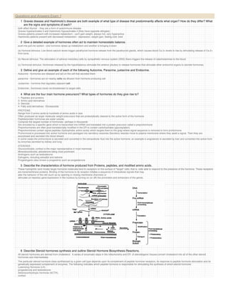
  • 3. Questions and Answers Exam 1 describe what is happening in this figure production of Testosterone leading to dihydrotestosterone from Cholesterol including the intermediate steps in synthesis 12 What are the Eicosanoids- how are they produced and what types of roles do they play? Prostaglandins & related compounds are collectively known as eicosanoids. (prostaglandins, prostacyclins, leukotrienes and thromboxanes) They are produced from arachidonic acid, polyunsaturated fatty acids. They have specific effects on target cells close to their site of formation. They are rapidly degraded, so they are not transported to distal sites within the body. 13 How do NSAIDs work in combination with Eicosanoids? Ibuprofen? Eicosanoids roles in inflammation, fever, regulation of blood pressure, blood clotting, control of reproductive processes & tissue growth, sleep/wake cycle regulation. Ibuprofen and related compounds block the hydrophobic channel by which arachidonate enters the cyclooxygenase active site. 14 Why is Aspirin a permanent effector of PGH2 synthesis? inhibit cyclooxygenase activity of PGH2 Synthase it is IRREVERSIBLE They inhibit formation of prostaglandins involved in fever, pain, & inflammation. They inhibit blood clotting by blocking thromboxane formation in blood platelets. 15 A hormonal stimulus typically produces one or more of what kinds changes in a cell. Alters plasma membrane permeability or membrane potential, or both, by opening or closing ion channels Stimulates synthesis of proteins or regulatory molecules such as enzymes within the cell Activates or deactivates enzymes Induces secretory activity Stimulates mitosis 16 Describe Second messenger systems. a hormone binds to a receptor on the plasma membrane. Of the second messengers, cyclic AMP is by far the best understood, so it will receive most of our attention. (a) Mechanisms that generate cyclic AMP by activation of adenylate cyclase are mediated by G proteins, which are activated when a hormone (first messenger, A in this case) binds to plasma membrane receptors. Cyclic AMP (the second messenger) acts intracellularly to activate protein kinase enzymes that mediate the cell's responses to the hormone. Notice that not all hormones provoke the activation of the effector enzyme. Sometimes they act to inhibit. 17 Describe how G proteins work. (1) The hormone, acting as the first messenger, binds to its receptor. This causes the receptor to change shape, allowing it to bind a nearby inactive G protein. (2) The G protein is activated as the guanosine diphosphate (GDP) bound to it is displaced by the high-energy compound guanosine triphosphate (GTP). The G protein behaves like a light switch; it is "off" when GDP is bound to it, and "on" when GTP is bound. (3) The activated G protein (moving along the membrane) binds to and activates the effector enzyme adenylate cyclase. At this point the GTP bound to the G protein is hydrolyzed to GDP and the G protein becomes inactive once again. (The G protein possesses GTPase activity and cleaves the terminal phosphate group off GTP in much the same way that ATPase enzymes hydrolyze ATP.) (4) The activated adenylate cyclase generates the second-messenger cAMP from ATP. (5) cAMP, which is free to diffuse throughout the cell, triggers a cascade of chemical reactions in which one or more enzymes, called protein kinases, are activated. The protein kinases phosphorylate (add a phosphate group to) various proteins, many of which are other enzymes. Because phosphorylation activates some of these proteins and inhibits others, a variety of reactions may occur in the same target cell at the same time. 18 Describe cyclic AMP signaling in terms of activation and repression. In this mechanism, three plasma membrane components interact to determine intracellular levels of cyclic AMP (cAMP): a hormone receptor, a signal transducer (a G protein), and an effector enzyme (adenylate cyclase). 19 Why is cAMP action short term? it is rapidly degraded by the intracellular enzyme phosphodiesterase 20 Outline the PIP-Calcium Signal Mechanism. intracellular calcium ions act as the final mediator Second-messenger mechanisms of amino acid-based hormones. Some mechanisms use Ca 2+ as an intracellular messenger. The activated G protein stimulates the effector molecule, phospholipase, to cleave PIP 2 into two fragments-inositol triphosphate (IP 3 ) and diacylglycerol (DAG), which act as intracellular second messengers to activate protein kinases and increase the cytoplasmic concentration of Ca 2+ . Ionic calcium also acts as a second messenger to modify the activity of cellular proteins.
  • 4. 20 Outline the PIP-Calcium Signal Mechanism. intracellular calcium ions act as the final mediator Second-messenger mechanisms of amino acid-based hormones. Some mechanisms use Ca 2+ as an intracellular messenger. Questions andprotein stimulates the effector molecule, phospholipase, to cleave PIP 2 into two fragments-inositol triphosphate (IP 3 ) and diacylglycerol (DAG), which act as The activated G Answers Exam 1 intracellular second messengers to activate protein kinases and increase the cytoplasmic concentration of Ca 2+ . Ionic calcium also acts as a second messenger to modify the activity of cellular proteins. 21 Describe the process of Steroid Hormone Direct Gene Activation. This interaction "turns on" a gene, that is, prompts transcription of DNA to produce a messenger RNA (mRNA). The mRNA is then translated on the cytoplasmic ribosomes, producing specific protein molecules. These proteins include enzymes that promote the metabolic activities induced by that particular hormone and, in some cases, promote synthesis of either structural proteins or proteins to be exported from the target cell. 22 Describe Target Cell Specificity . In order for a target cell to respond to a hormone, the cell must have specific protein receptors on its plasma membrane or in its interior to which that hormone can bind. For example, receptors for adrenocorticotropic hormone (ACTH) are normally found only on certain cells of the adrenal cortex. By contrast, thyroxine is the principal hormone stimulating cellular metabolism, and nearly all body cells have thyroxine receptors 23 What is meant by the concept of receptor and hormone affinity? Although binding of a hormone to a receptor is the crucial first step, target cell activation by hormone-receptor interaction depends equally on three factors: (1) blood levels of the hormone, (2) relative numbers of receptors for that hormone on or in the target cells, and (3) affinity (strength) of the bond between the hormone and the receptor. 24 Define Half-Life, Onset, and Duration of Hormone Activity. Hormones are potent chemicals, and they exert profound effects on their target organs at very low concentrations. Hormones circulate in the blood in two forms--free or bound to a protein carrier. In general, lipid-soluble hormones (steroids and thyroid hormone) travel in the bloodstream attached to plasma proteins. All others circulate unencumbered by carriers. The concentration of a circulating hormone in blood at any time reflects (1) its rate of release, and (2) the speed at which it is inactivated and removed from the body and (3) the amount and type of carrier molecules available. Carrier molecules also effect the function and bio availability of the hormone. Some hormones are rapidly degraded by enzymes in their target cells, but most are removed from the blood by the kidneys or liver, and their breakdown products are excreted from the body in urine or, to a lesser extent, in feces. As a result, the length of time a hormone remains in the blood, referred to as its half-life, is usually brief--from a fraction of a minute to 30 minutes, with the water-soluble hormones exhibiting the shortest half-lives The time required for hormone effects to appear varies greatly. Some hormones provoke target organ responses almost immediately, while others, particularly the steroid hormones, require hours to days before their effects are seen. Additionally, some hormones are secreted in a relatively inactive form and must be activated in the target cells. The duration of hormone action is limited, ranging from 10 seconds to several hours, depending on the hormone. Effects may disappear rapidly as blood levels drop, or they may persist for hours after very low hormone levels have been reached. Because of these many variations, hormonal blood levels must be precisely and individually controlled to meet the continuously changing needs of the body. 25 Define and give an example of Permissiveness interactions. Permissiveness is the situation when one hormone cannot exert its full effects without another hormone being present. For example, the development of the reproductive system is largely regulated by reproductive system hormones, as we might expect. However, thyroid hormone is necessary (has a permissive effect) for normal timely development of reproductive structures; without thyroid hormone, reproductive system development is delayed. 26 Define and give an example of Synergism interactions. Synergism of hormones occurs in situations where more than one hormone produces the same effects at the target cell and their combined effects are amplified. For example, both glucagon (produced by the pancreas) and epinephrine cause the liver to release glucose to the blood; when they act together, the amount of glucose released is about 150% of what is released when each hormone acts alone. 27 Define and give an example of Antagonism interactions. When one hormone opposes the action of another hormone, the interaction is called antagonism. For example, insulin, which lowers blood sugar levels, is antagonized by the action of glucagon, which acts to raise blood sugar levels. Antagonists may compete for the same receptors, act through different metabolic pathways, or even, as noted in the progesterone- estrogen interaction at the uterus, cause down-regulation of the receptors for the antagonistic hormone. 28 Describe in detail the three main forms of Endocrine Gland Stimuli and give example of each. Some endocrine glands secrete their hormones in direct response to changing blood levels of certain ions and nutrients. These stimuli are called humoral stimuli to distinguish them from hormonal stimuli, which are also blood borne chemicals. The term humoral harks back to the ancient use of the term humor to refer to various body fluids (blood, bile, and others). This is the simplest of the endocrine control systems. For example, cells of the parathyroid glands monitor blood Ca 2+ levels, and when they detect a decline from normal values, they secrete parathyroid hormone (PTH). Because PTH acts by several routes to reverse that decline, blood Ca 2+ levels soon rise, ending the initiative for PTH release. Other hormones released in response to humoral stimuli include insulin, produced by the pancreas, and aldosterone, one of the adrenal cortex hormones Neural stimuli In a few cases, nerve fibers stimulate hormone release. The classic example of neural stimuli is sympathetic nervous system stimulation of the adrenal medulla to release catecholamines (norepinephrine and epinephrine) during periods of stress. Hormonal stimuli Finally, many endocrine glands release their hormones in response to hormones produced by other endocrine organs, and the stimuli in these cases are called hormonal stimuli. For example, release of most anterior pituitary hormones is regulated by releasing and inhibiting hormones produced by the hypothalamus, and many anterior pituitary hormones in turn stimulate other endocrine organs to release their hormones). 29 Describe Nervous System Modulation. Both "turn on" factors (hormonal, humoral, and neural stimuli) and "turn off" factors (feedback inhibition and others) may be modified by the nervous system. Without this added safeguard, endocrine system activity would be strictly mechanical, much like a household thermostat. A thermostat can maintain the temperature at or around its set value, but it cannot sense that your grandmother visiting from Florida feels cold at that temperature and reset itself accordingly. You must make that adjustment. This is not usually the case in your body, however, where the nervous system can, in certain cases, override normal endocrine controls as needed to maintain homeostasis. For example, the action of insulin and several other hormones normally keeps blood sugar levels in the range of 90-110 mg glucose per 100 ml of blood. However, when the body is under severe stress, blood sugar levels rise because the hypothalamus and sympathetic nervous system centers are strongly activated. This ensures that body cells have sufficient fuel for the more vigorous activity required during such periods.
  • 5. 29 Describe Nervous System Modulation. Both "turn on" factors (hormonal, humoral, and neural stimuli) and "turn off" factors (feedback inhibition and others) may be modified by the nervous system. Without this added safeguard, endocrine system activity would be strictly mechanical, much like a household thermostat. A thermostat can maintain the temperature at or around its set value, but it cannot sense that your grandmother visiting from Florida feels cold at that temperature and reset itself accordingly. You must make that adjustment. QuestionsusuallyAnswers your body, however, where the nervous system can, in certain cases, override normal endocrine controls as needed to maintain homeostasis. For This is not and the case in Exam 1 example, the action of insulin and several other hormones normally keeps blood sugar levels in the range of 90-110 mg glucose per 100 ml of blood. However, when the body is under severe stress, blood sugar levels rise because the hypothalamus and sympathetic nervous system centers are strongly activated. This ensures that body cells have sufficient fuel for the more vigorous activity required during such periods. 30 Describe the spectrum of autoimmune endocrine diseases. Autoimmune diseases form a spectrum ranging from organ-specific conditions in which one organ only is affected to systemic diseases in which the pathology is diffused throughout the body. The extremes of this spectrum result from quite distinct underlying mechanisms, but there are many conditions in which there are components of both organ-specific and systemic damage. . Some pathologies work by shutting a system down – others by hyper stimulating it. 31 Define and describe 3 factors that can lead to Autoimmunity. Endocrine Most autoimmune disease do not occur with equal frequency in males and females. For example Graves' and Hashimoto's are 4-5 times, and SLE 10 times, more common in females while Ankylosing Spondylitis is 3-4 × more frequent in males. These differences are believed to be the result of hormonal influences A second well documented hormonal effect is the marked reduction in disease severity seen in many autoimmune conditions during pregnancy. Rheumatoid arthritis is perhaps the classic example of this effect. In some cases there is also a rapid exacerbation (rebound) after giving birth. Environmental However, it is clear that environmental factors also play a role in autoimmune disease. If you examine how frequently identical twins both develop a disease (the concordance rate), it is only about 20-40% for common autoimmune diseases such as diabetes, SLE and rheumatoid arthritis. This makes it highly likely that environmental factors must also be important. While we might expect factors such as diet to play a role, we can postulate that infectious organisms are the most significant environmental factor. 32 Contrast the mechanisms of Grave’s disease and Hashimoto’s syndrome. Graves disease thyroid is hyperactive autoimmune disease that can be transfered witn IgG antibodies Hashimoto disease thyroid is hypoactive mononuclear cellular infiltrate and autoantibodies targeted at thyroglobulin and thyroid peroxidase 33 Briefly outline the components of the immune system. Made up of two cellular systems humoral or circulating antiBody system - B cells cell mediaTed immunity - T cells Both work by identifying antigens (foreign proteins or polysaccharides) either as part of a virus or bacterium or as a partially degraded byproduct Also recognizes human antigens not made by the individual resulting in graft rejection The humoral antiBody system produces secreted antibodies (proteins) which bind to antigens and identify the antigen complex for destruction. Antibodies act on antigens in the serum and lymph. B-cell produced antibodies may either be attached to B-cell membranes or free in the serum and lymph. The cell mediaTed system acts on antigens appearing on the surface of individual cells. T-cells produce T-cell receptors which recognize specific antigens bound to the antigen presenting structures on the surface of the presenting cell. 34 Describe 3 Autoimmune Polyendocrine Syndromes in detail. Hirata The insulin autoimmune syndrome, associated with Graves’ disease and methimazole therapy (or other sulfhydryl containing medications) is of particular interest due to a remarkably strong association with a specific HLA haplotype . Such patients with elevated titers of anti-insulin autoantibodies frequently present with hypoglycemia. The disease in Japan is essentially confined to DR4-positive individuals with DRB1*0406. In Hirata syndrome the anti-insulin autoantibodies are polyclonal. Some patients have monoclonal anti-insulin autoantibodies that also induce hypoglycemia. For these patients there is no HLA association with their disease. Poems POEMS (Plasmacytoma, endocrinopathy, monoclonal gammopathy, and skin changes) patients usually present with a sensory motor polyneuropathy, diabetes mellitus (50%), primary gonadal failure (70%), and a plasma cell dyscrasia with sclerotic bony. T emporary remission may result following radiotherapy directed at the plasmacytoma. The syndrome is assumed to be secondary to circulating immunoglobulins but patients have excess vascular endothelial growth factor as well as elevated IL1-b, IL-6, and TNF-a. Thymic Tumor Thymomas and thymic hyperplasia are associated with a series of autoimmune diseases. The most common autoimmune diseases are myasthenia gravis and red cell aplasia. Graves’ disease, type 1 diabetes, and Addison’s disease may also be associated with thymic tumors. Unique anti-acetylcholine receptor autoantibodies may be present with thymoma and disease may be initiated by transcription of molecules within the tumor related to acetylcholine receptors. 35 Describe the location and embryology of the Hypothalamus and pituitary gland. Hypothalamus is located in the middle of the base of the brain, and encapsulates the ventral portion of the third ventricle The pituitary gland, also known as the hypophysis, is a roundish organ that lies immediately beneath the hypothalamus, resting in a depression of the base of the skull called the sella turcica ("Turkish saddle").
  • 6. 35 Describe the location and embryology of the Hypothalamus and pituitary gland. The pituitary gland, also known as the hypophysis, is a roundish organ that lies immediately beneath the hypothalamus, resting in a depression of the base of the skull called the sella Questions and saddle"). Exam 1 turcica ("Turkish Answers The pituitary gland develops as a fusion of two groups of cells. There is an upgrowth of ectodermal cells from the roof of the primitive pharynx (known as Rathke's pouch), and a down- growth of neural tissue cells from the hypothalamus. These two distinct areas form the anterior (the adenohypophysis) and posterior (the neurohypophysis) lobes respectively. 36 What hormones are produced in the Hypothalamus and which of them go directly to the blood stream? Hypothalamus produces releasing factors that stimulate production of anterior pituitary hormone which act on peripheral endocrine gland to stimulate release of third hormone 37 Describe vascularization in the hypothalamus and pituitary- why is it this way? How does it improve function? hypophyseal artery A branch of the hypophyseal artery ramifies into a capillary bed in the lower hypothalamus, and hypothalmic hormones destined for the anterior pituitary are secreted into that capillary blood. Blood from those capillaries drains into hypothalamic-hypophyseal portal veins. Portal veins are defined as veins between two capillary beds; the hypothalamic-hypophyseal portal veins branch again into another series of capillaries within the anterior pituitary. Capillaries within the anterior pituitary, which carry hormones secreted by that gland, coalesce into veins that drain into the systemic venous blood. Those veins also collect capillary blood from the posterior pituitary gland. The utility of this unconventional vascular system is that minute quantities of hypothalamic hormones are carried in a concentrated form directly to their target cells in the anterior pituitary, and are not diluted out in the systemic circulation. 38 What is Insulin Autoimmune Syndrome (Hirata Syndrome) and how does it work? The insulin autoimmune syndrome, associated with Graves’ disease and methimazole therapy (or other sulfhydryl containing medications) is of particular interest due to a remarkably strong association with a specific HLA haplotype . Such patients with elevated titers of anti-insulin autoantibodies frequently present with hypoglycemia. The disease in Japan is essentially confined to DR4-positive individuals with DRB1*0406. In Hirata syndrome the anti-insulin autoantibodies are polyclonal. Some patients have monoclonal anti-insulin autoantibodies that also induce hypoglycemia. For these patients there is no HLA association with their disease. 39 Describe the relationship between the pituitary gland and hypothalamus in great detail. Hypothalamic neurons in the supraoptic and paraventricular nuclei synthesize ADH and Oxytocin, which are transported along the hypothalamic-hypophyseal tract to the neurohypophysis for storage. Neurons in the ventral hypothalamus have short axons that discharge releasing and inhibiting hormones to the capillaries of the hypophyseal portal system, which runs to the adenohypophysis. These factors influence the adenohypophyseal secretory cells to release (or not release) their hormones. Environmental stimuli (stressor, temperature, light-dark, etc.) ↓ Higher brain centers (e.g., cerebral cortex) ↓ Neural pathways using neurotransmitters/neuromodulators Hypothalamus ↓ Hypothalamic (a.k.a. hypophysiotropic) hormones Pituitary gland ↓ Hypophyseal hormones Peripheral endocrine glands ↓ Peripheral hormones Target tissues
  • 7. Environmental stimuli (stressor, temperature, light-dark, etc.) ↓ Higher brain centers (e.g., cerebral cortex) ↓ Neural pathways using neurotransmitters/neuromodulators Hypothalamus Questions and Answers Exam 1 ↓ Hypothalamic (a.k.a. hypophysiotropic) hormones Pituitary gland ↓ Hypophyseal hormones Peripheral endocrine glands ↓ Peripheral hormones Target tissues 40 What is GnRH- what does it do – where is it produced- how much is made- what does it regulate? Is it the same in males and females? What is Kallman’s? How is it involved in endometriosis? The vertebrate gonadotropin-releasing hormone (GnRH) is a decapeptide involved in regulating reproduction. The release of GnRH from the hypothalamus regulates the production of gonadotropins in the pituitary and these gonadotropins are responsible for gonadal development and growth in vertebrates. In addition to the hypothalamic GnRH (also called GnRH1 or GnRH-I), many vertebrate species have been found to express other GnRH forms. These include a midbrain GnRH (called GnRH-II or GnRH2) and a telencephalic GnRH (called GnRH-III or GnRH3). 10 aa. Samuel M. McCann, 1960 present in most vertebrate classes Pulsatile secretion!! Hypothalamic site of production: medial preoptic area Extrahypothalamic production: milk, placenta, frog sympathetic nervous system   Effects: FSH↑↑ LH↑↑ mating behavior, sexual drive in animals   Secretion: in utero from puberty → Gonadotropin-releasing hormone (GnRH) plays a key role in the regulation of the reproductive system. GnRH has a similar structure in all animals. It is a decapeptide, meaning that it consists of a chain of 10 amino acids. GnRH acts primarily to stimulate the anterior pituitary gland to synthesise and secrete the gonadotropins FSH and LH. It exerts three principal actions on the anterior pituitary gland: Activation: the movement of gonadotropins from the reserve pool to a pool ready for direct action Immediate release (direct secretion) of gonadotropins      DOPAMINE / SEROTONIN supress production of GnRH KALLMAN'S Kallman’s is a rare condition affecting the hypothalamus, which secretes hormones to the pituitary gland. The hypothalamus is dysfunctional and cannot secret GnRH to the pituitary gland which, as a result, is unable to send either Luteinizing Hormone (LH) or Follicle Simulating Hormone, (FSH) to the ovaries, (women) and testes (men). Symptoms of undiagnosed KS are: no sense of smell (anosmia), failure to enter puberty, lack of sexual drive and infertility (non-ovulation in women and little or no sperm-count in men). Other symptoms may include mood swings, depression, anxiety, fatigue and insomnia. If undiagnosed, the condition can lead to osteoporosis in later life. The treatment for diagnosed patients is either estrogen or testosterone replacement therapy and fertility treatment if a KS patient desires to have a child. Regular bone-density scans, an MRI scan, (magnetic resonance imaging) and blood-tests may be required. ENDOMETRIAL - the mucous membrane lining the uterus, which thickens during the menstrual cycle in preparation for possible implantation of an embryo. ENDOMETRIOSIS - a condition resulting from the appearance of endometrial tissue outside the uterus and causing pelvic pain. Gonadotropin releasing hormone (GnRH) agonists are the most effective hormone treatments for endometriosis. They are able to block the release of the reproductive hormones LH (luteinizing hormone) and FSH (follicular-stimulating hormone). As a result, the ovaries stop ovulating and no longer produce estrogen. They relieve pain in most patients by the second or third month. 41 What is TrH- what does it do – where is it produced- how much is made- what does it regulate? Is it the same in males and females? Thyrotropin-releasing hormone - Present in most vertebrate classes Hypothalamic site of production: PVN Extrahypothalamic production: other brain regions retina pancreas skin of frogs Effects: TSH↑↑ PRL ↑↑ (GH ↑-?) general stimulatory effects (TRH) is a tripeptide hormone that stimulates the release of TSH and prolactin by the anterior pituitary. TRH is produced by the hypothalamus and travels across the median eminence to the pituitary via the pituitary portal system. In addition to the brain, TRH can also be detected in other areas of the body including the gastrointestinal system and pancreatic islets. Medical preparations of TRH are used in diagnostic tests of thyroid disorders and in acromegaly. The sequence of TRH was first determined and the hormone synthesized by Roger Guillemin and Andrew V. Schally in 1969. Its molecular weight is 359.5 Da and its structure is:
  • 8. (TRH) is a tripeptide hormone that stimulates the release of TSH and prolactin by the anterior pituitary. TRH is produced by the hypothalamus and travels across the median Questions andpituitary via the pituitary portal system. eminence to the Answers Exam 1 In addition to the brain, TRH can also be detected in other areas of the body including the gastrointestinal system and pancreatic islets. Medical preparations of TRH are used in diagnostic tests of thyroid disorders and in acromegaly. The sequence of TRH was first determined and the hormone synthesized by Roger Guillemin and Andrew V. Schally in 1969. Its molecular weight is 359.5 Da and its structure is: (pyro)Glu-His-Pro-NH2 42 Somatostatin what does it do – where is it produced- how much is made- what does it regulate? What is the functional difference between the two major forms of it? Why so many receptors? 2 isoforms: 28 aa. (GIT) and 14 aa. (CNS) present in most vertebrate classes Hypothalamic site of production: periventricular nuclei Extrahypothalamic production: pancreas and other GIT structures Effects: GH↓↓ TSH↓ PRL↓   Systemic effects: general inhibitory effects insulin↓ glucagon↓ PP↓ renin↓ PTH↓ calcitonin↓ Somatostatin was first discovered in hypothalamic extracts and identified as a hormone that inhibited secretion of growth hormone. Subsequently, somatostatin was found to be secreted by a broad range of tissues, including pancreas, intestinal tract and regions of the central nervous system outside the hypothalamus. Somatostatin is secreted not only by cells of the hypothalamus but also by so called delta cells of stomach, intestine and pancreas. All actions of the hormone are inhibitory. Somatostatin's main actions are: * Inhibits the release of growth hormone (GH) * Inhibits the release of thyroid stimulating hormone (TSH) * Suppresses the release of gastrointestinal hormones o Gastrin o Cholecystokinin (CCK) o Secretin o Motilin o Vasoactive intestinal peptide (VIP) o Gastric inhibitory polypeptide (GIP) o Enteroglucagon (GIP) * Prolongs gastric emptying, gall bladder contraction and intestinal motility * Suppresses the release of pancreatic hormones o Inhibits the release of insulin o Inhibits the release of glucagon * Suppresses the exocrine secretory action of pancreas Somatostatin antagonizes the effects of Growth Hormone Releasing Hormone (GHRH) Two forms of somatostatin are synthesized. They are referred to as SS-14 and SS-28, reflecting their amino acid chain length. Both forms of somatostatin are generated by proteolytic cleavage of prosomatostatin, which itself is derived from preprosomatostatin. Two cysteine residules in SS-14 allow the peptide to form an internal disulfide bond. Each of the receptors activates distinct signaling mechanisms within cells, although all inhibit adenylyl cyclase. 43 What is GHRH- what does it do – where is it produced- how much is made- what does it regulate? Is it the same in males and females? Growth hormone-releasing hormone 44 aa Pulsatile secretion!! present in most vertebrate classes Hypothalamic site of production: arcuate nucleus Extrahypothalamic production: GIT structures Effects: GH↑↑ (not in >40 yr old) a 44-amino acid peptide hormone produced in the arcuate nucleus of the hypothalamus. It stimulates the production and secretion of growth hormone (GH) from the anterior pituitary gland, and like GH, it is released in a pulsatile manner. Additionally, GHRH also promotes non-REM(rapid eye movement) sleep directly 44 What is CRH- what does it do – where is it produced- how much is made- what does it regulate? Is it the same in males and females? Corticotropin-releasing hormone a 41 aa Hypothalamic site of production: PVN Extrahypothalamic production: very extensive! other brain regions lung GIT placenta Effects: POMC↑↑ (ACTH ↑↑, opioids↑↑, aMSH↑↑ ) general activating effects
  • 9. a 41 aa Hypothalamic site of production: PVN Extrahypothalamic production: very extensive! other brain regions lung GIT Questions and Answers Exam 1 placenta Effects: POMC↑↑ (ACTH ↑↑, opioids↑↑, aMSH↑↑ ) general activating effects (CRH), also called corticotrophin-releasing factor (CRF) or corticoliberin, is a polypeptide hormone involved in the stress response. It is produced by the hypothalamus and stimulates corticotropic cells of the anterior lobe of the pituitary to produce ACTH and other biologically active substances (for example β-endorphin). CRH is also synthesized by the placenta and seems to determine the duration of pregnancy. CRH is a hypophysiotropin that has behavioral effects at the paraventricular nucleus of the hypothalamus, the central nucleus of the amygdala, the bed nucleus of the stria terminalis, and at the locus coeruleus. Release of CRH is affected by serum levels of cortisol and leptin, by stress and by the sleep/wake cycle. 45 Define SET POINTS AND FEEDBACK LOOPS in HYPOTHALAMIC SECRETION. Set point: the “normal” hormone level that the organism tries to maintain at a certain time and under a certain condition.   The hormone levels maintained at the level of set point by feedback mechanisms.   The set point is adjusted according to the needs of the organism, e.g., set point may change across the day (circadian rhythm) or in response to environmental challenges (altered ambient temperature, stress, etc.). Feedback loops: a controlled variable (e.g., blood hormone level) determines the rate of the secretion of the hormone.   Negative feedback loops Positive feedback loops Long feedback loops Short feedback loops Ultrashort feedback loops 46 What are Zeitgebers- how do they affect you on a daily- monthly yearly basis? endogenous (spontaneous) periodic changes.  Endogenous rhythms are generated within the body but are synchronized to the environmental rhythms by environmental stimuli (a.k.a. Zeitgebers). Zeitgebers are environmental cues that usually help keep the circadian cycle. Zeitgebers include sunlight, noise, social interactions, and alarm clocks. Light/ Dark Cycles 47 What do TSH, FSH and LH have in common and why do they need it? – Thyroid-stimulating hormone, also known as thyrotropin, is secreted from cells in the anterior pituitary called thyrotrophs, finds its receptors on epithelial cells in the thyroid gland, and stimulates that gland to synthesize and release thyroid hormones. Follicle stimulating hormone (FSH) – Stimulates follicle development and estrogen secretion in females and sperm production in males – Suppressed by INHIBIN Leutinizing hormone (LH) – Causes ovulation and progestin production in females and androgen production in males – Progestins – Androgens (testosterone) Luteinizing hormone (LH) and follicle-stimulating hormone (FSH) are called gonadotropins because stimulate the gonads - in males, the testes, and in females, the ovaries. They are not necessary for life, but are essential for reproduction. As described for thyroid-simulating hormone, LH and FSH are large glycoproteins composed of alpha and beta subunits. The alpha subunit is identical in all three of these anterior pituitary hormones, while the beta subunit is unique and endows each hormone with the ability to bind its own receptor. 48 What are the functions of FSH and LH in males and females? Be specific! Do they work the same? How are they regulated? What Disease States are associated with LH and FSH? How does manipulating them work in contraception? In both sexes, LH stimulates secretion of sex steroids from the gonads. In the testes, LH binds to receptors on Leydig cells, stimulating synthesis and secretion of testosterone. Theca cells in the ovary respond to LH stimulation by secretion of testosterone, which is converted into estrogen by adjacent granulosa cells. Physiologic effects of the gonadotrophins are known only in the ovaries and testes. Together, then regulate many aspects of gonadal function in both males and females. In females, ovulation of mature follicles on the ovary is induced by a large burst of LH secretion known as the preovulatory LH surge.
  • 10. associated with LH and FSH? How does manipulating them work in contraception? Questions ovulation of matureExam 1 the ovary is induced by a large burst of LH secretion known as the preovulatory LH surge. In females, and Answers follicles on As its name implies, FSH stimulates the maturation of ovarian follicles. Administration of FSH to humans and animals induces "superovulation", or development of more than the usual number of mature follicles and hence, an increased number of mature gametes. FSH is also critical for sperm production. It supports the function of Sertoli cells, which in turn support many aspects of sperm cell maturation. The principle regulator of LH and FSH secretion is gonadotropin-releasing hormone or GnRH (also known as LH-releasing hormone). GnRH is a ten amino acid peptide that is synthesized and secreted from hypothalamic neurons and binds to receptors on gonadotrophs. In a classical negative feedback loop, sex steroids inhibit secretion of GnRH and also appear to have direct negative effects on gonadotrophs. This regulatory loop leads to pulsatile secretion of LH and, to a much lesser extent, FSH. The number of pulses of GnRH and LH varies from a few per day to one or more per hour. In females, pulse frequency is clearly related to stage of the cycle. the gonads secrete at least two additional hormones - inhibin and activin - which selectively inhibit and activate FSH secretion from the pituitary DISEASE STATE Diminished secretion of LH or FSH can result in failure of gonadal function (hypogonadism). This condition is typically manifest in males as failure in production of normal numbers of sperm. In females, cessation of reproductive cycles is commonly observed. Elevated blood levels of gonadotropins usually reflect lack of steroid negative feedback. Removal of the gonads from either males or females, as is commonly done to animals, leads to persistent elevation in LH and FSH. In humans, excessive secretion of FSH and/or LH most commonly the result of gonadal failure or pituitary tumors. In general, elevated levels of gonadotropins per se have no biological effect. CONTRACEPTION manipulation Normal patterns of gonadotropin secretion are absolutely required for reproduction, and interfering particularly with LH secretion is a widely-used strategy for contraception. Oral contraceptive pills contain a progestin (progesterone-mimicking compound), usually combined with an estrogen. As discussed above, progesterone and estrogen inhibit LH secretion, and oral contraceptives are effective because they inhibit the LH surge that induces ovulation. Another route to suppressing gonadotropin secretion is to block the GnRH receptor. GnRH receptor antagonists have potent contraceptive effects in both males and females, but have not been widely deployed for that purpose. 49 Adrenocorticotropic hormone- how is it made? What is made with it? What does it do? Adrenocorticotropic hormone, as its name implies, stimulates the adrenal cortex. More specifically, it stimulates secretion of glucocorticoids such as cortisol, and has little control over secretion of aldosterone, the other major steroid hormone from the adrenal cortex. Another name for ACTH is corticotropin. ACTH is secreted from the anterior pituitary in response to corticotropin-releasing hormone from the hypothalamus. corticotropin-releasing hormone is secreted in response to many types of stress, which makes sense in view of the "stress management" functions of glucocorticoids. Corticotropin-releasing hormone itself is inhibited by glucocorticoids, making it part of a classical negative feedback loop. Within the pituitary gland, ACTH is produced in a process that also generates several other hormones. A large precursor protein named proopiomelanocortin (POMC, "Big Mama") is synthesized and proteolytically chopped into several fragments as depicted below. Not all of the cleavages occur in all species and some occur only in the intermediate lobe of the pituitary. The major attributes of the hormones other than ACTH that are produced in this process are summarized as follows: * Lipotropin: Originally described as having weak lipolytic effects, its major importance is as the precursor to beta-endorphin. * Beta-endorphin and Met-enkephalin: Opioid peptides with pain-alleviation and euphoric effects. * Melanocyte-stimulating hormone (MSH): Known to control melanin pigmentation in the skin of most vertebrates. 50 What is GH or somatotropin? What is related to? What forms of it are there? Why can a mutation in one receptor allele make such a big difference? (GH or somatotropin) – Stimulates cell growth and replication through release of somatomedins or IGF Growth-hormone releasing hormone (GH-RH) Growth-hormone inhibiting hormone (GH-IH) HYPOTHALMUS metabolized in the liver declines after birth for 2-3 weeks increases during growth spurts polypeptide, 2 isoforms; 4-10% of pituitary weight GH RECEPTORS   Structure:   Member of the cytokine receptor superfamily.   Enzyme-linked receptor: - EC ligand-binding domain - single transmembrane domain - IC domain (= Janus kinase)   Ligand binding → Receptor dimerization Receptor-ligand complex internalized  
  • 11.   Member of the cytokine receptor superfamily.   Enzyme-linked receptor: - EC ligand-binding domain - single transmembrane domain Questions and Answers Exam 1 - IC domain (= Janus kinase)   Ligand binding → Receptor dimerization Receptor-ligand complex internalized   Second messenger mechanisms: Janus kinase (JAK) ↓ phosphorylates STAT proteins 51 What are the effects of growth hormone? Differentiate between direct and indirect actions of growth hormone. How does it partner with IGF-I for these effects? Direct effects are the result of growth hormone binding its receptor on target cells. Fat cells (adipocytes), for example, have growth hormone receptors, and growth hormone stimulates them to break down triglyceride and supresses their ability to take up and accumulate circulating lipids. Indirect effects are mediated primarily by a insulin-like growth factor-1 (IGF-1), a hormone that is secreted from the liver and other tissues in response to growth hormone. A majority of the growth promoting effects of growth hormone is actually due to IGF-1 acting on its target cells. Keeping this distinction in mind, we can discuss two major roles of growth hormone and its minion IGF-1 in physiology. The major role of growth hormone in stimulating body growth is to stimulate the liver and other tissues to secrete IGF-1. IGF-1 stimulates proliferation of chondrocytes (cartilage cells), resulting in bone growth. Growth hormone does seem to have a direct effect on bone growth in stimulating differentiation of chondrocytes. IGF-1 also appears to be the key player in muscle growth. It stimulates both the differentiation and proliferation of myoblasts. It also stimulates amino acid uptake and protein synthesis in muscle and other tissues. 52 Detail growth hormones metabolic actions. Protein metabolism: In general, growth hormone stimulates protein anabolism in many tissues. This effect reflects increased amino acid uptake, increased protein synthesis and decreased oxidation of proteins. Fat metabolism: Growth hormone enhances the utilization of fat by stimulating triglyceride breakdown and oxidation in adipocytes. Carbohydrate metabolism: Growth hormone is one of a battery of hormones that serves to maintain blood glucose within a normal range. Growth hormone is often said to have anti- insulin activity, because it supresses the abilities of insulin to stimulate uptake of glucose in peripheral tissues and enhance glucose synthesis in the liver. Somewhat paradoxically, administration of growth hormone stimulates insulin secretion, leading to hyperinsulinemia.   1.  Metabolic actions: conserves glucose for glucose-dependent tissues (brain) and increases lean body mass   a.  mobilizes fat stores as a major energy source: triglycerides breakdown (lipolysis) ↑ → plasma FFA↑ b.  suppresses glucose uptake by muscles (muscle will utilize FFA for energy) and stimulates glucose secretion by liver → plasma glucose↑ (diabetogenic action) c.   stimulates protein synthesis (cell a.a. uptake ↑→ plasma a.a level↓) and suppresses protein degradation (→ plasma urea levels decrease) →→ anabolic action on protein metabolism (positive nitrogen balance) EC collagen ↑ (→ positive phosphorus balance)
  • 12.   1.  Metabolic actions: conserves glucose for glucose-dependent tissues (brain) and increases lean body mass   a.  mobilizes fat stores as a major energy source: triglycerides breakdown (lipolysis) ↑ → plasma FFA↑ Questions and Answers Exam 1 (muscle will utilize FFA for b.  suppresses glucose uptake by muscles energy) and stimulates glucose secretion by liver → plasma glucose↑ (diabetogenic action) c.   stimulates protein synthesis (cell a.a. uptake ↑→ plasma a.a level↓) and suppresses protein degradation (→ plasma urea levels decrease) →→ anabolic action on protein metabolism (positive nitrogen balance) EC collagen ↑ (→ positive phosphorus balance) d.  Electrolyte balance: Na+ and K+ excretion↓ calcium absorption from GIT ↑ 53 Describe the Dual effector theory As blood levels of the hormones produced by the final target glands increase, they inhibit the release of anterior pituitary hormones and thus their own release. This hypothalamic- pituitary-target endocrine organ feedback loop lies at the very core of endocrinology, and it will come up many times in this chapter. Hormonal stimuli promote rhythmic hormone release, with hormone blood levels rising and falling in a specific pattern. Dual effector theory GH primes undifferentiated stem cells → stem cells start dividing and differentiating slowly, produce IGF-1 locally Local IGF-1 from primed stem cells (paracrine-autocrine) and circulating IGF-1 from liver (endocrine) → further proliferation and hypertrophy of the cells 54 Outline the regulation of growth hormone production and secretion. REGULATION OF GH SECRETION   Hypothalamic control: GHRH: stimulates the synthesis and release of GH episodic (phasic) secretion   SST: Somatostatin general “turn off” signal; inhibits neuronal electrical activity, secretory processes and cell proliferation tonic secretion Feedback regulation: IGFs stimulate SST and inhibit GH GH stimulates SST and inhibits GHRH Factors that stimulate GH secretion:   1.    Actual or threatened decrease in the substrate for energy production hypoglycemia fasting exercise   2.    Increase in plasma levels of amino acids protein-rich meal infusion of arginine or other a.a.   3.    Stress fever psychological stress 55 What are the Normal Changes in the Growth Hormone Axis with Aging and what do they mean?
  • 13. Questions andthe Normal Exam 1 in the Growth Hormone Axis with Aging and what do they mean? 55 What are Answers Changes The rate of GH secretion from the anterior pituitary is highest around puberty, and declines progressively thereafter. This age-related decline in GH secretion involves a number of changes in the GH axis, including decreased serum levels of insulin-like growth factor-1 (IGF-1) and decreased secretion of growth hormone-releasing hormone from the hypothalamus. The cause of the normal age-related decrease in GH secretion is not well understood, but is thought to result, in part, from increased secretion of somatostatin, the GH-inhibiting hormone. Normal aging is accompanied by a number of catabolic effects, including a decrease in lean mass, increase in fat mass, and decrease in bone density. Associated with these physiologic changes is a clinical picture often referred to as the somatopause: frailty, muscle atrophy, relative obesity, increased frequency of fractures and disordered sleep. These clinical signs of aging are, without doubt, the manifestation of a very complex set of changes which involve, at least in part, the GH-axis. Naturally, this has spurred considerable interest in administering supplemental GH as a "treatment" for aging in humans, and the availability of recombinant human GH has made such studies feasible 56 What disease states are associated with growth hormone production? How do they differ from hypothyroid states? Why is there an age factor? What are the sources of over production? What are some of the symptoms of late onset growth hormone onset? Clinically, deficiency in growth hormone or receptor defects are as growth retardation or dwarfism. The manifestation of growth hormone deficiency depends upon the age of onset of the disorder and can result from either heritable or acquired disease. I. The innate hypofunction of the GHRH-GH-IGF axis: dwarfism   Etiology (= the cause of the disease): - hypothalamic dysfunction - pituitary dysfunction - defective GH receptor → GH insensitivity → deficient secretion of IGF (= Laron dwarfism)   The effect of excessive secretion of growth hormone is also very dependent on the age of onset and is seen as two distinctive disorders: Giantism is the result of excessive growth hormone secretion that begins in young children or adolescents. It is a very rare disorder, usually resulting from a tumor of somatotropes Acromegaly results from excessive secretion of growth hormone in adults. The onset of this disorder is typically insidious. Clinically, an overgrowth of bone and connective tissue leads to a change in appearance that might be described as having "coarse features". The excessive growth hormone and IGF-1 also lead to metabolic derangements, including glucose intolerance. II. Excess secretion of GH Etiology: - Intrapituitary GH-secreting tumor (eutopic hormone production) - Extrapituitary GH-secreting tumor (ectopic hormone production) - Hypothalamic GHRH-secreting tumor Symptoms:   1. Childhood onset: gigantism: excessive linear growth   2. Onset after adolescence: acromegaly (“acro” = end, tip, peak; “megaly” = increased size) Effects of GH overproduction: - thickening of the skull bones (protruding lower jaw, overgrowth of other facial bones, enlargement of supraorbital ridges) - enlargement of the feet and hands - enlargement of ears, nose and lips - visceral enlargement - thickening of the skin - increased body hair - arthritis 57 Describe the structure of prolactin. Where is it made and how does it work? Is it the same in males and females? Why would a fish need prolactin? Prolactin is a single-chain protein hormone closely related to growth hormone. It is secreted by so-called lactotrophs in the anterior pituitary. It is also synthesized and secreted by a broad range of other cells in the body, most prominently various immune cells, the brain and the decidua of the pregnant uterus. Prolactin is synthesized as a prohormone. Following cleavage of the signal peptide, the length of the mature hormone is between 194 and 199 amino acids, depending on species. Hormone structure is stabilized by three intramolecular disulfide bonds. Chemical structure: 199 a.a. polypeptide (23 kD)  Source: acidophil lactotrop cells in the pituitary pregnant uterus, mammary gland, neurons Plasma: molecular heterogeneity (dimers, tetramers, glycosylated and phosphorylated isoforms) Receptor: related to GH receptor, belongs to cytokine superfamily dimerization JAK/STAT pathway 1.  Promotes mammary growth and development 2.  Stimulates milk secretion in estrogen and progesterone primed mammary gland; effect only on alveolar epithelial cells (no effect on myoepithelial cells, therefore PRL does not stimulate milk ejection) 3.  Males and females: suppresses hypothalamic GnRH secretion → LH↓↓ and FSH↓↓ → prevents ovulation in pregnant and lactating women 4.  Stimulates lymphocyte proliferation 5.  Nonhuman species: regulation of water and salt balance (e.g., salmon’s adjustment from salt to fresh water); premigratory fattening of birds, etc. 58 How is prolactin production and secretion regulated? How does this change during the course of a day- With aging and with immune function? II. Regulation of PRL secretion   Hypothalamic control of PRL secretion:   Inhibition : PRL secretion is under tonic inhibition by dopamine (the hypothalamus continuously keeps a brake on PRL secretion)   Stimulation: The most important stimulatory effect is the removal of the dopaminergic inhibition (i.e., removal of the hypothalamic
  • 14. 58 How is prolactin production and secretion regulated? How does this change during the course of a day- With aging and with immune function? II. Regulation of PRL secretion   Questions and Answerssecretion: 1 Hypothalamic control of PRL Exam   Inhibition : PRL secretion is under tonic inhibition by dopamine (the hypothalamus continuously keeps a brake on PRL secretion)   Stimulation: The most important stimulatory effect is the removal of the dopaminergic inhibition (i.e., removal of the hypothalamic brake) Additional hormones that stimulate PRL secretion: - TRH (T4 suppresses PRL secretion by causing the downregulation of TRH receptors) - VIP - estrogens (cause the upregulation of TRH receptors) - PRF (?)   Feedback: PRL stimulates hypothalamic DA secretion Plasma PRL levels change as a function of a.  age b.  time of the day (circadian rhythm, sleep) c.   pregnancy (increase) d.  stress – exercise (increase) e.  nursing causes burst increases in PRL secretion by activating a neuroendocrine reflex: receptors: mechanoreceptors in nipple afferents: sensory afferents from the nipple center: hypothalamus (inhibition of dopaminergic cells) effector: anterior pituitary (increased PRL secretion) 59 Describe hyperprolactinemia. III. Prolactin overproduction: hyperprolactinemia The most common anterior pituitary disorder, it is present in 25% of infertile women   Etiology: PRL-secreting pituitary tumor (microadenomas or macroadenoma) 60 What is the difference between the anterior and posterior pituitary glands? The posterior pituitary secretes two hormones- Oxytocin which is involved in contraction and Arginine vasopressin (ADH) involved in water reabsorption. These are closely related hormones both made up of nine amino acids- 6 in a ring structure and three extending outwards as a tail. SON (supraoptic nuclei ) makes much of the vasopressin PVN (paraventricular nuclei) makes much of the oxytocin. The neurohypophysis is known also as the pars nervosa - posterior pituitary 61 What are Herring’s bodies and what do they do? What would happen if they weren’t there? HERRING BODIES are dilated areas or bulges in the terminal portion of axons that contain clusters of neurosecretory granules. The granules contain oxytocin or antidiuretic hormone, along with their associated neurophysins. Herring bodies often are seen in association with capillaries Hormones relased from Hypothalamus into post pit, are synthesised while traveling down the neuron axons. Thus, if the herring bodies were not present, the hormones would never be fully synthesized. 62 Describe the Vasopressin-Neurophysin and Oxytocin-Neurophysin genes and their products. How are they regulated? The Vasopressin-Neurophysin and Oxytocin-Neurophysin genes The genes encoding VP and OT are in tandem array on chromosome 20 in Man, separated by 8 Kb of DNA. Each has 3 exons, and encodes a polypeptide precursor with a modular structure: an amino-terminal signal peptide; the VP or OT peptide; a hormone-specific mid-molecule peptide termed a neurophysin (NPI and NPII for OT and VP respectively); and a carboxyl-terminal peptide known as co-peptin . There is considerable homology between the NP sequences of the VP-NP and OT-NP genes, positions 10-74 of the NP sequences being highly conserved at the amino acid level.
  • 15. The Vasopressin-Neurophysin and Oxytocin-Neurophysin genes The genes encoding VP and OT are in tandem array on chromosome 20 in Man, separated by 8 Kb of DNA. Each has 3 exons, and encodes a polypeptide precursor with a modular structure: an amino-terminal signal peptide; the VP or OT peptide; a hormone-specific mid-molecule peptide termed a neurophysin (NPI and NPII for OT and VP respectively); and a carboxyl-terminal peptide known as co-peptin . There is considerable homology between the NP sequences of the VP-NP and OT-NP genes, positions 10-74 of the NP sequences being Questions and at the amino acid level. highly conserved Answers Exam 1 Post-translational processing of the Vasopressin-neurophysin II gene product. Sequential modification of the AVP-NPII preprohormone in endoplasmic reticulum and golgi lead to trafficking through the regulated secretory pathway and ultimately release from neurosecretory vesicles in the posterior pituitary. A small amount of partially processed precursor is released through the constitutive secretory pathway. OT is processed in a similar manner. VP and OT circulate unbound to plasma proteins, though VP does bind to specific receptors on platelets. VP concentrations in platelet-rich plasma are 5-fold higher than in platelet- depleted plasma (4). VP and OT have short circulating half-lives of 5-15 minutes. Several endothelial and circulating endo- and amino-peptidases degrade the peptides. A specific placental cysteine amino-peptidase degrades VP and OT rapidly during pregnancy and the peri-partum period. 63 What are the Neurophysins and what are their roles? A group of small, soluble proteins secreted by the hypothalamus. They serve as binding proteins for oxytocin and vasopressin during their transport to the posterior pituitary. They are secreted with the hormones and serveng as a carrier. 64 What are the effects of oxytocin on lactation- parturition and behavior? What does it do in males? Oxytocin in a nine amino acid peptide that is synthesized in hypothalamic neurons and transported down axons of the posterior pituitary for secretion into blood. Oxytocin is also secreted within the brain and from a few other tissues, including the ovaries and testes. Oxytocin differs from antidiuretic hormone in two of the nine amino acids. Both hormones are packaged into granules and secreted along with carrier proteins called neurophysins. Oxytocin and lactation In the rat, stimulation of vagal sensory afferents in the nipple by the act of suckling triggers reflex synchronized firing of oxytotic magnocellular neurons in the neurohypophysis, and corresponding pulsatile OT release. OT acts on OT-Rs on smooth muscle cells lining the milk ducts of the breast, initiating milk ejection. OT is essential for completion of this milk ejection reflex in rodent. Mice lacking OT fail to transfer milk to their suckling young. This deficit is corrected by injection of OT. In contrast, women lacking posterior pituitary function can breast-feed normally, illustrating that OT is not necessary for lactation in man. Pituitary lactotrophs express OT-R mRNA, and OT released into the hypophyseal portal blood supply from the median eminence can stimulate prolactin release. However, the role of OT in the physiology of prolactin release remains unclear Oxytocin and parturition OT is a uterotonic agent. In many mammals there is both an increase in OT secretion and an increase in uterine responsiveness to OT during parturition. These data suggest a key role for the hormone in the initiation and progression of labour. It is believed that falling progesterone concentrations toward the end of pregnancy lead to up-regulation of uterine myometrial OT-Rs, enhanced contractility, and increased sensitivity to circulating OT. Stretching of the 'birth canal' during parturition leads to the stimulation of specific autonomic afferents, reflex firing of oxytotic neurons and OT release. Males synthesize oxytocin in the same regions of the hypothalamus as in females, and also within the testes and perhaps other reproductive tissues. Pulses of oxytocin can be detected during ejaculation. Current evidence suggests that oxytocin is involved in facilitating sperm transport within the male reproductive system and perhaps also in the female, due to its presence in seminal fluid. It may also have effects on some aspects of male sexual behavior. 3. Sperm transport↑   Females: contractions of the non-pregnant uterus Males: contractions of the vas deferens   4. Maternal behavior↑, mating behavior↑   6. Memory↓   5. ACTH↓, PRL↑ 65 What regulates secretion of oxytocin?
  • 16. Questions and Answers Exam oxytocin? 65 What regulates secretion of 1 The most important stimulus for release of hypothalamic oxytocin is initiated by physical stimulation of the nipples or teats. The act of nursing or suckling is relayed within a few milliseconds to the brain via a spinal reflex arc. These signals impinge on oxytocin-secreting neurons, leading to release of oxytocin. tactile stimulation - stimulates production stress - inhibits production Both the production of oxytocin and response to oxytocin are modulated by circulating levels of sex steroids. The burst of oxytocin released at birth seems to be triggered in part by cervical and vaginal stimulation by the fetus, but also because of abruptly declining concentrations of progesterone. Another well-studied effect of steroid hormones is the marked increase in synthesis of uterine (myometrial) oxytocin receptors late in gestation, resulting from increasing concentrations of circulating estrogen. 66 How does Vasopressin work as a neurotransmitter? What is its receptor? Antidiuretic hormone, also known as vasopressin, is a nine amino acid peptide secreted from the posterior pituitary. Within hypothalamic neurons, the hormone is packaged in secretory vesicles with a carrier protein called neurophysin, and both are released upon hormone secretion. ADH secretion by the magnocellular neurons: synthesis in the rER (in the cell body; hypothalamus) packed into secretory granules axonal transport to the axon terminal (in the neurohypophysis) when action potentials arrive at the terminal → exocytosis of the granules hormone release into the blood stream   In the absence of adequate stimuli: ADH-secreting neurons low discharge rate Adequate stimulus: phasic bursting of the ADH-secreting neurons → ADH release ↑ Vasopressin as a neurotransmitter/neuromodulator in the brain: Suprachiasmatic nucleus PVN neurons projecting to the medial eminence, co-expressed with CRH in parvocellular neurons, facilitates the effects of CRH PVN neurons projecting to the brain stem PVN neurons projecting to brain regions associated with memory   Extraneuronal sources of vasopressin: gonads adrenal cortex platelets Receptors: heptahelical, G protein-coupled   V1A (V1): IP3/DAG, smooth muscle in mesenteric arteries V1B (V3): IP3/DAG, anterior pituitary V2 : cAMP, in the renal collecting ducts, mediate the principle physiological action of ADH   67 Describe in detail how ADH or vasopressin works in the kidney and in the vascular system. Effects on the Kidney The single most important effect of antidiuretic hormone is to conserve body water by reducing the output of urine. A diuretic is an agent that increases the rate of urine formation. Injection of small amounts of antidiuretic hormone into a person or animal results in antidiuresis or decreased formation of urine, and the hormone was named for this effect. Effects on vascular system In many species, high concentrations of antidiuretic hormone cause widespread constriction of arterioles, which leads to increased arterial pressure. It was for this effect that the name vasopressin was coined. In healthy humans, antidiuretic hormone has minimal pressor effects. 68 Describe the affects of ADH. How does it relate to the aquaporins? Low pressure receptors? Osmoreceptors? ADH facilitates water retention by increasing the permeability of tubular wall (main effect) and by increasing the osmolarity of the kidney interstitium (secondary effect). Main Effects - urine volume ↓ - urine becomes more concentrated - the osmotic concentration of body fluids ↓ Accomplished by: Increasing the water permeability of the collecting ducts Increasing the osmolarity of the kidney interstitium Vasopressin and renal water handling Although VP has multiple actions, its principle physiological effect is in the regulation of water resorption in the distal nephron, the structure and transport processes of which allow the kidney to both concentrate and dilute urine in response to the prevailing circulating VP concentration. Active transport of solute out of the thick ascending loop of Henle generates an osmolar gradient in the renal interstitium, which increases from renal cortex to inner medulla, a gradient through which distal parts of the nephron pass en route to the collecting system. The presence of selective water channel proteins (aquaporins) in the wall of the distal nephron allows resorption of water from the duct lumen along an osmotic gradient, and excretion of concentrated urine.
  • 17. Vasopressin and renal water handling Although VP has multiple actions, its principle physiological effect is in the regulation of water resorption in the distal nephron, the structure and transport processes of which allow the kidney to both concentrate and dilute urine in response to the prevailing circulating VP concentration. Active transport of solute out of the thick ascending loop of Henle generates an osmolar gradient in the renal interstitium, which increases from renal cortex to inner medulla, a gradient through which distal parts of the nephron pass en route to the collecting system. The presence of selective water channel proteins (aquaporins) in the wall of the distal nephron allows resorption of water from the duct lumen along an osmotic gradient, and excretion of Questions and Answers Exam 1 concentrated urine. Binding of VP to cell-surface V2-Rs in the distal nephron triggers a signal transduction cascade leading to a biphasic increase in water channel expression on the luminal surface cell membrane. There is increased assembly of pre-synthesized AQP2 monomers to functional tetramers, and enhanced cell surface expression. In addition, AQP2 gene expression is stimulated. V2 → cAMP → PKA → CREB → AP2 formation ↑ V2 → cAMP → PKA → phosphorylation of AP2 ↑ → translocation of AP2 ↑ Increases blood pressure  V1A receptor mediated   smooth muscle contraction in arterial walls↑↑ ↓ vasoconstriction ↓ resistance in the circulation ↑ ↓ arterial blood pressure ↑   Importance: to maintain blood pressure in severe hemorrhage or dehydration   ACTH secretion ↑ V1B receptor mediated  Vasopressin as a neurotransmitter/neuromodulator in the brain - antipyresis - memory consolidation and learning - analgesia - CNS control of blood pressure   69 How do you Control Antidiuretic Hormone Secretion? The most important variable regulating antidiuretic hormone secretion is plasma osmolarity, or the concentration of solutes in blood. Osmolarity is sensed in the hypothalamus by neurons known as an osmoreceptors, and those neurons, in turn, simulate secretion from the neurons that produce antidiuretic hormone. When plasma osmolarity is below a certain threshold, the osmoreceptors are not activated and antidiuretic hormone secretion is suppressed. When osmolarity increases above the threshold, the ever-alert osmoreceptors recognize this a the cue to stimulate the neurons that secrete antidiuretic hormone. As seen the the figure at the right , antidiuretic hormone concentrations rise steeply and linearly with increasing plasma osmolarity. 70 How does Decreased blood volume/blood pressure control ADH?   When the volume of extracellular fluids decreases, three major compensatory mechanisms will be activated to restore EC volume: 1. water retention increases (ADH) 2. sodium retention increases (aldosterone) 3. water intake increases   ADH secretion: neuroendocrine reflex 71 What other factors influence ADH secretion? How does it relate to thirst? VP is produced and regulated by the neurohypophysis, and is modulated by sensory signals chiefly reflecting osmotic status and blood pressure/circulating volume. The relationships of the SON and PVN with the autonomic afferents and central nervous system nuclei responsible for osmo- and baroregulation are key to the physiological regulation of VP.
  • 18. VP is produced and regulated by the neurohypophysis, and is modulated by sensory signals chiefly reflecting osmotic status and blood pressure/circulating volume. The relationships of Questions and Answers Examafferents and central nervous system nuclei responsible for osmo- and baroregulation are key to the physiological regulation of VP. the SON and PVN with the autonomic 1 1. Increased plasma osmolarity stimulates ADH secretion   At normal plasma osmotic levels (285 mosmol/kg and below): no ADH release At increased osmotic levels: ADH↑↑ 2. Decreased blood volume/blood pressure stimulates ADH secretion   blood volume ↓ ↓ blood pressure ↓, if the decrease is greater than 10%, then low-pressure receptors in the circulation are activated (great veins, atria of the heart, pulmonary vessels) ↓ neurons carry the signal from the receptors to the CNS (vagus nerve) ↓ the activity of hypothalamic vasopressinergic neurons↑↑ ↓ plasma ADH levels↑↑ Secretion of antidiuretic hormone is also simulated by decreases in blood pressure and volume, conditions sensed by stretch receptors in the heart and large arteries. 3. Other factors that affect ADH secretion  -        stress, pain, emotions → endogenous opioid↑ → ADH↑ -        postural changes -        angiotensin induces thirst and also stimulates ADH -        estrogen and progesterone stimulate ADH -        nicotine stimulates ADH -        increased body temperature stimulates ADH   -        alcohol: ADH↓ 72 How do Defects in the production or action of VP manifest? Defects in the production or action of VP manifest as clinical problems in maintaining plasma sodium concentration and fluid balance, reflecting the key role of the hormone in these processes. A further group of related clinical conditions reflect primary defects in thirst. In some cases, the two may coincide, reflecting the close anatomical and functional relationship of both processes. There are no clinical consequences resulting from defects in OT production or action. 73 Describe the types of Diabetes Insipidus- how do they come about? What are its symptoms? Diabetes insipidus (DI) is characterized by production of dilute urine in excess of 3l/24 hours (>40 ml/kg/24 hours in adults, >100 ml/kg/24 hours in infants). DI arises through one of three mechanisms. Deficiency of VP: hypothalamic diabetes insipidus (HDI). Renal resistance to the antidiuretic action of VP: nephrogenic diabetes insipidus (NDI). Inappropriate, excessive water drinking: dipsogenic diabetes insipidus (DDI). 1. ADH deficiency: diabetes insipidus   diabetes insipidus diabetes mellitus   Forms: 1. Central diabetes insipidus (ADH is not produced) 2. Nephrogenic diabetes insipidus (defects of the V2 receptors) Congenital: - mutation of ADH gene -        mutation of V2 receptor gene -        mutation of AQP2 gene   Acquired: - Head trauma -  Tumors that destroy the posterior pituitary -  Lithium therapy (V2 receptors↓↓) -  Chemotherapies (AQP2↓↓)   Symptoms: polyuria (12-20 l/day) compensatory (secondary) polydipsia 74 What happens if you make too much ADH? Syndrome of “inappropriate” hypersecretion of ADH (SIADH) ADH hypersecretion in the absence of adequate osmotic or volume stimuli.  Etiology: diverse, ectopic production of ADH, e.g., chronic lung diseases (cc)  Symptoms: water retention↑↑ dilutional hyponatremia (plasma sodium concentration decreases because of the retained water) “water intoxication” (cells shrink) urine is inappropriately concentrated 75 What are some ways in which the normal relationship between plasma osmolality and VP concentration breaks down? There are situations where the normal relationship between plasma osmolality and VP concentration breaks down. Rapid changes of plasma osmolality: rapid increases in plasma osmolality result in exaggerated VP release. During the act of drinking: drinking rapidly suppresses VP release, through afferent pathways originating in the oropharynx.  Pregnancy: the osmotic threshold for VP release is lowered in pregnancy. Aging: plasma VP concentrations increase with age, together with enhanced VP responses to osmotic stimulation. Age-related changes in VP production can be accompanied by blunting of thirst appreciation, reduced fluid intake, decreased ability to excrete a free water load, and reduced renal concentrating capacity. These changes predispose the elderly to both hyper- and hyponatraemia. Baroregulation of Vasopressin Reductions in circulating volume stimulate VP release through activation of mechanoreceptors in the cardiac atria and central veins. Hypotension stimulates VP release independently through aortic arch and carotid sinus afferents. Falls in arterial blood pressure of 5 to 10 per cent are necessary to increase circulating VP concentrations in man. Progressive reduction in blood pressure produces an exponential increase in plasma VP, in contrast to the linear increases of osmoregulated VP release. Baroregulated VP responses can be modified by other neurohumoral influences triggered as part of the coordinated neurohumoral response to changes in circulating volume and blood pressure. Atrial natriuretic peptide (ANP) inhibits, while norepinephrine augments baroregulated VP release.
  • 19. Baroregulation of Vasopressin Reductions in circulating volume stimulate VP release through activation of mechanoreceptors in the cardiac atria and central veins. Hypotension stimulates VP release independently through aortic arch and carotid sinus afferents. Falls in arterial blood pressure of 5 to 10 per cent are necessary to increase circulating VP concentrations in man. Questions Progressive reduction in blood pressure produces an exponential increase in plasma VP, in contrast to the linear increases of osmoregulated VP release. Baroregulated VP and Answers Exam 1 responses can be modified by other neurohumoral influences triggered as part of the coordinated neurohumoral response to changes in circulating volume and blood pressure. Atrial natriuretic peptide (ANP) inhibits, while norepinephrine augments baroregulated VP release. Other ways Nausea and emesis. Manipulation of abdominal contents 76 What are the cardiovascular effects of VP? Its actions on the pituitary? Cardiovascular Effects VP is a potent pressor agent; its effects mediated through a specific receptor (V1-R) expressed by vascular smooth muscle cells. Though systemic effects on arterial blood pressure are only apparent at high concentrations, VP is important in maintaining blood pressure in mild volume depletion. The most striking vascular effects of VP are in the regulation of regional blood flow. The sensitivity of vascular smooth muscle to the pressor effects of VP varies according to the vascular bed. Vasoconstriction of splancnic, hepatic and renal vessels occur at VP concentrations close to the physiological range. Furthermore, there are differential pressor responses within a given vascular bed, selective effects on intrarenal vessels resulting in redistribution of renal blood flow from medulla to cortex. Baroregulated VP release thus constitutes one of the key physiological mediators of an integrated haemodynamic response to volume depletion. Effects on the Pituitary VP is an ACTH secretagogue, acting through pituitary corticotroph-specific V3-Rs. Though the effect is weak in isolation, VP and CRF act synergistically. VP and CRF co-localize in neurohypophyseal parvicellular neurons projecting to the median eminence and the neurohypophyseal portal blood supply of the anterior pituitary. Levels of both VP and CRF in these neurons are inversely related to glucocorticoid levels, consistent with a role in feedback regulation. 77 Describe the relationship between ADH and thirst. Thirst Thirst and drinking are key processes in the maintenance of fluid and electrolyte balance. Thirst perception and the regulation of water ingestion involve complex, integrated neural and neurohumoral pathways. The osmoreceptors regulating thirst are situated in the circumventricular AV3V region of the hypothalamus, distinct from those mediating VP release. As with VP release, there are also specific physiological situations in which the relationship between plasma osmolality and thirst breaks down. The act of drinking: reduces osmotically stimulated thirst. Extracellular volume depletion: this stimulates thirst through volume-sensitive cardiac afferents and the generation of circulating and intracerebral Angiotensin II, a powerful dipsogen. Aging: both thirst appreciation and fluid intake can be blunted in the elderly 78 Describe the role of the placenta and syncytiotrophoblast in endocrine production in pregnancy. The endocrine tissue of the placenta is the syncytiotrophoblast, which covers the chorionic villi, and arises from the fusion of the cytotrophoblasts. Placental hormones are required for the establishment and maintenance of pregnancy, adaptation of the maternal organism to pregnancy, fetal growth and well being, and development of the mechanisms involved in parturition. The placenta is a unique, autonomous and transient organ. It ensures maternal-fetal exchanges and is also involved in maternal tolerance of feto-paternal antigens. The human placenta is characterized by the major invasion of the trophoblast, which comes in contact with the maternal blood, and by the intensity and the specificity of its endocrine functions. One of the first jobs of the syncytiotrophoblast is to signal the mother that she is pregnant.  It does this by producing a hormone that is virtually identical to LH.  This is called human (in the case of the human) chorionic gonadotropin.  The hCG rescues the corpus luteum which becomes the corpus luteum of pregnancy.  This continues for about the first 2 months of pregnancy, until the placenta can produce sufficient estrogen and progesterone to maintain itself and the uterine lining.  This hCG signal can be detected as soon as the syncytiotrophoblast is formed (before the mother misses her first period).  Current pregnancy tests may be sensitive enough to detect hCG in the blood after 11-12 days after ovulation.  However, usually the new parent is advised to wait until the time of the missed period (or a few days after).  Recall that pituitary LH is shut down by the high progesterone and estrogen from the corpus luteum.  So, any hCG found signals the presence of a functional placenta. As it develops, the placenta produces other hormones that are similar to those in the pituitary including chorionic thyrotropin, and somatomammotropin.  These may have specific developmental functions for the fetus.  In its production of estrogens, the placenta is somewhat like the granulosa cells.  It requires androgen precursors from the fetal adrenal before it can make estrogens.  Thus, a partnership is formed between the fetal adrenal and the placenta that produces a significant rise in maternal estrogen levels.  If the fetus fails to develop a brain (anencephalic), there is a third trimester drop in adrenal androgens, because the fetal pituitary has not continued to provide stimulation to the adrenal.  This results in a precipitous drop in estrogens and is often diagnostic of anencephaly. 79 Describe progestin production during pregnancy – where it happens – what regulates it and what the hormones do at different times. Progestins: Progestins are molecules that bind to the progesterone receptor. Progesterone itself is often called the hormone of pregnancy because of the critical role it plays in supporting the endometrium and hence on survival of the conceptus. In some species, placenta produces enough progestin so that the ovaries / corpota lutea can be removed without affecting the pregnancy. In other species, placenta does not produce enough progesting - so ovaries / corpota lutea are important organs synthesizing the needed progesterone throughout pregnancy ROLES OF PROGESTIN Support of the endometrium to provide an environment conducive to fetal survival. If the endometrium is deprived of progestins, the pregnancy will inevitably be terminated. Suppression of contractility in uterine smooth muscle, which, if unchecked, would clearly be a disaster. This is often called the "progesterone block" on the myometrium. Toward the end of gestation, this myometrial-quieting effect is antagonized by rising levels of estrogens, thereby facilitating parturition. Progesterone and other progestins also potently inhibit secretion of the pituitary gonadotropins luteinizing hormone and follicle stimulating hormone. This effect almost always prevents ovulation from occuring during pregnancy. 80 Describe estrogen and its related compounds production during pregnancy – where it happens – what regulates it and what the hormones do at different times. The placenta produces several distinct estrogens. In women, the major estrogen produced by the placenta is estriol, and the equine placenta synthesizes a unique group of estrogens not seen in other animals. Depending on the species, placental estrogens are derived from either fetal androgens, placental progestins, or other steroid precursors. Female Horse urine contains high concentrations of estrogens during the second and third trimesters of pregnancy. source of production of Premarin, hormone replacement therapy ROLES OF ESTROGEN Stimulate growth of the myometrium and antagonize the myometrial-suppressing activity of progesterone. In many species, the high levels of estrogen in late gestation induces myometrial oxytocin receptors, thereby preparing the uterus for parturition. Stimulate mammary gland development. Estrogens are one in a battery of hormones necessary for both ductal and alveolar growth in the mammary gland. Like progestins, estrogens suppress gonadotropin secretion from the pituitary gland. In species like humans and horses, where placental estrogens are synthesized from androgens produced by the fetus, maternal estrogen levels are often a useful indicator of fetal well being.