Biodiesel from microalgae production methods - a review
Manipulating Diatom Silica Cell Walls for Nanotechnology Applications
1. ABSTRACT
Diatoms are unicellular photosynthetic eukaryotes within the class Bacillariophyceae whose peculiarity amongst other
microalgae is their siliceous cell wall, called frustule. In nature, diatoms are widely distributed, diverse and abundant.
There are about 250 genera and more than 100.000 species. Thus, large variety of structures of silicified cell walls on
diatoms offers a promising natural source of materials suitable for nanotechnological applications. Each species posses a
frustule with unique regular architectural nanometric features on their surface which is suitable to use it on catalysis, as a
support for biomedical implants, as a biosensor, etc. Diatom silica can also be converted into other materials, with
maintenance of the detailed morphology of the frustule. To explore different uses of frustules in nanotechnology we
started axenic cultures of Thalassiosira pseudonana, a centric microalgae which has a frustule around 5 µm diameter and
a “petri dish” shape. Our culturing conditions in the laboratory are: f/2 on sterile sea water, 800-1000 Lux on a 12/12 light
cycle at 18-20 ºC. Due to more controlled conditions of unialgal culture we obtain homogeneous populations of frustules
on shape and size after removal of organic matter that cover the silica diatoms. In the future, our goal is to explore
methods of manipulating diatom silica structure, by non-genetic and genetic means.
METHODS
Culture Conditions. Axenic cultures of centric diatom T. Pseudonana (Hustedt)
Hasle and Heimdal (Provasoli-Guillard National Center for Culture of Marine
Phytoplankton, CCMMP1335
Frustules cleaning process. Diatoms are soak 16 hrs on piranha solution
(0.34M K2Cr2O7, 30% H2O2 on H2SO4) at RT. Washed on milliQ water quality
to neutral pH.
For the AFM images, a Digital Instruments equipment consisting in a
Nanoscope IIIa Digital Instruments controller and a Molecular
Imaging head was used at Tapping Mode to achieve high resolution without
inducing destructive frictional forces.
MANIPULATING DIATOM´S SILICA CELL WALL.
Marcia Cortés-Gutiérrez, Rodrigo del Río* & Patricio Vélez
Centro de Neurociencia Celular y Molecular de Valparaíso, Facultad de
Ciencias, Universidad de Valparaíso.
*Instituto de Química, Electroquímica, Universidad Católica de Valparaíso.
CNV
Centro de NeurocienciaCentro de Neurociencia
Celular y Molecular deCelular y Molecular de
ValparaísoValparaíso
Picture BPicture A
10 µm
Cleaning of diatom frustules by piranha solution (PS) treatment. A before PS, dark dots may
represent organelles (i.e. Chloroplasts) B. After PS, complete and clean frustules petri dish
shapes and also “lids” can be seen. 10 µm Bar
Picture C
C. Disassembling of clean frustules by sonication
compared to a SEM image of an intact frustule.
Some details shown in the SEM image can be seen
under light microscope
End product. A dry white
powder, aproximaly 500µl from
a starting 3 liter culture during 3
days.
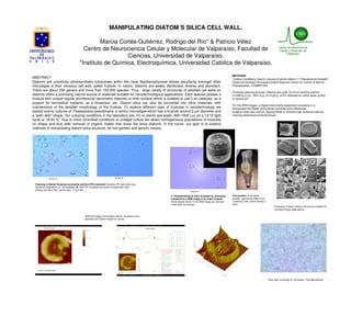
AFM 3d images and analysis section showing a 4µm
diameter and 330nm height for valves.
Examples of large variety of structures suitable for
nanotechnology applications.
Next step is arrange on monolayer. First approaches.