
Recomendados
Más contenido relacionado
La actualidad más candente
La actualidad más candente (10)
Similar a 104 O Haver Handout
Similar a 104 O Haver Handout (20)
Más de MedicineAndDermatology
Más de MedicineAndDermatology (20)
Último
Último (20)
104 O Haver Handout
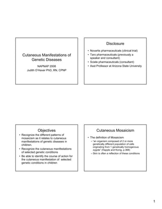
- 1. Disclosure • Novartis pharmaceuticals (clinical trial) • Taro pharmaceuticals (previously a Cutaneous Manifestations of speaker and consultant) Genetic Diseases • Sciele pharmaceuticals (consultant) • Asst Professor at Arizona State University NAPNAP 2008 Judith O’Haver PhD, RN, CPNP Objectives Cutaneous Mosaicism • Recognize the different patterns of • The definition of Mosaicism mosaicism as it relates to cutaneous – “an organism composed of 2 or more manifestations of genetic diseases in genetically different population of cells children. originating from 1 genetically homogenous • Recognize the cutaneous manifestations zygote” (Happle and Konig, p.368) of selected genetic conditions – Skin is often a reflection of these conditions • Be able to identify the course of action for the cutaneous manifestation of selected genetic conditions in children 1
- 2. Types of Cutaneous Mosaicism Lines of Blaschko • Blaschko lines • The lines can be narrow or broad – Checkerboard pattern • The configuration is based on the outgrowth of embryonic cells from the – Phylloid pattern neural crest – Patchy pattern without midline separation – Lateralization • There is a characteristic pattern that arises – It is probable that others will be defined in the as a result of the growth of the embryo in future conjunction with this proliferation of cells May be further defined…. Case 1 • Functional X chromosome mosaicism • 4 year old male is referred to your clinic with a history of brown adherent scaly skin • Autosomal epigenetic mosaicism that waxes and wanes depending on the • Genomic mosaicism weather • The child was born by C-section after failure to progress • Reported cradle cap as an infant 2
- 3. X linked Recessive Ichthyosis Diagnostic features in children • There is an absence of steroid sulfatase due to a • Brown adherent scale gene deletion (Xp22.32) or contiguous gene • May affect the eyes (comma shaped deletion syndrome corneal opacities) • In female carriers this absence leads to perinatal problems such as delayed onset of labor and • Males may have cryptorchidism (20%) failure to progress • Cutaneous involvement waxes and wanes • May be diagnosed prenatally throughout life-may be worse in the dry • The neonates often present with a diffuse weather erythema and exfoliation • Usually present at 2-6 weeks of age Management Case 2 • Newborns • A female is born with a rash that is – -monitor fluid and electrolytes, protein intake partially vesicular on an erythematous and temperature base – -more at risk for infection • It is primarily in a linear pattern on the • Dermatology referral (usually managed child’s leg with topical treatment) • The child was born after a term healthy • If needed refer to pediatric urology pregnancy and does not appear ill • Referral to pediatric opthalmology • Advise OB-GYN in future pregnancies 3
- 4. Incontinentia Pigmenti 4 stages (Bloch-Sulzberger syndrome) • I-Vescicular • This is an X linked dominant disorder that – occurs in the first few weeks to about 4 months of life is usually lethal to males – may present at birth – -male survivors may also have Klinefelter – Appears as blisters or pustules on an erythematous syndrome base or erythematous macules and papules in the • Presents in a Blashko line distribution Blashko line distribution which distinguishes it from other newborn • II-Verrucous rashes – Usually seen about 1 month of age and consists of warty lesions that are usually red brown and scaly – Resolves by 4-6 months Diagnosis and Treatment • III-Hyperpigmentation • Biopsy confirms the diagnosis – Usually 3-6 months • Consult ophthalmology and genetics and eventually dental – Linear and whorls and swirls hyperpigmentation that may persist for years • Additional evals pending symptoms along Blaschko lines • Symptomatic treatment as needed for the • IV-Hypopigmentation lesions – Hypopigmented atrophic streaks (adulthood) • Prognosis is a normal life span may be associated with or without follicular atrophy 4
- 5. Case 3 Neurofibromatosis • Child presents to clinic for evaluation of • There are 3 types of NF increasing numbers of café au lait macules • Type 1 and 2 are autosomal dominant and • Family history suggests that no other occur spontaneously 50% of the time members of the family have multiple • Types 3-8 appear to be partial forms of lesions type 1 and 2 • Child is otherwise well and developmentally normal Neurofibromatosis Neurofibromatosis Type 1 • Type 1 (von Recklinghausen Disease) • Clinical features: • The gene responsible is on chromosome 17 – Café au lait macules (increase in size and number in (17q11.2) and encodes the protein the first 5 years of life) neurofibromin (90%) – Two or more neurofibromas (any type) or 1 or more – Usually appears in young children plexiform neurofibromas • Type 2 is (Acoustic Neuroma Syndrome) – Axillary or inguinal freckling (90%) • The gene responsible is located on the long arm – Bilateral Optic nerve glioma of chromosome 22 (22 q 11-13.1)and encodes the protein merlin – 2 or more Lisch nodules (iris hamartomas) – Usually appears in teens and young adults – Osseous lesions (skeletal dysplasia) – Bilateral vestibular Schwannomas – First degree relative with NF 5
- 6. Neurofibromatosis type 1 Diagnosis and Treatment other features • Macrocephaly • Made on clinical criteria • Short stature • AAP has issued a policy statement on NF that may assist in providing care for these children • Scoliosis and offer guidance for health maintenance • Hypertension • Management depends on symptoms but • Learning disabilities children should be referred to genetics and • ADHD ophthalmology, neurology and other specialists as needed for SX • Mental retardation (rare about 5%) • Prognosis is dependant on involvement • Increased risk of malignancy Monitor for…. Case 4 • Pain • Infant presents to clinic with multiple hypopigmented macules and multiple café • Plexiform neurofibromas au lait macules • Changes in visual acuity • Family history is non-contributory • HA • Hypertension • Infant is otherwise healthy and appears normal • Scoliosis • Abnormalities of long bones • Refer as needed 6
- 7. Tuberous Sclerosis Complex Manifestations (Bourneville’s Syndrome) • Neurocutaneous syndrome • Hypomelanotic macules appear usually • Mutations on 2 genes early in life (ash leaf macule) – TSC1 on chromosome 9 (9q34) • Facial angiofibromas usually in nasolabial – TSC2 on chromosome 16 (16pl3) • Transmitted as an autosomal dominant trait, folds, cheeks or chin may be a spontaneous mutation and parent may • Shagreen patch be asymptomatic • May be diagnosed prenatally if the mutation is • Fibrous facial plaques known • Periungual fibromas • May also note rhabdomyoma if fetal echo is performed • Café au lait macules Other manifestations Diagnosis and Treatment • Requires the presence of 1 major and 2 minor • Neurological component features • Eyes (retinal hamartomas) • Treat symptomatically (ie. Sz) • Kidney • Genetic counseling • Includes multiple specialties • Cardiac (rhabdomyoma) – Neurology (MRI), neurodevelopmental testing • Oral – Opthalmology • Musculoskeletal – Cardiology (EKG, ECHO) – Renal U/S • lungs – Pulmonology if symptomatic 7
- 8. Prognosis Case 5 • Depends on organ involvement • Child presents to clinic with sparse hair, abnormal dentition, frontal bossing, saddle • CNS, cardiac and renal complications are nose, supraorbital ridging and absent the leading cause of morbidity and eyebrows, hyperpigmentation in the orbital mortality area and thick lips. On history mother reports this child does not sweat much. Ectodermal dysplasia Anhidrotic Ectodermal Dysplasia • More than one type • Usually X linked recessive (ectodysplasin – Hypohidrotic ectodermal dysplasia (Christ- (EDA) gene (Xq12-q13) Siemens-Touraine Syndrome) • May be diagnosed prenatally – Hidrotic ectodermal dysplasia (Clouston • Ectodysplasin –defective regulation in the syndrome) ectodermal structures – EEC syndrome (Ectrodactyly-Ectodermal Dysplasia-Cleft lip palate syndrome) • Hypohidrotic ED – AEC syndrome (Ankyloblepharon filiforme – Autosomal dominant adenatum-Ectodermal Dysplasia-Cleft Palate- – 2q11-q13 Hay Wells Syndrome 8
- 9. Multiple structures affected Prognosis and nursing care • Skin • Very important to avoid overheating and treat infections and temps • Hair • May have dry mucosa and skin-treat with • Nails emollients • Craniofacial • Referrals to dental and plastic surgery • Teeth • ENT-prn • Wig • Usually have normal life span Case 6 Epidermolysis Bullosa • Rare genetic skin disease • Nursery consult of an infant born with large blisters after a vaginal delivery • Vesicles and bullae in response to friction • 3 general categories • Negative for infectious disease – Epidermolysis bullosa simplex • Not currently ill or on any medications • Weber-Cockayne • Köebner • Dowling-Meara – Junctional EB (JEB) – Dystrophic EB 9
- 10. Epidermolysis Bullosa Simplex Junctional EB • Autosomal dominant • Herlitz • Defect in the keratin gene (K5 12q, K14 • Junctional EB with pyloric atresia 17q) • Non—Herlitz JEB • Weber-Cockayne • Other variants • Koebner • Dowling-Meara Dystrophic EB Diagnosis and Treatment • Biopsy to make diagnosis-sent for • Dominant (Cockayne-Touraine’s Disease) histopathology, immunomapping and electron • Recessive (Hallopeau-Siemens variant) microscopy • DNA analysis • Prenatal diagnosis is possible • Treatment is supportive – Pain control – Wound management – Nutritional support – Social support • Prognosis depends on the type 10
- 11. Summary References • Dermatlas.com • Role of genetics in recognizing cutaneous • Fitzpatrick, T.B., Johnson, R.A., Wolff, K., & Suurmond, manifestations of selected conditions D. (2001). Color Atlas and Synopsis of Clinical Dermatology (4th ed.) New York: McGraw Hill. • Suggestion of resources for further • Happle, R. Cutaneous Mosaicism (2003). In L.A. information Schachner & R.C. Hansen (eds.) Pediatric Dermatology (3rd edition). Edinburgh: Mosby. • johaver@phoenixchildrens.com • Itin, P.H., Burgdor, W.H.C., Happle, R., Paller, A., Konig, A., Pierini, A., Lenane, P., Krafchik, B., Korf, B., Orlow, S., Mallory, S., Eady, R., & McGrath, J. (2003). Genodermatosis. In L.A. Schachner & R.C. Hansen (eds.) Pediatric Dermatology (2003). (3rd edition). Edinburgh: Mosby. More references… • Krowchuk, D.P. & Mancini, A.J. (Eds.), Pediatric Dermatology: A Quick Reference Guide. American Academy of Pediatrics. • Richard, C., Moss, C., Traupe, H., Pittelkow, M., Lautenschlager, S., Konig, A., Happle, R., & Iten, P. (2003). Ichthyosis and Disorders of Cronification. In L.A. Schachner & R.C. Hansen (eds.) Pediatric Dermatology (3rd edition). Edinburgh: Mosby. • Spitz, J.L. (2005). Genodermatoses (2nd ed.). Philadelphia: Lippincott Williams & Wilkins. • Tidman, M.J. & Garzon, M.C. (2003). Vesiculobullous Disease. In L.A. Schachner & R.C. Hansen (eds.) Pediatric Dermatology (3rd edition). Edinburgh: Mosby. 11