

Recomendados
Más contenido relacionado
La actualidad más candente
La actualidad más candente (20)
Destacado
Destacado (20)
Similar a Approach to anemia
Similar a Approach to anemia (20)
Approach to anemia
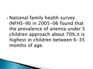
- 1. National family health survey (NFHS-III) in 2005-06 found that the prevalence of anemia under 5 children approach about 70%.it is highest in children between 6-35 months of age.
- 2. PRESENTED BY DR. VIJAY KR. SINGH(DNB PED) UNDRE GUIDENCE OF DR. T. K. MONDAL MD(PED) M R BANGUR HOSPITAL TOLLYGUNJ KOLKATA
- 3. Decreased erythropoesis either due to deficiency of nutrition or defect in erythipoesis Increased blood loss. Diminished red cell survival as a result of immune disorder and chronic diseases. Inadequate iron supply at birth like- prenatal or maternal nutrition deficiency, prematurity, low birth weight, multiple birth.
- 4. Inadequate absorption of iron- Gluten induced entrepathy, atrophic gastritis, post – gasterectomy, presence of phytats in diet and calcium, presence of recurrent diarrhea.
- 5. A. disorder of red cell production in which the rate of red cell production is less than expected. 1. marrow faillure - Aplatic anemia( congenital and aquired) 2. Pure red cell aplasia congenital Diamond- Blackfan Syndrome
- 6. Aese Symdrome Transient erthroblastopenia of childhood C. Bone marrow replacement malignancy Ostipetrosis Myelofibrosis Chronic renal disesas Vitamin D
- 7. Chronic renal disease Hypothyriodism Chronic infection Protein energy malnutrition Hemoglobin mutant with decreased affinity of O2
- 8. Abnormalities in cytoplasmic maturation Iron deficiency Thalassemia syndrome Lead poisoning Abnormalities in nuclear maturation Vitamin B12 folic acid Thiamin respond megaloblastic anemia hereditary abnormalities in folate metabolism Orotic aciduria
- 9. Iron deficiency anemia is the most wide spread micronutrient deficiency and affect nearly 1.5 billion people globally. Infant,pre school children, adolescent, and growing children are at greater risks. According to NFHS-III in 2005-06, the prevalence of anemia under -5 yrs of children is about 70%.
- 10. Normal infant have 75mg of iron of per kg body weight. Two third present in RBC. The majority of body iron is in the form of Hb with about 10% in iron containing protien like myoglobin ant cytochrome.
- 11. Pulses green leafy vegetable Dal Bajra, dates, Nuts, Jaggery Meat and fish Administration of Vitamin- C increased the absorption of iron
- 12. A. Decreased iron store Preterm Small for date baby Twins B. Decreased intake assimilation Delayed intake of complementary feeding Malnutrition Poor iron diet Mal absorption Chronic diarrhoea/ infection Gastero- intestinal surgery
- 13. C. Increased loss Gastro intestinal bleeding Malaria Hook worm infestation, trichuris trichuria. H. pylori. Peptic ulcer. Diveticulutis, bleeding diathesis, feto- femoral haemorrahagem, repeated venous sampling.
- 14. Prematurity LBW Baby Recovery from mal nutrition Adolescence
- 15. Mostly are asymptomatic. Pallor is most important clinical sign but usually not visible until Hb fall to 7-8gm/ dl. Initial manifestation irritability, anorexia, pallor, hyper dynamic circulation leads to palpitation. Shortness of breath, easy fatigability, exercise intorelerance, heart failure. Sign- koilonychias, glossittis, angular chellitis, these are more common in long standing Anemia.
- 16. Hb 6 month-5yrs < 11gm/dl 6- 11yrs <11.5gm/dl non pregnant <12gm/dl pregnant < 11gm/DL MCV <82 Transferring saturation <16% Serum ferritin < 5yrs<12 >5yrs 15 in infection < 30 erythrocytes Zinc protoporphyrin < 5yrs >70 > 5yrs >80
- 17. Serum ferritin decreased Serum iron decreased Iron binding capacity increased
- 18. RDW – Increased>19 RBC- Decreased Peripheral smear shows- Hypochoromic microcytic red cell with sustacial variation in cell size, elliptocytic or cigar shaped red cell are seen.
- 20. Study Iron deficiency anemia thalassemia Anemia of chronic diseases Hemoglobin Decreased Decreased decreased MCV Decreased Decreased Decreased- Normal RDW Increased Decreased Normal- Decreased RBC Decreased Normal- Increased Normal- Decreased Serum ferritin Decreased Normal Increased Total iron binding capacity Increased Normal Dcreased Reticulocyte Decreased Normal Normal -
- 21. Daily doses of 3-6 mg of elemental iron per kg of body weight in two or three divided doses Parental iron prepation is usually used only in malabsorption,poor compliance. Parental iron sucrose and gluconoate complex have a lower risk of serious reaction than dextran. Doses of parental iron-2.3x body weight( 15- patient Hb in gm/dl) +500 to 1000 mg, given in divided doses. Excessive intake of milk should be avoided
- 22. Repeat blood count after 4weeks Hb begins to increased 0.1 to 0.4 gm/ dl per day. Iron medication should be continue for 8weeks after blood value normalized to re- establishment a response to therapy.
- 23. Time after iron administration Response 12-24 hrs Replacement of intracellular iron enzyme , subjective improvement, decreased irritability, increased appetite 36- 48 hrs Initial bone marrow response, erythroid hyperplasia 48-72hrs Reticulocytisis, peaking at 5-7 days 4- 30 days Increased in Hb level 1- 3 months Repletion of store
- 24. Poor compliance. Incorrect dose or administration. Malabsorption of absorbed iron. On going blood loss. Concurrent vitamin B 12 deficiency or fIoate deficiency. Diagnosis other than iron deficiency like Thalassemias Hb C and E disorder Anemia of chronic diseases lead poisoning, sickle cell anemia
- 25. Sideroblastic anemia result from disorder of heme synthesis. Characterized by microcytic, mixed with normal RBC a picture of dimorphic anemia with high RDW, serum iron elevated and trasferrin saturation increased. Elevated iron deposited in mitochondria.
- 26. Megaloblastic anemia is characterized by macrocytic red cells and erthroid precursor which show nuclear dymaturity. The common causes are Vitamin B12 and Folic acid. Incidence incidence of folate deficiency as 6.8% and vitamin B12 32% and combined 20%.
- 27. Megaloblastic anemia affect all hem poetic cell line resulting anemia, thrombocytopenia, and leucopenia. DNA synthesis impaired because of lack of methytetrahydrofolate, vit B12 plays an important role as co factor which is necessary for DNA synthesis.
- 28. Folate deficiency caused by Decreased ingestion celiac disease Malabsorption state Drugs like Methotrexate 6- mercaptopurine Trimithprine, azathiprine, phynatoin and other anticonvulsent and increased requirement infancy, hyperthyroidism, chronic hemolytic disease. Vitamin B 12 deficiency causesd by Decreased ingestion intestinal parasite
- 29. Intritrisic intestinal diseas intrinsic factor deficiency Oritic aciduria HIV infection H. pylori infection
- 30. Pallor Anorexia Irritability Thrombocytopenia, neurtopenia, Glossitis, stomatitis, hyper pigmentation over skin on knuckles Enlarged liver and spleen in 30- 40% cases. loss of position sense, vibration sensation, memory loss, confusion, and neuron psychiatric symptoms.
- 31. MCV>110 Hyper segmented neutrophil. Bone marrow shows one or more cell line affected Bone marrow is cellular and nuclear – cytoplasmic asynchrony. Elevated lactate and billirubin.
- 32. Folate- 1-5 mg/day for 3-4 weeks Vitamin B 12 500-1000micro gram for 2-3 weeks then 100-250 micro gram once in a month to prevent recurrence. Treatment with folic acid alone can produce hematological response in vitamin B 12 deficiency but does not currect neurological impairment caused by Vitamin B12
- 33. Difinition- A term infant have higher hemoglobin and hematocrit and larger RBC than older child and adult, within the 1st week of life a progressive decline of Hb level begins then persist for 6-8wks of life called physiological anemia of infancy. Causes – there is gradual , normal development switch from Hb F to adult Hb after birth which is capable of delivering of more oxygen to the tissue.The increased in blood oxygen content and delivery result in the down regulation of erythropoietin production leading to suppression of eryththropoesis. Tratment- No therapy required
- 34. Hb may decreased to 8gm /dl at 4-8 weeks of life in premature neonate with birth weight less than 1.5 kg. Factors which contributes development of anemia Reduced hematopoietic activity as evident by decreased reticulocytes counts Reduced red cell survival. Treatment – recombinant human erythropoietin with or without transfusion. with adequate protein vitamin E, iron to achieve full benefit of medication.
- 35. DEFINITION A plastic anemia is defined by as presence of any two peripheral blood criteria ANC(Absolute neutrophil count) < 1500/mm platelet count < 40,000/cumm.
- 36. Congenital Fanconi anemia Dyskeratotic congenita Reticular dysgenesis Shwachman- Diamond syndrome Acquired Idiopathic Radiation Viruses- hepatitis, epstein – Bar virus, Parvo virus pregnancy,Hypogamaglubenumia, thymoma, Enophilic fascitis,pre- leukemic state Chemical- benzene,sniffing insectiside
- 37. Infection hepatitis, parvo virus,Epsttien bar virus Drugs sulfa, anticancer drugs chlotramphenical,gold,carbamazepi n .indomethacin . Radiation.
- 38. Thrombocytopenia, with bleeeding in skin, mucasal, GI tract, hematuria, menorrhagia. Neutropenia leads to fever, infection. Anemia appears last with breathlessness, puffiness of face, edema of feet, congestive cardiac failure
- 39. Peripheral smear- normocytic normochromic RBC,leukopenia, decreased ANC, Thrombocytopenia. Coagulation parametre will be normal. Bone marrow biopsy hypocellular marrow with empty spicule, increased fat space, hypoplasia.
- 40. Curative-stem cell transplant Criteria for stem cell transplant 1. Patient who are young 2. Severe a plastic anemia 3. Matched related sibling donor Other treatment Antithymocyte globin(ATG) G-CSF CyclosporineA
- 41. condition Inheritan cce Associated features Risk of malignancy Falconi anemia AR Absent thumb,absent radius, microcephaly,renal anomalies,short stature,skin pigmentation AML, Myelodysplasia,o ral and liver cancer Dyskerato sis congenita X– linked, AD, AR Nail dustrophy, leukoplakia, Squamous cell calcinoma, myelodysplasia Diamond Blackman AD, AR Short stuture,congenital anomalies , elevated Hb F, Raised adenosine deaminase, Leukemia, myelodypkasia
- 42. Diamond blackman syndrome is isolated erythroid hypoplasia occurs in early child hood and associated with congenital anomalies like stabismus, webbed neck, deformed thumb, bony abnormalities of finger and thumb, double ureter, hydronephrosis. Bone marrow shows profound erythroid hypoplasia, macrocytosis. Inceased Hb F Treatment-transfusion therapy, and steroid
- 43. A rare disorder characterized by diminised megakaryocytic tith stem cell defect without associated physical defect or abnormal defect.
- 44. A variety of cells are abnormally sensitive by complement, due to defect in glycosylphosphatidylinositol anchor, which bind protein to the cell membrane. Chronic hemolytic is more common than sleep- induced hemoglobinuria. Diagnosis – molecular testing of CD-56
- 45. It is characterized by presence of vacuoles in bone marrow precursor cells along with sideroblastic anemia with pancreatic exocrine abnormalities.
- 46.