SlideShare una empresa de Scribd logo
1 de 12
Descargar para leer sin conexión
J Oral Maxillofac Surg
69:142-153, 2011


               Classification of Maxillary Central
             Incisors—Implications for Immediate
                  Implant in the Esthetic Zone
                          Sze Lok Lau, BDS(HK), MDS(OMS)(HK), AdvDipOMS(HK), MOSRCS(Edin),
                                          FHKAM(DS), FCDSHK(OMS),*
                         James Chow, MBBS(HK), BDS(HK), MDS(OMS)(HK), FHKAM(DS),
                                      FCDSHK(OMS), FDSRCS(Eng), FRCD(C),†
                          William Li, BDS(HK), MDS(OMS)(HK), AdvDipOMS(Hk), MOSRCS(Edin),
                                   FRACDS, FHKAM(DS), FCDSHK(OMS),‡ and
                          Lop Keung Chow, BDS(HK), MDS(OMS)(HK), MOSRCS(Edin), FHKAM(DS),
                                                FCDSHK(OMS)§

     Purpose: This is the first study to analyze the positions and angulations of the central maxillary incisors
     with reference to the alveolus, providing data for clinicians to achieve good esthetic results for immediate
     implant placement in the esthetic zone.
     Materials and Methods: A total of 300 cone beam images were selected randomly. Five aspects were
     measured: the thickness of the palatal and buccal bone at their mid-root and apical level and the apical
     bone height. A classification was established according to the positions and angulations of the tooth.
     Results: The data from 170 cone beam images were included in the present study. The mean thickness
     of the buccal bone at the mid-root level was 0.9 0.4 mm and at the apical level was 2.04 1.01 mm.
     The mean thickness of the palatal bone at the mid-root level was 3.76 1.37 mm and at the apical level
     was 8.51      2.54 mm. The mean apical bone height was 9.53           2.76 mm. The proportion of incisors
     positioned more buccally (type B) was 78.8%, 19.4%, and 1.8% positioned midway (type M) and more
     palatally (type P), respectively. Regarding the angulation, 49.9% were classified as type 2 (toward buccal),
     34.7% as type 3 (toward buccal, with the long axis anterior to the A point), and 15.4% were categorized
     as type 1 (toward palatal or parallel to the alveolus).
     Conclusions: We recommend that clinicians appreciate the socket in 3 dimensions to achieve a good
     outcome. According to the difficulty of achieving good results, the cases were categorized as levels I to
     III and recommendations were given.
     © 2011 American Association of Oral and Maxillofacial Surgeons
     J Oral Maxillofac Surg 69:142-153, 2011


Throughout the past 40 years, dental implantation has                    by a dental implant has become the reference stan-
gradually changed our thoughts about the line be-                        dard. Traditional guidelines have suggested a 2- to
tween saving a tooth and extraction, as well as tooth                    3-month healing period after dental extraction to al-
replacement therapy. With rapid advances in grafting                     low sufficient bone infilling and remodeling before
materials and refinements in the surgical and pros-                       dental implant placement,1,2 after which, a 3- to
thetic protocols, the restoration of a single tooth gap                  6-month healing period should be allowed before the

Received from Branemark Osseointegration Centre, Hong Kong,              ter, 99 Queen’s Road Central, Hong Kong, People’s Republic of
People’s Republic of China.                                              China; e-mail: alfred.lau@disc-hk.com
  *Specialist in Oral and Maxillofacial Surgery.                         © 2011 American Association of Oral and Maxillofacial Surgeons
  †Specialist in Oral and Maxillofacial Surgery.                         0278-2391/11/6901-0020$36.00/0
  ‡Specialist in Oral and Maxillofacial Surgery.                         doi:10.1016/j.joms.2010.07.074
  §Specialist in Oral and Maxillofacial Surgery.
   Address correspondence and reprint requests to Dr Lau: Brane-
mark Osseointegration Centre, Hong Kong, Room 1901, The Cen-


                                                                   142
LAU ET AL                                                                                                        143


implant is exposed and loaded with a fixed prosthe-            thetic results. The thicknesses of the bone at the
sis.1,2 However, the negative effect of tooth extrac-         buccal and palatal side were evaluated, as was the
tion on the volumetric hard and soft tissue changes in        bone height at the apical tooth region. A new classi-
the buccolingual and vertical dimensions has been             fication is introduced to categorize the different tooth
reported extensively in published studies. Using diag-        positions and angulations, providing a reference to
nostic casts,3-5 radiographs,6-8 and direct measure-          help avoid compromising the buccal bone thickness
ments9 to evaluate the magnitude of bone resorption,          and to prevent fenestration and perforation during
it has been reported that during the first 4 months            implant placement. Different types of teeth were also
after extraction, the mean bone reduction was 5 to 7          ranked according to their difficulties for achieving
mm buccolingually8,9 and 2 to 4.5 mm vertically.6             good long-term results, and general recommendations
Because such bone resorption will strongly affect the         for their management were given.
implant placement and, thus, the esthetics outcome,
clinicians started to insert the implant directly into
                                                              Materials and Methods
the extraction socket immediately after extraction,
under the assumption that this would reduce the                  A series of 300 cone beam (CB) images of the
bone resorption. The first case report of this tech-           maxilla taken from April 2006 to August 2006 were
nique was published in 1976.10 Since then, numerous           randomly selected from the computer record at the
studies have been published regarding the refinement           Dental Implant and Maxillofacial Centre in Hong
of this technique.11-13 Additional clinical research has      Kong. All CB images were taken with the same ma-
shown that the outcomes of immediate implant place-           chine (I-CAT cone beam volumetric tomography and
ment, delayed implant placement, and implant place-           panoramic dental imaging system; Imaging Science
ment in healed socket sites were all comparable.14            International, Hatfield, PA). The head positions were
   The multiple advantages of immediate implant place-        standardized by aligning the patient’s head with hor-
ment are well known, especially in the esthetic zone,         izontal and vertical reference lines, constructed using
such as the maxillary central incisors. These advantages      a beam of light. The patient’s head was positioned
include a reduction in the number of surgeries, a reduc-      such that the horizontal reference beam was at the
tion of the total treatment time, the preservation of         level of the patient’s eyes and the vertical reference
alveolar bone,15-17 the maintenance of a good soft tissue     beam was passing through the patient’s facial midline.
profile,18 and the reduction of the patients’ psychologi-      Once the ideal head position had been located, the
cal trauma owing to the loss of a front tooth.19 However,     patient’s head was fixed with a head frame. The CB
such therapy is not without drawbacks. The greatest           images were viewed and measured by the software
problem for clinicians is the unpredictability of the long-   provided. Each image was examined to identify a fully
term soft tissue stability. The problem of soft tissue        formed, intact, and healthy permanent central upper
recession is usually exacerbated in patients with rela-       right incisor (tooth 11) for analysis. If the central
tively thin buccal bone and thin gingival biotypes.20         upper right incisor was missing, the upper left central
Because the gingival biotype cannot be changed, the           incisor was selected for analysis. The CB imaging data
critical aspect of achieving esthetic success depends on      were excluded if any radiographically detectable
the ideal 3-dimensional implant position21 and the main-      enamel or dentine defect, caries, apical pathologic
tenance of adequate buccal bone thickness over the            features, periodontal alveolar bone loss, restoration of
implant buccal surface.22 Immediate implant therapy           any kind, or fracture was present or when both cen-
has been reported to be very technically demanding14          tral incisors were missing.
and requires an understanding of all these aspects and           Landmarks were identified and marked in the com-
more. Failure to do so will result in an adverse outcome,     puter before the measurements were done. The sag-
defeating the initial objectives of the therapy itself. Im-   ittal section of the chosen incisor was viewed at the
mediate implantation is never easy, especially when the       center of its mesial-distal dimension. As shown in
demand for long-term stable esthetic results is high;         Figure 1, both the palatal line (line 1) and the buccal
thus, case selection is of the utmost importance.             line (line 2) were marked by a line best-fit to the
   The criteria for implant success have been chang-          palatal alveolar surface and the buccal alveolar sur-
ing, and interest in the esthetic outcome is becoming         face, respectively. The alveolar line (line 3) was
a major concern. Implant dentistry has gradually              marked by bisecting the palatal and buccal lines. This
evolved from a bone-driven surgical protocol to a             indicated the angulation of the alveolar process in the
restorative and biologically driven protocol. The             sagittal plane. The angulation of the tooth root was
present study is the first to analyze the positions and        indicated by its long axis (line 4), and this axis was
angulations of the central maxillary incisors with ref-       marked by the midpoint of a line drawn from the buccal
erence to the alveolar bone. Our results will provide         enamel-dentine junction to its palatal counterpart, the
data for clinicians to achieve the best long-term es-         apex of the root. The present study involved measure-
144                                                                 CLASSIFICATION OF MAXILLARY CENTRAL INCISORS


                                                                 ward the buccal bone surface. The thickness of the
                                                                 buccal bone at the mid-root level (measurement I) was
                                                                 measured from a point at the buccal root surface to the
                                                                 buccal bone surface along this line. The palatal bone
                                                                 thickness at the mid-root level (measurement II) was
                                                                 measured from a point at the palatal root surface toward
                                                                 the palatal bone along this same line. The thickness of
                                                                 labial bone at the apical level (measurement III) was
                                                                 defined as a line perpendicular to the long axis of the
                                                                 root, from the apex of the root toward the buccal bone
                                                                 surface. The thickness of the palatal bone at the apical
                                                                 level (measurement IV) was defined as the distance
                                                                 of the line perpendicular to the long axis of the root
                                                                 from the apex of the root toward the palatal bone
                                                                 surface. The apical bone height (measurement V) was


           FIGURE 1. Landmarks for measurement.
Lau et al. Classification of Maxillary Central Incisors. J Oral
Maxillofac Surg 2011.


ment of the residual bone thickness and angulations of
the extraction sockets. The long axis of the tooth root,
instead of the long axis of the whole tooth, was used,
because the range of the crown-root angle is 25.5° for
maxillary central incisors.23 From these reference lines,
4 measurements were performed, using the built-in mea-
suring function of the computer software. The measure-           FIGURE 2. Measurements of bone thickness at different aspects
ments are shown in Figure 2.                                     (measurements I to V).
   A line was drawn from the mid-point of the long axis          Lau et al. Classification of Maxillary Central Incisors. J Oral
of the root perpendicularly from the palatal bone to-            Maxillofac Surg 2011.
LAU ET AL                                                                                                                 145


measured along the long axis of the root from the root                    All measurements and classifications were per-
apex toward the superior bone surface.                                 formed by 2 investigators. Reliability tests were also
   From the CB measurements, the positions and angu-                   performed to check the consistency and accuracy
lations of the tooth roots were classified with reference               using the Statistical Package for Social Sciences, ver-
to the alveolar process. By comparing the buccal and                   sion 11.0, software (SPSS, Chicago, IL). Studies of this
palatal bone thickness at the mid-root level, their posi-              type are exempt from approval by the local institu-
tions with reference to the mid-alveolar line were de-                 tional review board.
fined and classified as follows (Fig 3): type B (closer to
the buccal alveolar surface); type M (midway between
                                                                       Results
the buccal and palatal alveolar surface); and type P
(closer to the palatal alveolar surface). Comparing the                  The CB imaging data from a series of 300 randomly
angulations of the alveolar process with the long axis of              selected patients were accessed initially. The images
the roots, the angulations were classified as follows (Fig              were all examined to identify useful data according to
3): type 1 (root apex angulated toward the palatal side                the defined inclusion and exclusion criteria. The CB
or parallel to the alveolus); type 2 (root apex angulated              imaging data from 170 patients were included in the
toward the buccal side with the long axis passing pos-                 final analysis. Of the excluded 130 patients, 109 had a
terior to point A); and type 3 (root apex angulated                    defective incisor (eg, caries, infections, restorations,
toward the buccal side with the long axis passing ante-                periodontal bone loss, or fracture) and 21 had both
rior to point A).                                                      central incisors missing. The mean patient age was 47


                                    FIGURE 3. Classification according to position and angulation.
Lau et al. Classification of Maxillary Central Incisors. J Oral Maxillofac Surg 2011.
146                                                                          CLASSIFICATION OF MAXILLARY CENTRAL INCISORS


 Table 1. MEASUREMENT RESULTS

                               Thickness at Mid-Root
                                      Level                                 Thickness at Apical Level
  Measurement               Buccal Bone            Palatal Bone       Buccal Bone               Palatal Bone       Apical Bone Height
     (mm)                 (Measurement I)        (Measurement II)   (Measurement III)         (Measurement IV)      (Measurement V)

Mean                           0.90                   3.76               2.04                       8.51                   9.53
Median                         0.89                   3.69               1.96                       8.40                   9.57
Standard deviation             0.40                   1.37               1.01                       2.54                   2.76
Maximum                        1.99                  10.10               7.72                      22.41                  17.20
Minimum                        0.10                   0.50               0.10                       2.56                   2.15
Lau et al. Classification of Maxillary Central Incisors. J Oral Maxillofac Surg 2011.


years (range 13 to 85). Of the 170 patients, 76 (45%)                    root to the nasal floor, almost one half of the patients
were males and 94 were females (55%).                                    (47.6%) had at least 10 mm of bone height; 97.6% had
   The mean thickness of the buccal bone at the                          4 mm or more of bone height and 71.2% of patients
mid-root level (measurement I) was 0.9 mm (range                         had 8 mm or more of bone height (Table 6).
0.1 to 1.99), and the mean thickness of the palatal                         A classification of the positions and angulations of
bone at the mid-root level (measurement II) was 3.76                     the central incisors with reference to the alveolar
1.37 mm (range 0.5 to 10.1). The mean thickness of                       bone was established. Most of the central incisors
the buccal bone at the apical level (measurement III)                    (78.8%) were positioned more buccally within the
was 2.04      1.01 mm (range 0.1 to 7.72), and the                       alveolar bone (type B), 19.4% were positioned in
mean thickness of the palatal bone at the apical level                   midway (type M), and 1.8% were positioned more
(measurement IV) was 8.51 2.54 mm (range 2.56 to                         palatally (type P; Table 7). Regarding the incisor an-
22.41). The mean apical bone height (measurement
                                                                         gulations, almost one half (49.9%) of the central inci-
V) was 9.53     2.76 mm (range 2.15 to 17.2). These
                                                                         sors were classified as type 2, 34.7% were classified as
measurements are listed in Table 1.
                                                                         type 3, and 15.4% (26 cases) were categorized as type
   Additional analysis of the measurements revealed
that 57% of the patients had a buccal bone thickness                     1. An analysis that combined both the position and
at the mid-root level of less than 1 mm (Table 2), and                   the angulation type showed that most were type B2
62.4% of patients had a palatal bone thickness at the                    (38.2%). The incidence of type B3 (34.7%) was only
mid-root level of less than 4 mm—up to 86.5% of                          slightly less than that of type B2, followed by type M2
patients had a thickness of less than 5 mm (Table 3).                    (11.7%). No type P2, P3, or M3 was found in our
Similarly, the buccal bone at the apical level was                       patient series. The rest of the types were few and are
much thinner than its palatal counterpart. The buccal                    listed in Table 7.
bone thickness at the apical level was less than 5 mm                       A reliability test was done to determine the accu-
in 98.8% of the patients, and more than one half of the                  racy of the measurements and classifications between
patients (51.8%) had a thickness of less than 2 mm                       the 2 investigators. The correlation coefficients for
(Table 4). In contrast, 96.5% of patients had a palatal                  the different measurements are listed in Table 8 and
bone thickness at the apical level of 5 mm or more,                      reflected the good consistency between the 2 inves-
and more than one half of the patients (55.9%) had a                     tigators.
thickness of 8 mm or more (Table 5). Regarding the
apical bone height, measured from the apex of the

                                                                           Table 3. THICKNESS OF PALATAL BONE AT MID-
 Table 2. THICKNESS OF BUCCAL BONE AT MID-                                 ROOT LEVEL (MEASUREMENT II)
 ROOT LEVEL (MEASUREMENT I)
                                                                                        Thickness (mm)           Patients (n)
           Thickness (mm)                   Patients (n)
                                                                                               2                   8 (4.7)
                   0.5                       23 (13.5)                                         3                  40 (23.5)
                   1.0                       97 (57)                                           4                 106 (62.4)
                   1.5                      158 (92.9)                                         5                 147 (86.5)
                   2.0                      170 (100)                                          6                 159 (93.5)
Data in parentheses are percentages.                                     Data in parentheses are percentages.
Lau et al. Classification of Maxillary Central Incisors. J Oral           Lau et al. Classification of Maxillary Central Incisors. J Oral
Maxillofac Surg 2011.                                                    Maxillofac Surg 2011.
LAU ET AL                                                                                                                147


 Table 4. THICKNESS OF BUCCAL BONE AT APICAL                      Table 6. APICAL BONE HEIGHT (MEASUREMENT V)
 LEVEL (MEASUREMENT III)
                                                                            Thickness (mm)             Patients (n)
           Thickness (mm)             Patients (n)
                                                                                     4                  166 (97.6)
                   1                    17 (10.0)                                    6                  153 (90.0)
                   2                    88 (51.8)                                    8                  121 (71.2)
                   3                   152 (89.4)                                   10                   81 (47.6)
                   4                   164 (96.5)
                   5                   168 (98.8)                Data in parentheses are percentages.
                                                                 Lau et al. Classification of Maxillary Central Incisors. J Oral
Data in parentheses are percentages.                             Maxillofac Surg 2011.
Lau et al. Classification of Maxillary Central Incisors. J Oral
Maxillofac Surg 2011.

                                                                 evaluate the root position 3 dimensionally before
Discussion                                                       placement.
                                                                    Although it might be worthwhile information, we
   The present study aimed to provide data to aid the            purposely did not include the measurements of the
treatment planning of immediate implant in the es-               buccal and palatal bone thickness at the crestal re-
thetic zone using a CB imaging technique. With rapid             gion, because of the uncontrollable high percentage
advances in surgical and imaging techniques, the de-             of error owing to its relative thinness. Although mag-
mand for accuracy in 3-dimensional and volumetric                nification is possible in the computer, the resolution
measurements is ever increasing. CB imaging has be-              will be too low for measurement. Moreover, a beam
come the standard for implant planning, especially in            hardening effect is always present over the edge,
the highly esthetically demanding areas. Numerous                making measuring the bone thickness at the crestal
studies have been published to support the routine               level worthless.
use of CB imaging in implant dentistry, including the               As previously mentioned, the main drawback of
advantages of convenience, accuracy, and relatively
                                                                 immediate implant placement into the extraction
low radiation dosage.24-27 Increasingly, more clini-
                                                                 sockets has been the lack of predictability of the
cians have been incorporating this into daily practice.
                                                                 long-term soft tissue profile, especially on the buccal
   Only the roots of the maxillary central incisors
                                                                 aspect. Thus, the position of implant placement is
were evaluated in the present study, instead of the
                                                                 critical and is a limiting variable. Although it was
whole tooth. Neither were the crown-to-root relation-
                                                                 previously believed that the bone remodeling and soft
ships measured. This was because it was reported by
                                                                 tissue recession over an extraction socket would be
Bryant et al23 that only a mean of 1.74° is present
                                                                 arrested by inserting a dental implant into the socket,
between the long axis of the root and the crown of
                                                                 recent studies have failed to support this.28-30 It has
normal central maxillary incisors. With priority given
                                                                 been confirmed that, irrespective of the placement of
to a restorative driven concept, if implants could be
                                                                 a dental implant, bone resorption after extraction will
placed into the extraction sockets exactly at the same
                                                                 still occur, leading to a loss of bone volume.28-30 It has
angulations as the roots inside the alveolar bone, it
                                                                 also been reported that the buccal bone resorbs more
would provide an ideal 3-dimensional position for the
                                                                 than the lingual or palatal bone after extraction, be-
prosthetic crown and only a simple straight stock
                                                                 cause it is composed of bundle bone alone. In con-
abutment would be needed. Thus, it is important to
                                                                 trast, lingual or palatal bone is composed of cortical
                                                                 bone, at least at the outermost surface; thus, the
                                                                 resistance to resorption is better.31 The mid-buccal
 Table 5. THICKNESS OF PALATAL BONE AT APICAL                    recession of an immediate implant placed into a fresh
 LEVEL (MEASUREMENT IV)
                                                                 extraction socket has been reported to be 0.55 to
           Thickness (mm)             Patients (n)               0.75 mm at 1 year of follow-up.32,33 However, long-
                                                                 term data are still not available to determine whether
                    5                  164 (96.5)
                                                                 additional resorption occurs. Kohal et al34 have
                    6                  153 (90)
                    7                  126 (74.1)                shown that pressure of the inserted implant on the
                    8                   95 (55.9)                bony wall can result in microfractures, leading to
                    9                   61 (35.9)                crestal bone loss. Therefore, the ideal placement of an
                   10                   38 (22.4)                immediate implant should be aimed toward obtaining
Data in parentheses are percentages.                             maximum bone-to-implant contact to achieve good
Lau et al. Classification of Maxillary Central Incisors. J Oral   primary stability and promote greater osseointegra-
Maxillofac Surg 2011.                                            tion.35,36 However, the key to long-term good esthetic
148                                                                        CLASSIFICATION OF MAXILLARY CENTRAL INCISORS


 Table 7. CLASSIFICATION

                                                                               Type
Type                             B                                 M                             P                            Total

 1                           5.9 (10)                          7.7 (13)                       1.8 (3)                      15.4 (26)
 2                          38.2 (65)                         11.7 (20)                         0 (0)                      49.9 (85)
 3                          34.7 (59)                            0 (0)                          0 (0)                      34.7 (59)
Total                       78.8 (134)                        19.4 (33)                       1.8 (3)                       100 (170)
Data are presented as parentheses, with numbers in parentheses.
Lau et al. Classification of Maxillary Central Incisors. J Oral Maxillofac Surg 2011.


results is to avoid exerting pressure on the crestal                   giva, thus minimizing mid-buccal recession and
bony wall, particularly on the buccal aspect.                          shrinkage of the papillae. However, only 7.7% of type
   With respect to the classification reported in the                   M1 were reported (Table 7).
present study, both the position and the angulation                       Although type P1 is not as perfect as type M1, these
types were categorized into different ranks according                  types are still good, because they will have enough
to their difficulty for achieving ideal esthetic results in             buccal bone to support the overlying soft tissue. Rel-
immediate implant cases. The classification levels are                  atively less bone will be present at the palatal side;
level I to level III (Fig 4).                                          however, the palatal soft tissue is very thick and the
                                                                       palatal bone can resist resorption much better. The
  LEVEL I                                                              recession of palatal tissue will be less significant, if
   Level I (M1, P1) indicates that the implant could be                any occurs, and the esthetic demand is usually not
placed with the same angulation as the extraction                      high on the palatal side. Similar to those with type M1,
socket, without compromising the primary stability                     those with type P1 were in the minority, with only 3
and long-term esthetic outcome because of the com-                     patients (1.8%) classified as having type P1 (Table 7).
mon features of a relatively thicker buccal bone. Be-
cause no modification of the drilling angle would be                       LEVEL II
required, a straight stock abutment could be used.                       Level II (B1, B2, M2, M3, P2, P3) is more technically
These types are the most straightforward for both                      demanding. The angulation of the implant should be
surgery and its restoration of all the levels. However,
only 9.5% of patients had extraction sockets catego-
rized as this level (Fig 4).
   Type M1 should be ideal for immediate implant
cases. This type of tooth lies in the middle of the
alveolar bone and root apex, angulated away from the
buccal wall. When placing an implant into the extrac-
tion socket, enough bone will be present to support
the implant, thereby achieving good primary stability
and enough bone-to-implant contact for good os-
seointegration. Also, the implant angulation will be
perfect for the superstructure. Moreover, enough
buccal bone is present to support the overlying gin-


 Table 8. INTERINVESTIGATOR RELIABILITY TEST

        Investigation             Correlation Coefficients

    Measurement I                           0.955
    Measurement II                          0.805
    Measurement III                         0.830
    Measurement IV                          0.876
    Measurement V                           0.898                      FIGURE 4. Levels according to difficulty in achieving good long-
    Classifications                          0.922                      term esthetic results for immediate implant.
Lau et al. Classification of Maxillary Central Incisors. J Oral         Lau et al. Classification of Maxillary Central Incisors. J Oral
Maxillofac Surg 2011.                                                  Maxillofac Surg 2011.
LAU ET AL                                                                                                            149


changed to avoid thinning of the buccal bone to               Table 9. APICAL DIMENSIONS OF COMMONLY
maintain a long-term stable esthetic outcome, be-             USED IMMEDIATE IMPLANTS IN ESTHETIC ZONE
cause it has the common disadvantage of a relatively
thin buccal plate. The angle of the implant should be                                           Apical Diameter    Radius
                                                                     Implant Brand                   (mm)          (mm)
placed more palatally to avoid compressing or drilling
the buccal bone, minimizing the chance of perfora-           Ankylos (A type), Densply
tions and fenestrations. Because a discrepancy exists          Friadent                                3.5           1.75
between the implant angle to the original tooth, an          OsseoSpeed (4.5), Astra Tech
                                                               Dental                                  3.5           1.75
angled abutment should be chosen to obtain good              Tapered Internal (3.8 mm),
esthetics. More than one half of the cases (55.8%)             BioHorizons                             2.8           1.40
were categorized as level II (Table 7).                      ITI (Bone Level), Straumann               3.5           1.75
   Regarding the positions of the maxillary central          Replace Tapered Groovy (RP),
incisors, most were type B (78.8%); thus, most of the          Nobel Biocare                           2.7           1.35
                                                             Tapered Screw-Vent (3.7
teeth lie more buccally. Type B1 (5.9%) is not ideal for       mmD), Zimmer Dental                     3.1           1.55
immediate implantation, because it is close to the
buccal wall, indicating relatively thinner buccal bone.      Lau et al. Classification of Maxillary Central Incisors. J Oral
                                                             Maxillofac Surg 2011.
However, type B1 is not the most challenging of this
level because it does not angulate toward the natural
depression of the buccal alveolus. The chance of             the side of the implant drill should not be cutting on
additional thinning of the buccal wall or fenestration       the buccal plate during osteotomy, especially for type
will be reduced.                                             B, because it is expected to be thinner than other
   The least favorable types of maxillary central inci-      types. Otherwise, the risk of perforation or thinning
sors for immediate implant are those positioned near         down the already thin buccal plate will be high. Table
the buccal wall with the apex pointing toward the            9 provides a reference for the apical radius of com-
buccal side (types B2 and B3; type B3 was categorized        monly used implant brands for immediate implant in
as difficult [level III] and was included in the next         the esthetic zone, which ranges from 1.35 to 1.75
section). A type B2 tooth would naturally produce a          mm. More than one half of the patients had a buccal
thin buccal plate at both the crestal and the apical         bone thickness of less than 2 mm at the apical region,
region. For immediate implant placement, good pri-           indicating that placing implant drills exactly along the
mary stability is mandatory, especially for those            axis of extraction sockets will result in a high risk of
scheduled to have immediate placement of a provi-            perforation, especially when placing the implant
sional crown. When achieving good primary stability,         deeper toward the apical bone to achieve primary
pressure is almost always present on the buccal bone,        stability.
either at the apical region or at both the apical and the       The mean thickness of the palatal bone at the apical
crestal regions. Because this type has a thin buccal         level (measurement IV) is much greater than its buc-
plate, any pressure exerted on it will significantly          cal counterpart. We found an average of 8.51 mm of
increase the risk of bone resorption, leading to the         palatal bone at this region, with a maximal thickness
loss of soft tissue in the long term. However, a major       of 22.41 mm (compared with an average of 2.04
proportion of patients will have these types (type B2,       1.01 mm). More than one half (55.9%) of the patients
38.2%). This explains the “physiologic” bone and soft        had a thickness of 8 mm or more and 96.5% of pa-
tissue loss on the buccal side in some immediate             tients had a thickness of 5 mm or more (Table 5). In
implant cases. Although it can be difficult to achieve        contrast, the thickness of the palatal bone at the
good primary stability, clinicians will still need to        mid-root level (measurement II) had a mean of only
avoid overcompression of the buccal plate in such            3.76 mm, and 76.5% of patients have a bone thickness
cases. This explains why these cases are so technique-       of less than 3 mm. This probably resulted from the
sensitive.                                                   contour of the palatal vault. Thus, the more apically
   Our data suggest that the buccal bone of maxillary        placed, the thicker the palatal bone. However, this is
incisors were, on average, thin. The mean thickness          not the same for the buccal aspect owing to the
of the buccal bone at the mid-root level (measure-           natural depression at point A and, sometimes, the
ment I) was only 0.9 mm, and the buccal bone at the          extreme root angulations, such as a type 3 tooth.
apical level (measurement III) was 2.04 mm. More             Thus, the implant should be placed more palatally in
than one half of the patients (57%) have a buccal bone       the extraction socket, because more bone will be
thickness at the mid-root level of less than 1 mm            present in that area to achieve good implant stability
(Table 2), and 51.8% of patients had a buccal bone           and to avoid thinning of the buccal wall. However,
thickness at the apical level of less than 2 mm (Table       the implant should also be placed more palatally at
4). It is critical not to drill over the buccal area— even   the apical area, pivoting around the mid-root level.
150                                                             CLASSIFICATION OF MAXILLARY CENTRAL INCISORS


This is because the palatal bone is relatively thicker at   angulation with reference to the extraction socket,
the apical area than at the mid-root region. Clinicians     and in this case, more palatally.
must also be careful not to angulate the apex of the
implant too palatally, otherwise, the coronal part of         LEVEL III
the implant will be tilted too buccally. This can lead to      The type B3 tooth is the most challenging case with
overcompression, or even perforation, on the buccal         respect to achieving good long-term esthetic out-
crestal wall, making the coronal platform point out-        comes because it not only will have a very thin buccal
ward buccally and compromising the esthetic out-            plate, but also the long axis of the tooth apex will be
come. A recent study confirmed that the buccal-lin-          angulated very buccally, passing anterior to the natu-
gual position of the implant shoulder is a very             ral contour of the maxillary alveolar bone (Fig 5A).
important factor determining the degree of buccal           This type is mostly seen in patients with maxillary
marginal tissue recession.37 It was reported that an        alveolar hyperplasia and angle Class II division 2 oc-
implant with a shoulder positioned at, or buccally, to      clusion. Because a large difference exists between the
a line drawn between the cervical margin of the             angulations of the alveolar bone and the tooth, the
adjacent teeth resulted in 3 times more recession than      implant position will be compromised, regardless of
an implant with a shoulder positioned lingually or          whether a bone-driven or restorative-driven protocol
palatally to this line.37 If an ideal angulation for good   is used (Figs 5A,B). Sometimes, traditional guidelines
apical stability without exerting excessive pressure        can be followed for such cases by extracting the
on the buccal wall and without placing the shoulder
buccal to this line cannot be achieved, clinicians can
always consider choosing an implant with a smaller
coronal diameter.
   Regarding the apical bone height (measurement V),
it was recorded that 97.6% of patients had a bone
height of 4 mm or more, and 90% of patients had a
bone height of 6 mm or more (Table 6). The general
recommendation for the placement of immediate im-
plant in an extraction socket in the apical dimension
is to engage the implant 3 to 5 mm beyond the apex
of the socket to achieve good primary stability.38,39
The results of the present study have shown adequate
bone is present at the apical area. Even when the
primary stability is not optimal, the implant could
be placed deeper to achieve this, because usually
more than enough bone is present at this region
(almost one half of the patients had 10 mm or more of
apical bone height). However, a suitable implant
length should carefully be chosen, because the apical-
coronal position of the implant shoulder is also an
important factor in determining the long-term soft
tissue profile and the esthetic outcome.21,40,41 The
length of the implant should be optimal, deep enough
to provide good apical stability, without perforation,
and the shoulder positioned with, or slightly apical to,
the buccal marginal bone crest (around 3 to 4 mm
from the buccal gingival margin38,42,43 or 3 mm apical
to the cementoenamel junctions of the periodontally
sound adjacent tooth44).
   Unlike type B2, types M2, M3, P2, and P3 have
similar characteristics, including a relatively thicker
buccal plate. However, the root apexes will point
toward the buccal side. If the implant were placed
                                                            FIGURE 5. Examples of level III socket. A, Large difference be-
exactly into the extraction socket with the same an-        tween angulations of original tooth and future implant. B, Extreme
gulation, the risk of perforation would be very high.       angulation requiring traditional treatment or socket transformation.
The general recommendation for all level II cases is        Lau et al. Classification of Maxillary Central Incisors. J Oral
that the implants should be inserted with a modified         Maxillofac Surg 2011.
LAU ET AL                                                                                                           151


tooth, with or without simultaneous grafting, and          supply from the periosteum to the buccal plate during
inserting the implant several months later. As such,       the flap raising. For level I cases, inserting the implant
although the advantages of immediate implantation          with the same angulation of the socket using a stan-
would not be achieved, the long-term soft tissue sta-      dard drilling protocol will be straightforward. For
bility will be more predictable.                           level II cases, the implant angulation should be
   If enough bone is present at the palatal and apical     changed to a more palatal aspect at the apical region,
aspects, an attempt could be made by placing the           pivoting around the mid-palatal area. It is recom-
implant both palatally and apically to avoid touching      mended to first use a round bur to create a step at the
the buccal plate. However, even if primary stability       palatal aspect of the apical area of the socket before
can be achieved, the difference will still be large        using a straight drill to ensure that the hardness of the
between the angle of the implant and prosthetic            cortical bone and natural contour of the socket does
crown. An angled abutment will always be needed.           not misguide the operator to drill parallel toward the
   One should be familiar with the implant system one      apical long axis (Fig 6). The implant should be in-
is using, whether an angulated temporary abutment is       serted with good primary stability, without exerting
available, and, if so, the different angles available if   pressure on the buccal wall. To ensure the absence of
planning an immediate provisional crown. The final          pressure on the buccal bone, the operator can leave a
restoration should also be considered before the im-       gap between the implant surface and the buccal
plant is inserted. Most often, for an abutment with a      bone, as long as the primary stability is not compro-
relatively big angle, a step of metal will be present on   mised. The gap can be treated with grafting materials
the buccal aspect. It can sometimes show through the       and/or a barrier membrane to achieve maximum os-
buccal gingiva, compromising the esthetic outcome.         seointergration and good soft tissue healing, depend-
Placing the implant further apically can be done to        ing on the gap size.12,14,45-48 The implant shoulder
solve this problem. Customized abutments or a ce-          should be optimally placed in terms of the depth and
ramic abutment can also be used according to the           buccal-palatal dimension. A provisional crown should
position and angulation of the implant. However, the im-   be considered whenever possible for soft tissue sup-
plant position will most often be controlled by the        port, favoring the long-term esthetic outcome,31 by
bone available for such cases. Thus, type B3 is very       selecting an appropriate abutment. For level III cases,
technically demanding for immediate implant, be-           extra precautions should be given to the prosthetic
cause it is both a bone-driven and a prosthetic-driven     piece owing to its extreme angulations. One should
procedure.                                                 be familiar with the different types of abutments avail-
   If the demand for a good esthetic outcome and           able, as well as their dimensions and shapes. The
immediate provisional restoration is very high, a          implant can be placed deeper apically to avoid buccal
socket transformation procedure can be done by             showing of the metal margin of an angulated abut-
grafting the buccal wall in the first stage, followed by
extraction of the problematic tooth and a normal
immediate implantation protocol several months
later. Thus, a type B3 (level III) socket could be
transformed iatrogenically to an easier type, such as
type B2 (level II) or, even, type M2 (level II), keeping
the advantages of immediate implant. However, more
evidence is needed to justify the long-term success of
this treatment concept.
   In conclusion, the bone availability in all dimen-
sions should be considered to achieve a good implant
esthetic. The angulation and position of the original
tooth should be well appreciated 3-dimensionally dur-
ing the planning stage. This is best performed by
measurement of a CB image. A suitable implant type
and the length and diameter at the apical, mid-body,
and coronal level should be customized to the original
socket as much as possible. Tapered-screw implants
are suggested, because they can fit to the extraction
socket better, and primary stability can be achieved
easily. After an atraumatic extraction, all granulation      FIGURE 6. Drilling with a round bur toward palatal aspect.
tissue should be removed. A flapless approach is rec-       Lau et al. Classification of Maxillary Central Incisors. J Oral
ommended to minimize the breakdown of the blood            Maxillofac Surg 2011.
152                                                                          CLASSIFICATION OF MAXILLARY CENTRAL INCISORS


ment. A traditional protocol can sometimes be fol-                           comparing different types of serial immediate implantation. Int
                                                                             J Oral Maxillofac Implants 10:561, 1995
lowed to achieve a more predictable long-term result                   18.   Werbitt MJ, Goldberg PV: The immediate implant: Bone pres-
by grafting or preserving the alveolar ridge before                          ervation and bone regeneration. Int J Periodontics Restorative
implant placement. However, if the esthetic require-                         Dent 12:206, 1992
ment is very high, a socket transformation procedure                   19.   Gelb DA: Immediate implant surgery: Three-year retrospective
                                                                             evaluation of 50 consecutive cases. Int J Oral Maxillofac Im-
should be considered.                                                        plants 8:388, 1993
  It is never easy to achieve long-term stable esthetic                20.   Kan JY, Rungcharassaeng K, Umezu K, et al: Dimensions of
results with immediate implantation. However, it is                          peri-implant mucosa: An evaluation of maxillary anterior single
                                                                             implants in humans. J Periodontol 74:557, 2003
not impossible, provided that a good case selection                    21.   Buser D, Martin W, Belser UC: Optimizing esthetics for implant
protocol is in place and surgery is performed by                             restorations in the anterior maxilla: Anatomic and surgical
experienced operators.                                                       considerations. Int J Oral Maxillofac Implants 19:43, 2004
                                                                             (suppl)
                                                                       22.   Grunder U, Gracis S, Capelli M: Influence of the 3-D bone-to-
                                                                             implant relationship on esthetics. Int J Periodontics Restorative
                                                                             Dent 25:113, 2005
References                                                             23.   Bryant RM, Sadowsky PL, Hazelrig JB: Variability in three mor-
 1. Albrektsson T, Branemark PI, Hansson HA, et al: Osseointe-               phologic features of the permanent maxillary central incisor.
    grated titanium implants: Requirements for ensuring a long-              Am J Orthod 86:25, 1984
    lasting, direct bone-to-implant anchorage in man. Acta Orthop      24.   Rugani P, Kirnbauer B, Arnetzl GV, et al: Cone beam comput-
    Scand 52:155, 1981                                                       erized tomography: Basics for digital planning in oral surgery
 2. Branemark PI: Osseointegration and its experimental back-                and implantology. Int J Comput Dent 12:131, 2009
    ground. J Prosthet Dent 50:399, 1983                               25.   Bamgbose BO, Adeyemo WL, Ladeinde AL, et al: Cone beam
 3. Lekovic V, Camargo PM, Klokkevold PR, et al: Preservation of             computed tomography (CBCT): The new vista in oral and
    alveolar bone in extraction sockets using bioabsorbable mem-             maxillofacial imaging. Nig Q J Hosp Med 18:32, 2008
    branes. J Periodontol 69:1044, 1998                                26.   Spector L: Computer-aided dental implant planning. Dent Clin
 4. Schropp L, Wenzel A, Kostopoulos L, et al: Bone healing and              North Am 52:761, 2008
    soft tissue contour changes following single-tooth extraction: A   27.   Mandelaris GA, Rosenfeld AL: The expanding influence of com-
    clinical and radiographic 12-month prospective study. Int J              puted tomography and the application of computer-guided
    Periodontics Restorative Dent 23:313, 2003                               implantology. Pract Proced Aesthet Dent 20:297, 2008
 5. Camargo PM, Lekovic V, Weinlaender M, et al: Influence of           28.   Araujo MG, Sukekava F, Wennstrom JL, et al: Ridge alterations
    bioactive glass on changes in alveolar process dimensions after          following implant placement in fresh extraction sockets: An
    exodontia. Oral Surg Oral Med Oral Pathol Oral Radiol Endod              experimental study in the dog. J Clin Periodontol 32:645, 2005
    90:581, 2000                                                       29.   Araujo MG, Lindhe J: Dimensional ridge alterations following
 6. Iasella JM, Greenwell H, Miller RL, et al: Ridge preservation            tooth extraction: An experimental study in the dog. J Clin
    with freeze-dried bone allograft and a collagen membrane com-            Periodontol 32:212, 2005
    pared to extraction alone for implant site development: A          30.   Botticelli D, Berglundh T, Lindhe J: Hard-tissue alterations fol-
    clinical and histologic study in humans. J Periodontol 74:990,           lowing immediate implant placement in extraction sites. J Clin
    2003                                                                     Periodontol 31:820, 2004
 7. Atwood da Coy WA: Clinical, cephalometric, and densitometric       31.   De Rouck T, Collys K, Cosyn J: Single-tooth replacement in the
    study of reduction of residual ridges. J Prosthet Dent 26:280,           anterior maxilla by means of immediate implantation and pro-
    1971                                                                     visionalization: A review. Int J Oral Maxillofac Implants 23:897,
 8. Johnson K: A study of the dimensional changes occurring in               2008
    the maxilla following tooth extraction. Aust Dent J 14:241,
                                                                       32.   Kan JY, Rungcharassaeng K, Lozada J: Immediate placement
    1969
                                                                             and provisionalization of maxillary anterior single implants:
 9. Johnson K: A study of the dimensional changes occurring in
                                                                             1-Year prospective study. Int J Oral Maxillofac Implants 18:31,
    the maxilla following closed face immediate denture treatment.
                                                                             2003
    Aust Dent J 14:370, 1969
                                                                       33.   Cornelini R, Cangini F, Covani U, et al: Immediate restoration
10. Schulte W, Kleineikenscheidt H, Lindner K, et al: [The Tu-
                                                                             of implants placed into fresh extraction sockets for single-tooth
    bingen immediate implant in clinical studies]. Dtsch Zahnarztl
    Z 33:348, 1978                                                           replacement: A prospective clinical study. Int J Periodontics
11. Barzilay I: Immediate implants: Their current status. Int J              Restorative Dent 25:439, 2005
    Prosthodont 6:169, 1993                                            34.   Kohal RJ, Hurzeler MB, Mota LF, et al: Custom-made root
12. Schwartz-Arad D, Chaushu G: The ways and wherefores of                   analogue titanium implants placed into extraction sockets: An
    immediate placement of implants into fresh extraction sites: A           experimental study in monkeys. Clin Oral Implants Res 8:386,
    literature review. J Periodontol 68:915, 1997                            1997
13. Chen ST, Wilson TG Jr, Hammerle CH: Immediate or early             35.   Wilson TG Jr, Schenk R, Buser D, et al: Implants placed in
    placement of implants following tooth extraction: Review of              immediate extraction sites: A report of histologic and histomet-
    biologic basis, clinical procedures, and outcomes. Int J Oral            ric analyses of human biopsies. Int J Oral Maxillofac Implants
    Maxillofac Implants 19:12, 2004 (suppl)                                  13:333, 1998
14. Ataullah K, Chee LF, Peng LL, et al: Implant placement in          36.   Lundgren D, Rylander H, Andersson M, et al: Healing-in of root
    extraction sockets: A short review of the literature and presen-         analogue titanium implants placed in extraction sockets: An
    tation of a series of three cases. J Oral Implantol 34:97, 2008          experimental study in the beagle dog. Clin Oral Implants Res
15. Shanaman RH: The use of guided tissue regeneration to facili-            3:136, 1992
    tate ideal prosthetic placement of implants. Int J Periodontics    37.   Evans CD, Chen ST: Esthetic outcomes of immediate implant
    Restorative Dent 12:256, 1992                                            placements. Clin Oral Implants Res 19:73, 2008
16. Denissen HW, Kalk W, Veldhuis HA, et al: Anatomic consider-        38.   Lazzara RJ: Immediate implant placement into extraction sites:
    ation for preventive implantation. Int J Oral Maxillofac Im-             Surgical and restorative advantages. Int J Periodontics Restor-
    plants 8:191, 1993                                                       ative Dent 9:332, 1989
17. Watzek G, Haider R, Mensdorff-Pouilly N, et al: Immediate and      39.   Bhola M, Neely al Kolhatkar S: Immediate implant placement:
    delayed implantation for complete restoration of the jaw fol-            Clinical decisions, advantages, and disadvantages. J Pros-
    lowing extraction of all residual teeth: A retrospective study           thodont 17:576, 2008
LAU ET AL                                                                                                                          153

40. Garber DA: The esthetic dental implant: Letting restoration be          placed in a fresh extraction socket. Int J Oral Maxillofac
    the guide. J Am Dent Assoc 126:319, 1995                                Implants 2:217, 1987
41. Kois JC: Predictable single tooth peri-implant esthetics: Five      46. Becker W, Lynch SE, Lekholm U, et al: A comparison of ePTFE
    diagnostic keys. Compend Contin Educ Dent 22:199, 2001                  membranes alone or in combination with platelet-derived
42. Becker W, Becker BE, Handelsman M, et al: Guided tissue                 growth factors and insulin-like growth factor I or demineral-
    regeneration for implants placed into extraction sockets: A             ized freeze-dried bone in promoting bone formation around
    study in dogs. J Periodontol 62:703, 1991                               immediate extraction socket implants. J Periodontol 63:929,
43. Cochran DL, Douglas HB: Augmentation of osseous tissue                  1992
    around nonsubmerged endosseous dental implants. Int J Peri-
                                                                        47. Wilson TG Jr: Guided tissue regeneration around dental
    odontics Restorative Dent 13:506, 1993
44. Langer B, Sullivan DY: Osseointegration: Its impact on the              implants in immediate and recent extraction sites: Initial
    interrelationship of periodontics and restorative dentistry. Part       observations. Int J Periodontics Restorative Dent 12:185,
    3. Periodontal prosthesis redefined. Int J Periodontics Restor-          1992
    ative Dent 9:240, 1989                                              48. Lang NP, Bragger U, Hammerle CH, et al: Immediate trans-
45. Todescan R Jr, Pilliar RM, Melcher AH: A small animal model for         mucosal implants using the principle of guided tissue regen-
    investigating endosseous dental implants: Effect of graft mate-         eration. I. Rationale, clinical procedures and 30-month re-
    rials on healing of endosseous, porous-surfaced implants                sults. Clin Oral Implants Res 5:154, 1994

Más contenido relacionado

La actualidad más candente

Prosthodontics Journal Club: Altered cast impression techniques
Prosthodontics Journal Club: Altered cast impression techniquesProsthodontics Journal Club: Altered cast impression techniques
Prosthodontics Journal Club: Altered cast impression techniquesNeerajaMenon4
 
Creating papilla implant (dentalxp) by Stuart
Creating papilla implant (dentalxp) by StuartCreating papilla implant (dentalxp) by Stuart
Creating papilla implant (dentalxp) by StuartMuaiyed Mahmoud Buzayan
 
Quinidine, Albino rats, Pentylenetetrazole, Gap junctions
Quinidine, Albino rats, Pentylenetetrazole, Gap junctionsQuinidine, Albino rats, Pentylenetetrazole, Gap junctions
Quinidine, Albino rats, Pentylenetetrazole, Gap junctionsiosrjce
 
Journal Club Presentation on Overlay Removable Partial Denture
Journal  Club Presentation on Overlay Removable Partial DentureJournal  Club Presentation on Overlay Removable Partial Denture
Journal Club Presentation on Overlay Removable Partial DentureNeerajaMenon4
 
Immediate Implant Placement and Loading in Esthetic Zone
Immediate Implant Placement and Loading in Esthetic ZoneImmediate Implant Placement and Loading in Esthetic Zone
Immediate Implant Placement and Loading in Esthetic ZoneAbu-Hussein Muhamad
 
Immediate Placement and Provisionalization of Maxillary Posterior Single Impl...
Immediate Placement and Provisionalization of Maxillary Posterior Single Impl...Immediate Placement and Provisionalization of Maxillary Posterior Single Impl...
Immediate Placement and Provisionalization of Maxillary Posterior Single Impl...Abu-Hussein Muhamad
 
Dentistry 1-1008
Dentistry 1-1008Dentistry 1-1008
Dentistry 1-1008drxray
 
Surgery first orthognathic approach
Surgery first orthognathic approach Surgery first orthognathic approach
Surgery first orthognathic approach Dr.Lekshmi Vijayan
 
2011 clinical outcome of dental implants placed with high insertion torques
2011   clinical outcome of dental implants placed with high insertion torques2011   clinical outcome of dental implants placed with high insertion torques
2011 clinical outcome of dental implants placed with high insertion torquesMuaiyed Mahmoud Buzayan
 
Surgery First Approach In Orthodontics
Surgery First Approach In OrthodonticsSurgery First Approach In Orthodontics
Surgery First Approach In OrthodonticsNguyễn Phan Tú Dung
 
Accuracy of computer aided implant placement
Accuracy of computer aided implant placementAccuracy of computer aided implant placement
Accuracy of computer aided implant placementAamir Godil
 
Immediate Implant Placement And Restoration With Natural Tooth In The Maxilla...
Immediate Implant Placement And Restoration With Natural Tooth In The Maxilla...Immediate Implant Placement And Restoration With Natural Tooth In The Maxilla...
Immediate Implant Placement And Restoration With Natural Tooth In The Maxilla...Abu-Hussein Muhamad
 
Corticotomy in the Modern Orthodontics
Corticotomy in the Modern OrthodonticsCorticotomy in the Modern Orthodontics
Corticotomy in the Modern OrthodonticsAbu-Hussein Muhamad
 
Periodontally Accelerated Osteogenic Orthodontics with Piezoelectric Surgery...
 Periodontally Accelerated Osteogenic Orthodontics with Piezoelectric Surgery... Periodontally Accelerated Osteogenic Orthodontics with Piezoelectric Surgery...
Periodontally Accelerated Osteogenic Orthodontics with Piezoelectric Surgery...Abu-Hussein Muhamad
 
journal club presentation on prosthodontics
journal club presentation on prosthodonticsjournal club presentation on prosthodontics
journal club presentation on prosthodonticsNAMITHA ANAND
 
prosthodontic management of maxillectomy/obturators part 2 final copy
prosthodontic management of maxillectomy/obturators part 2 final copyprosthodontic management of maxillectomy/obturators part 2 final copy
prosthodontic management of maxillectomy/obturators part 2 final copyNAMITHA ANAND
 

La actualidad más candente (20)

Prosthodontics Journal Club: Altered cast impression techniques
Prosthodontics Journal Club: Altered cast impression techniquesProsthodontics Journal Club: Altered cast impression techniques
Prosthodontics Journal Club: Altered cast impression techniques
 
Tilted implants cruz-2009
Tilted implants cruz-2009Tilted implants cruz-2009
Tilted implants cruz-2009
 
Creating papilla implant (dentalxp) by Stuart
Creating papilla implant (dentalxp) by StuartCreating papilla implant (dentalxp) by Stuart
Creating papilla implant (dentalxp) by Stuart
 
Quinidine, Albino rats, Pentylenetetrazole, Gap junctions
Quinidine, Albino rats, Pentylenetetrazole, Gap junctionsQuinidine, Albino rats, Pentylenetetrazole, Gap junctions
Quinidine, Albino rats, Pentylenetetrazole, Gap junctions
 
Journal Club Presentation on Overlay Removable Partial Denture
Journal  Club Presentation on Overlay Removable Partial DentureJournal  Club Presentation on Overlay Removable Partial Denture
Journal Club Presentation on Overlay Removable Partial Denture
 
Immediate Implant Placement and Loading in Esthetic Zone
Immediate Implant Placement and Loading in Esthetic ZoneImmediate Implant Placement and Loading in Esthetic Zone
Immediate Implant Placement and Loading in Esthetic Zone
 
132nd publication sjodr- 3rd name
132nd publication  sjodr- 3rd name132nd publication  sjodr- 3rd name
132nd publication sjodr- 3rd name
 
Immediate Placement and Provisionalization of Maxillary Posterior Single Impl...
Immediate Placement and Provisionalization of Maxillary Posterior Single Impl...Immediate Placement and Provisionalization of Maxillary Posterior Single Impl...
Immediate Placement and Provisionalization of Maxillary Posterior Single Impl...
 
Dentistry 1-1008
Dentistry 1-1008Dentistry 1-1008
Dentistry 1-1008
 
Surgery first orthognathic approach
Surgery first orthognathic approach Surgery first orthognathic approach
Surgery first orthognathic approach
 
5
55
5
 
2011 clinical outcome of dental implants placed with high insertion torques
2011   clinical outcome of dental implants placed with high insertion torques2011   clinical outcome of dental implants placed with high insertion torques
2011 clinical outcome of dental implants placed with high insertion torques
 
Jc ppt
Jc pptJc ppt
Jc ppt
 
Surgery First Approach In Orthodontics
Surgery First Approach In OrthodonticsSurgery First Approach In Orthodontics
Surgery First Approach In Orthodontics
 
Accuracy of computer aided implant placement
Accuracy of computer aided implant placementAccuracy of computer aided implant placement
Accuracy of computer aided implant placement
 
Immediate Implant Placement And Restoration With Natural Tooth In The Maxilla...
Immediate Implant Placement And Restoration With Natural Tooth In The Maxilla...Immediate Implant Placement And Restoration With Natural Tooth In The Maxilla...
Immediate Implant Placement And Restoration With Natural Tooth In The Maxilla...
 
Corticotomy in the Modern Orthodontics
Corticotomy in the Modern OrthodonticsCorticotomy in the Modern Orthodontics
Corticotomy in the Modern Orthodontics
 
Periodontally Accelerated Osteogenic Orthodontics with Piezoelectric Surgery...
 Periodontally Accelerated Osteogenic Orthodontics with Piezoelectric Surgery... Periodontally Accelerated Osteogenic Orthodontics with Piezoelectric Surgery...
Periodontally Accelerated Osteogenic Orthodontics with Piezoelectric Surgery...
 
journal club presentation on prosthodontics
journal club presentation on prosthodonticsjournal club presentation on prosthodontics
journal club presentation on prosthodontics
 
prosthodontic management of maxillectomy/obturators part 2 final copy
prosthodontic management of maxillectomy/obturators part 2 final copyprosthodontic management of maxillectomy/obturators part 2 final copy
prosthodontic management of maxillectomy/obturators part 2 final copy
 

Destacado

Morbidity of iliac crest
Morbidity of iliac crestMorbidity of iliac crest
Morbidity of iliac crestNader Elbokle
 
Nm petct clinical_perspectives_on_astonish_tf (1)
Nm petct clinical_perspectives_on_astonish_tf (1)Nm petct clinical_perspectives_on_astonish_tf (1)
Nm petct clinical_perspectives_on_astonish_tf (1)Jhon Arriaga Cordova
 
Morbidity of iliac crest
Morbidity of iliac crestMorbidity of iliac crest
Morbidity of iliac crestNader Elbokle
 
Virtual scanning total joint
Virtual scanning total jointVirtual scanning total joint
Virtual scanning total jointNader Elbokle
 
best friends forever 23
best friends forever 23best friends forever 23
best friends forever 23mrnham23
 
Best friends forever
Best friends foreverBest friends forever
Best friends forevermrnham23
 

Destacado (9)

Surgical navigation
Surgical navigationSurgical navigation
Surgical navigation
 
Morbidity of iliac crest
Morbidity of iliac crestMorbidity of iliac crest
Morbidity of iliac crest
 
Nm petct clinical_perspectives_on_astonish_tf (1)
Nm petct clinical_perspectives_on_astonish_tf (1)Nm petct clinical_perspectives_on_astonish_tf (1)
Nm petct clinical_perspectives_on_astonish_tf (1)
 
Morbidity of iliac crest
Morbidity of iliac crestMorbidity of iliac crest
Morbidity of iliac crest
 
Virtual scanning total joint
Virtual scanning total jointVirtual scanning total joint
Virtual scanning total joint
 
Report zalv
Report  zalvReport  zalv
Report zalv
 
Fmp storyboard
Fmp storyboardFmp storyboard
Fmp storyboard
 
best friends forever 23
best friends forever 23best friends forever 23
best friends forever 23
 
Best friends forever
Best friends foreverBest friends forever
Best friends forever
 

Similar a Central incisor implant

Selectionofpatientfordentalimplant 190308171548
Selectionofpatientfordentalimplant 190308171548Selectionofpatientfordentalimplant 190308171548
Selectionofpatientfordentalimplant 190308171548Dr. Shailee Swarup
 
Mandibular arch form the relationship between dental and basal anatomy
Mandibular arch form  the relationship between dental and basal anatomyMandibular arch form  the relationship between dental and basal anatomy
Mandibular arch form the relationship between dental and basal anatomyEdwardHAngle
 
The use of angulated abutments in
The use of angulated abutments inThe use of angulated abutments in
The use of angulated abutments inZardasht Bradosty
 
Two Treatment Approaches for Missing Maxillary Lateral Incisors: A Case
Two Treatment Approaches for Missing Maxillary Lateral Incisors: A CaseTwo Treatment Approaches for Missing Maxillary Lateral Incisors: A Case
Two Treatment Approaches for Missing Maxillary Lateral Incisors: A CaseAbu-Hussein Muhamad
 
Immediate implant placement following tooth extraction a case report
Immediate implant placement following tooth extraction a case reportImmediate implant placement following tooth extraction a case report
Immediate implant placement following tooth extraction a case reportAbu-Hussein Muhamad
 
Finishing stage in Orthodontics Treatment.pptx
Finishing  stage in Orthodontics Treatment.pptxFinishing  stage in Orthodontics Treatment.pptx
Finishing stage in Orthodontics Treatment.pptxMaen Dawodi
 
K-ortho-lec3-Diagnostic aids of orthodontics
K-ortho-lec3-Diagnostic aids of orthodonticsK-ortho-lec3-Diagnostic aids of orthodontics
K-ortho-lec3-Diagnostic aids of orthodonticsYahya Almoussawy
 
THE ART AND SCIENCE OF TREATMENT PLANNING ON ORTHODONTIC EXTRUSION
THE ART AND SCIENCE OF TREATMENT PLANNING ON ORTHODONTIC EXTRUSIONTHE ART AND SCIENCE OF TREATMENT PLANNING ON ORTHODONTIC EXTRUSION
THE ART AND SCIENCE OF TREATMENT PLANNING ON ORTHODONTIC EXTRUSIONAbu-Hussein Muhamad
 
The socket-shield technique (SST).pptx
The socket-shield technique (SST).pptxThe socket-shield technique (SST).pptx
The socket-shield technique (SST).pptxMehekBatra2
 
Modern Treatment for Congenitally Missing Teeth : A Multidisciplinary Appro...
Modern Treatment for Congenitally Missing Teeth   : A Multidisciplinary Appro...Modern Treatment for Congenitally Missing Teeth   : A Multidisciplinary Appro...
Modern Treatment for Congenitally Missing Teeth : A Multidisciplinary Appro...Abu-Hussein Muhamad
 
Histomorfometria mandibular si
Histomorfometria mandibular siHistomorfometria mandibular si
Histomorfometria mandibular siLuis Muñoz
 

Similar a Central incisor implant (20)

Selectionofpatientfordentalimplant 190308171548
Selectionofpatientfordentalimplant 190308171548Selectionofpatientfordentalimplant 190308171548
Selectionofpatientfordentalimplant 190308171548
 
Selection of patient for dental implant
Selection of patient for dental implantSelection of patient for dental implant
Selection of patient for dental implant
 
Mandibular arch form the relationship between dental and basal anatomy
Mandibular arch form  the relationship between dental and basal anatomyMandibular arch form  the relationship between dental and basal anatomy
Mandibular arch form the relationship between dental and basal anatomy
 
3rd publication JCDR-8th name.pdf
3rd publication JCDR-8th name.pdf3rd publication JCDR-8th name.pdf
3rd publication JCDR-8th name.pdf
 
Single tooth implants
Single tooth implantsSingle tooth implants
Single tooth implants
 
Socket shield
Socket shieldSocket shield
Socket shield
 
The use of angulated abutments in
The use of angulated abutments inThe use of angulated abutments in
The use of angulated abutments in
 
Two Treatment Approaches for Missing Maxillary Lateral Incisors: A Case
Two Treatment Approaches for Missing Maxillary Lateral Incisors: A CaseTwo Treatment Approaches for Missing Maxillary Lateral Incisors: A Case
Two Treatment Approaches for Missing Maxillary Lateral Incisors: A Case
 
Immediate implant placement following tooth extraction a case report
Immediate implant placement following tooth extraction a case reportImmediate implant placement following tooth extraction a case report
Immediate implant placement following tooth extraction a case report
 
Finishing stage in Orthodontics Treatment.pptx
Finishing  stage in Orthodontics Treatment.pptxFinishing  stage in Orthodontics Treatment.pptx
Finishing stage in Orthodontics Treatment.pptx
 
Yang2015
Yang2015Yang2015
Yang2015
 
2
22
2
 
K-ortho-lec3-Diagnostic aids of orthodontics
K-ortho-lec3-Diagnostic aids of orthodonticsK-ortho-lec3-Diagnostic aids of orthodontics
K-ortho-lec3-Diagnostic aids of orthodontics
 
THE ART AND SCIENCE OF TREATMENT PLANNING ON ORTHODONTIC EXTRUSION
THE ART AND SCIENCE OF TREATMENT PLANNING ON ORTHODONTIC EXTRUSIONTHE ART AND SCIENCE OF TREATMENT PLANNING ON ORTHODONTIC EXTRUSION
THE ART AND SCIENCE OF TREATMENT PLANNING ON ORTHODONTIC EXTRUSION
 
The socket-shield technique (SST).pptx
The socket-shield technique (SST).pptxThe socket-shield technique (SST).pptx
The socket-shield technique (SST).pptx
 
Oliveira2008
Oliveira2008Oliveira2008
Oliveira2008
 
Congenitally missing teeth
Congenitally missing teethCongenitally missing teeth
Congenitally missing teeth
 
1600
16001600
1600
 
Modern Treatment for Congenitally Missing Teeth : A Multidisciplinary Appro...
Modern Treatment for Congenitally Missing Teeth   : A Multidisciplinary Appro...Modern Treatment for Congenitally Missing Teeth   : A Multidisciplinary Appro...
Modern Treatment for Congenitally Missing Teeth : A Multidisciplinary Appro...
 
Histomorfometria mandibular si
Histomorfometria mandibular siHistomorfometria mandibular si
Histomorfometria mandibular si
 

Central incisor implant

  • 1. J Oral Maxillofac Surg 69:142-153, 2011 Classification of Maxillary Central Incisors—Implications for Immediate Implant in the Esthetic Zone Sze Lok Lau, BDS(HK), MDS(OMS)(HK), AdvDipOMS(HK), MOSRCS(Edin), FHKAM(DS), FCDSHK(OMS),* James Chow, MBBS(HK), BDS(HK), MDS(OMS)(HK), FHKAM(DS), FCDSHK(OMS), FDSRCS(Eng), FRCD(C),† William Li, BDS(HK), MDS(OMS)(HK), AdvDipOMS(Hk), MOSRCS(Edin), FRACDS, FHKAM(DS), FCDSHK(OMS),‡ and Lop Keung Chow, BDS(HK), MDS(OMS)(HK), MOSRCS(Edin), FHKAM(DS), FCDSHK(OMS)§ Purpose: This is the first study to analyze the positions and angulations of the central maxillary incisors with reference to the alveolus, providing data for clinicians to achieve good esthetic results for immediate implant placement in the esthetic zone. Materials and Methods: A total of 300 cone beam images were selected randomly. Five aspects were measured: the thickness of the palatal and buccal bone at their mid-root and apical level and the apical bone height. A classification was established according to the positions and angulations of the tooth. Results: The data from 170 cone beam images were included in the present study. The mean thickness of the buccal bone at the mid-root level was 0.9 0.4 mm and at the apical level was 2.04 1.01 mm. The mean thickness of the palatal bone at the mid-root level was 3.76 1.37 mm and at the apical level was 8.51 2.54 mm. The mean apical bone height was 9.53 2.76 mm. The proportion of incisors positioned more buccally (type B) was 78.8%, 19.4%, and 1.8% positioned midway (type M) and more palatally (type P), respectively. Regarding the angulation, 49.9% were classified as type 2 (toward buccal), 34.7% as type 3 (toward buccal, with the long axis anterior to the A point), and 15.4% were categorized as type 1 (toward palatal or parallel to the alveolus). Conclusions: We recommend that clinicians appreciate the socket in 3 dimensions to achieve a good outcome. According to the difficulty of achieving good results, the cases were categorized as levels I to III and recommendations were given. © 2011 American Association of Oral and Maxillofacial Surgeons J Oral Maxillofac Surg 69:142-153, 2011 Throughout the past 40 years, dental implantation has by a dental implant has become the reference stan- gradually changed our thoughts about the line be- dard. Traditional guidelines have suggested a 2- to tween saving a tooth and extraction, as well as tooth 3-month healing period after dental extraction to al- replacement therapy. With rapid advances in grafting low sufficient bone infilling and remodeling before materials and refinements in the surgical and pros- dental implant placement,1,2 after which, a 3- to thetic protocols, the restoration of a single tooth gap 6-month healing period should be allowed before the Received from Branemark Osseointegration Centre, Hong Kong, ter, 99 Queen’s Road Central, Hong Kong, People’s Republic of People’s Republic of China. China; e-mail: alfred.lau@disc-hk.com *Specialist in Oral and Maxillofacial Surgery. © 2011 American Association of Oral and Maxillofacial Surgeons †Specialist in Oral and Maxillofacial Surgery. 0278-2391/11/6901-0020$36.00/0 ‡Specialist in Oral and Maxillofacial Surgery. doi:10.1016/j.joms.2010.07.074 §Specialist in Oral and Maxillofacial Surgery. Address correspondence and reprint requests to Dr Lau: Brane- mark Osseointegration Centre, Hong Kong, Room 1901, The Cen- 142
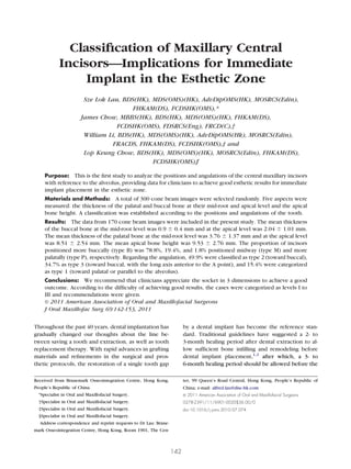
  • 2. LAU ET AL 143 implant is exposed and loaded with a fixed prosthe- thetic results. The thicknesses of the bone at the sis.1,2 However, the negative effect of tooth extrac- buccal and palatal side were evaluated, as was the tion on the volumetric hard and soft tissue changes in bone height at the apical tooth region. A new classi- the buccolingual and vertical dimensions has been fication is introduced to categorize the different tooth reported extensively in published studies. Using diag- positions and angulations, providing a reference to nostic casts,3-5 radiographs,6-8 and direct measure- help avoid compromising the buccal bone thickness ments9 to evaluate the magnitude of bone resorption, and to prevent fenestration and perforation during it has been reported that during the first 4 months implant placement. Different types of teeth were also after extraction, the mean bone reduction was 5 to 7 ranked according to their difficulties for achieving mm buccolingually8,9 and 2 to 4.5 mm vertically.6 good long-term results, and general recommendations Because such bone resorption will strongly affect the for their management were given. implant placement and, thus, the esthetics outcome, clinicians started to insert the implant directly into Materials and Methods the extraction socket immediately after extraction, under the assumption that this would reduce the A series of 300 cone beam (CB) images of the bone resorption. The first case report of this tech- maxilla taken from April 2006 to August 2006 were nique was published in 1976.10 Since then, numerous randomly selected from the computer record at the studies have been published regarding the refinement Dental Implant and Maxillofacial Centre in Hong of this technique.11-13 Additional clinical research has Kong. All CB images were taken with the same ma- shown that the outcomes of immediate implant place- chine (I-CAT cone beam volumetric tomography and ment, delayed implant placement, and implant place- panoramic dental imaging system; Imaging Science ment in healed socket sites were all comparable.14 International, Hatfield, PA). The head positions were The multiple advantages of immediate implant place- standardized by aligning the patient’s head with hor- ment are well known, especially in the esthetic zone, izontal and vertical reference lines, constructed using such as the maxillary central incisors. These advantages a beam of light. The patient’s head was positioned include a reduction in the number of surgeries, a reduc- such that the horizontal reference beam was at the tion of the total treatment time, the preservation of level of the patient’s eyes and the vertical reference alveolar bone,15-17 the maintenance of a good soft tissue beam was passing through the patient’s facial midline. profile,18 and the reduction of the patients’ psychologi- Once the ideal head position had been located, the cal trauma owing to the loss of a front tooth.19 However, patient’s head was fixed with a head frame. The CB such therapy is not without drawbacks. The greatest images were viewed and measured by the software problem for clinicians is the unpredictability of the long- provided. Each image was examined to identify a fully term soft tissue stability. The problem of soft tissue formed, intact, and healthy permanent central upper recession is usually exacerbated in patients with rela- right incisor (tooth 11) for analysis. If the central tively thin buccal bone and thin gingival biotypes.20 upper right incisor was missing, the upper left central Because the gingival biotype cannot be changed, the incisor was selected for analysis. The CB imaging data critical aspect of achieving esthetic success depends on were excluded if any radiographically detectable the ideal 3-dimensional implant position21 and the main- enamel or dentine defect, caries, apical pathologic tenance of adequate buccal bone thickness over the features, periodontal alveolar bone loss, restoration of implant buccal surface.22 Immediate implant therapy any kind, or fracture was present or when both cen- has been reported to be very technically demanding14 tral incisors were missing. and requires an understanding of all these aspects and Landmarks were identified and marked in the com- more. Failure to do so will result in an adverse outcome, puter before the measurements were done. The sag- defeating the initial objectives of the therapy itself. Im- ittal section of the chosen incisor was viewed at the mediate implantation is never easy, especially when the center of its mesial-distal dimension. As shown in demand for long-term stable esthetic results is high; Figure 1, both the palatal line (line 1) and the buccal thus, case selection is of the utmost importance. line (line 2) were marked by a line best-fit to the The criteria for implant success have been chang- palatal alveolar surface and the buccal alveolar sur- ing, and interest in the esthetic outcome is becoming face, respectively. The alveolar line (line 3) was a major concern. Implant dentistry has gradually marked by bisecting the palatal and buccal lines. This evolved from a bone-driven surgical protocol to a indicated the angulation of the alveolar process in the restorative and biologically driven protocol. The sagittal plane. The angulation of the tooth root was present study is the first to analyze the positions and indicated by its long axis (line 4), and this axis was angulations of the central maxillary incisors with ref- marked by the midpoint of a line drawn from the buccal erence to the alveolar bone. Our results will provide enamel-dentine junction to its palatal counterpart, the data for clinicians to achieve the best long-term es- apex of the root. The present study involved measure-
  • 3. 144 CLASSIFICATION OF MAXILLARY CENTRAL INCISORS ward the buccal bone surface. The thickness of the buccal bone at the mid-root level (measurement I) was measured from a point at the buccal root surface to the buccal bone surface along this line. The palatal bone thickness at the mid-root level (measurement II) was measured from a point at the palatal root surface toward the palatal bone along this same line. The thickness of labial bone at the apical level (measurement III) was defined as a line perpendicular to the long axis of the root, from the apex of the root toward the buccal bone surface. The thickness of the palatal bone at the apical level (measurement IV) was defined as the distance of the line perpendicular to the long axis of the root from the apex of the root toward the palatal bone surface. The apical bone height (measurement V) was FIGURE 1. Landmarks for measurement. Lau et al. Classification of Maxillary Central Incisors. J Oral Maxillofac Surg 2011. ment of the residual bone thickness and angulations of the extraction sockets. The long axis of the tooth root, instead of the long axis of the whole tooth, was used, because the range of the crown-root angle is 25.5° for maxillary central incisors.23 From these reference lines, 4 measurements were performed, using the built-in mea- suring function of the computer software. The measure- FIGURE 2. Measurements of bone thickness at different aspects ments are shown in Figure 2. (measurements I to V). A line was drawn from the mid-point of the long axis Lau et al. Classification of Maxillary Central Incisors. J Oral of the root perpendicularly from the palatal bone to- Maxillofac Surg 2011.
  • 4. LAU ET AL 145 measured along the long axis of the root from the root All measurements and classifications were per- apex toward the superior bone surface. formed by 2 investigators. Reliability tests were also From the CB measurements, the positions and angu- performed to check the consistency and accuracy lations of the tooth roots were classified with reference using the Statistical Package for Social Sciences, ver- to the alveolar process. By comparing the buccal and sion 11.0, software (SPSS, Chicago, IL). Studies of this palatal bone thickness at the mid-root level, their posi- type are exempt from approval by the local institu- tions with reference to the mid-alveolar line were de- tional review board. fined and classified as follows (Fig 3): type B (closer to the buccal alveolar surface); type M (midway between Results the buccal and palatal alveolar surface); and type P (closer to the palatal alveolar surface). Comparing the The CB imaging data from a series of 300 randomly angulations of the alveolar process with the long axis of selected patients were accessed initially. The images the roots, the angulations were classified as follows (Fig were all examined to identify useful data according to 3): type 1 (root apex angulated toward the palatal side the defined inclusion and exclusion criteria. The CB or parallel to the alveolus); type 2 (root apex angulated imaging data from 170 patients were included in the toward the buccal side with the long axis passing pos- final analysis. Of the excluded 130 patients, 109 had a terior to point A); and type 3 (root apex angulated defective incisor (eg, caries, infections, restorations, toward the buccal side with the long axis passing ante- periodontal bone loss, or fracture) and 21 had both rior to point A). central incisors missing. The mean patient age was 47 FIGURE 3. Classification according to position and angulation. Lau et al. Classification of Maxillary Central Incisors. J Oral Maxillofac Surg 2011.
  • 5. 146 CLASSIFICATION OF MAXILLARY CENTRAL INCISORS Table 1. MEASUREMENT RESULTS Thickness at Mid-Root Level Thickness at Apical Level Measurement Buccal Bone Palatal Bone Buccal Bone Palatal Bone Apical Bone Height (mm) (Measurement I) (Measurement II) (Measurement III) (Measurement IV) (Measurement V) Mean 0.90 3.76 2.04 8.51 9.53 Median 0.89 3.69 1.96 8.40 9.57 Standard deviation 0.40 1.37 1.01 2.54 2.76 Maximum 1.99 10.10 7.72 22.41 17.20 Minimum 0.10 0.50 0.10 2.56 2.15 Lau et al. Classification of Maxillary Central Incisors. J Oral Maxillofac Surg 2011. years (range 13 to 85). Of the 170 patients, 76 (45%) root to the nasal floor, almost one half of the patients were males and 94 were females (55%). (47.6%) had at least 10 mm of bone height; 97.6% had The mean thickness of the buccal bone at the 4 mm or more of bone height and 71.2% of patients mid-root level (measurement I) was 0.9 mm (range had 8 mm or more of bone height (Table 6). 0.1 to 1.99), and the mean thickness of the palatal A classification of the positions and angulations of bone at the mid-root level (measurement II) was 3.76 the central incisors with reference to the alveolar 1.37 mm (range 0.5 to 10.1). The mean thickness of bone was established. Most of the central incisors the buccal bone at the apical level (measurement III) (78.8%) were positioned more buccally within the was 2.04 1.01 mm (range 0.1 to 7.72), and the alveolar bone (type B), 19.4% were positioned in mean thickness of the palatal bone at the apical level midway (type M), and 1.8% were positioned more (measurement IV) was 8.51 2.54 mm (range 2.56 to palatally (type P; Table 7). Regarding the incisor an- 22.41). The mean apical bone height (measurement gulations, almost one half (49.9%) of the central inci- V) was 9.53 2.76 mm (range 2.15 to 17.2). These sors were classified as type 2, 34.7% were classified as measurements are listed in Table 1. type 3, and 15.4% (26 cases) were categorized as type Additional analysis of the measurements revealed that 57% of the patients had a buccal bone thickness 1. An analysis that combined both the position and at the mid-root level of less than 1 mm (Table 2), and the angulation type showed that most were type B2 62.4% of patients had a palatal bone thickness at the (38.2%). The incidence of type B3 (34.7%) was only mid-root level of less than 4 mm—up to 86.5% of slightly less than that of type B2, followed by type M2 patients had a thickness of less than 5 mm (Table 3). (11.7%). No type P2, P3, or M3 was found in our Similarly, the buccal bone at the apical level was patient series. The rest of the types were few and are much thinner than its palatal counterpart. The buccal listed in Table 7. bone thickness at the apical level was less than 5 mm A reliability test was done to determine the accu- in 98.8% of the patients, and more than one half of the racy of the measurements and classifications between patients (51.8%) had a thickness of less than 2 mm the 2 investigators. The correlation coefficients for (Table 4). In contrast, 96.5% of patients had a palatal the different measurements are listed in Table 8 and bone thickness at the apical level of 5 mm or more, reflected the good consistency between the 2 inves- and more than one half of the patients (55.9%) had a tigators. thickness of 8 mm or more (Table 5). Regarding the apical bone height, measured from the apex of the Table 3. THICKNESS OF PALATAL BONE AT MID- Table 2. THICKNESS OF BUCCAL BONE AT MID- ROOT LEVEL (MEASUREMENT II) ROOT LEVEL (MEASUREMENT I) Thickness (mm) Patients (n) Thickness (mm) Patients (n) 2 8 (4.7) 0.5 23 (13.5) 3 40 (23.5) 1.0 97 (57) 4 106 (62.4) 1.5 158 (92.9) 5 147 (86.5) 2.0 170 (100) 6 159 (93.5) Data in parentheses are percentages. Data in parentheses are percentages. Lau et al. Classification of Maxillary Central Incisors. J Oral Lau et al. Classification of Maxillary Central Incisors. J Oral Maxillofac Surg 2011. Maxillofac Surg 2011.
  • 6. LAU ET AL 147 Table 4. THICKNESS OF BUCCAL BONE AT APICAL Table 6. APICAL BONE HEIGHT (MEASUREMENT V) LEVEL (MEASUREMENT III) Thickness (mm) Patients (n) Thickness (mm) Patients (n) 4 166 (97.6) 1 17 (10.0) 6 153 (90.0) 2 88 (51.8) 8 121 (71.2) 3 152 (89.4) 10 81 (47.6) 4 164 (96.5) 5 168 (98.8) Data in parentheses are percentages. Lau et al. Classification of Maxillary Central Incisors. J Oral Data in parentheses are percentages. Maxillofac Surg 2011. Lau et al. Classification of Maxillary Central Incisors. J Oral Maxillofac Surg 2011. evaluate the root position 3 dimensionally before Discussion placement. Although it might be worthwhile information, we The present study aimed to provide data to aid the purposely did not include the measurements of the treatment planning of immediate implant in the es- buccal and palatal bone thickness at the crestal re- thetic zone using a CB imaging technique. With rapid gion, because of the uncontrollable high percentage advances in surgical and imaging techniques, the de- of error owing to its relative thinness. Although mag- mand for accuracy in 3-dimensional and volumetric nification is possible in the computer, the resolution measurements is ever increasing. CB imaging has be- will be too low for measurement. Moreover, a beam come the standard for implant planning, especially in hardening effect is always present over the edge, the highly esthetically demanding areas. Numerous making measuring the bone thickness at the crestal studies have been published to support the routine level worthless. use of CB imaging in implant dentistry, including the As previously mentioned, the main drawback of advantages of convenience, accuracy, and relatively immediate implant placement into the extraction low radiation dosage.24-27 Increasingly, more clini- sockets has been the lack of predictability of the cians have been incorporating this into daily practice. long-term soft tissue profile, especially on the buccal Only the roots of the maxillary central incisors aspect. Thus, the position of implant placement is were evaluated in the present study, instead of the critical and is a limiting variable. Although it was whole tooth. Neither were the crown-to-root relation- previously believed that the bone remodeling and soft ships measured. This was because it was reported by tissue recession over an extraction socket would be Bryant et al23 that only a mean of 1.74° is present arrested by inserting a dental implant into the socket, between the long axis of the root and the crown of recent studies have failed to support this.28-30 It has normal central maxillary incisors. With priority given been confirmed that, irrespective of the placement of to a restorative driven concept, if implants could be a dental implant, bone resorption after extraction will placed into the extraction sockets exactly at the same still occur, leading to a loss of bone volume.28-30 It has angulations as the roots inside the alveolar bone, it also been reported that the buccal bone resorbs more would provide an ideal 3-dimensional position for the than the lingual or palatal bone after extraction, be- prosthetic crown and only a simple straight stock cause it is composed of bundle bone alone. In con- abutment would be needed. Thus, it is important to trast, lingual or palatal bone is composed of cortical bone, at least at the outermost surface; thus, the resistance to resorption is better.31 The mid-buccal Table 5. THICKNESS OF PALATAL BONE AT APICAL recession of an immediate implant placed into a fresh LEVEL (MEASUREMENT IV) extraction socket has been reported to be 0.55 to Thickness (mm) Patients (n) 0.75 mm at 1 year of follow-up.32,33 However, long- term data are still not available to determine whether 5 164 (96.5) additional resorption occurs. Kohal et al34 have 6 153 (90) 7 126 (74.1) shown that pressure of the inserted implant on the 8 95 (55.9) bony wall can result in microfractures, leading to 9 61 (35.9) crestal bone loss. Therefore, the ideal placement of an 10 38 (22.4) immediate implant should be aimed toward obtaining Data in parentheses are percentages. maximum bone-to-implant contact to achieve good Lau et al. Classification of Maxillary Central Incisors. J Oral primary stability and promote greater osseointegra- Maxillofac Surg 2011. tion.35,36 However, the key to long-term good esthetic
  • 7. 148 CLASSIFICATION OF MAXILLARY CENTRAL INCISORS Table 7. CLASSIFICATION Type Type B M P Total 1 5.9 (10) 7.7 (13) 1.8 (3) 15.4 (26) 2 38.2 (65) 11.7 (20) 0 (0) 49.9 (85) 3 34.7 (59) 0 (0) 0 (0) 34.7 (59) Total 78.8 (134) 19.4 (33) 1.8 (3) 100 (170) Data are presented as parentheses, with numbers in parentheses. Lau et al. Classification of Maxillary Central Incisors. J Oral Maxillofac Surg 2011. results is to avoid exerting pressure on the crestal giva, thus minimizing mid-buccal recession and bony wall, particularly on the buccal aspect. shrinkage of the papillae. However, only 7.7% of type With respect to the classification reported in the M1 were reported (Table 7). present study, both the position and the angulation Although type P1 is not as perfect as type M1, these types were categorized into different ranks according types are still good, because they will have enough to their difficulty for achieving ideal esthetic results in buccal bone to support the overlying soft tissue. Rel- immediate implant cases. The classification levels are atively less bone will be present at the palatal side; level I to level III (Fig 4). however, the palatal soft tissue is very thick and the palatal bone can resist resorption much better. The LEVEL I recession of palatal tissue will be less significant, if Level I (M1, P1) indicates that the implant could be any occurs, and the esthetic demand is usually not placed with the same angulation as the extraction high on the palatal side. Similar to those with type M1, socket, without compromising the primary stability those with type P1 were in the minority, with only 3 and long-term esthetic outcome because of the com- patients (1.8%) classified as having type P1 (Table 7). mon features of a relatively thicker buccal bone. Be- cause no modification of the drilling angle would be LEVEL II required, a straight stock abutment could be used. Level II (B1, B2, M2, M3, P2, P3) is more technically These types are the most straightforward for both demanding. The angulation of the implant should be surgery and its restoration of all the levels. However, only 9.5% of patients had extraction sockets catego- rized as this level (Fig 4). Type M1 should be ideal for immediate implant cases. This type of tooth lies in the middle of the alveolar bone and root apex, angulated away from the buccal wall. When placing an implant into the extrac- tion socket, enough bone will be present to support the implant, thereby achieving good primary stability and enough bone-to-implant contact for good os- seointegration. Also, the implant angulation will be perfect for the superstructure. Moreover, enough buccal bone is present to support the overlying gin- Table 8. INTERINVESTIGATOR RELIABILITY TEST Investigation Correlation Coefficients Measurement I 0.955 Measurement II 0.805 Measurement III 0.830 Measurement IV 0.876 Measurement V 0.898 FIGURE 4. Levels according to difficulty in achieving good long- Classifications 0.922 term esthetic results for immediate implant. Lau et al. Classification of Maxillary Central Incisors. J Oral Lau et al. Classification of Maxillary Central Incisors. J Oral Maxillofac Surg 2011. Maxillofac Surg 2011.
  • 8. LAU ET AL 149 changed to avoid thinning of the buccal bone to Table 9. APICAL DIMENSIONS OF COMMONLY maintain a long-term stable esthetic outcome, be- USED IMMEDIATE IMPLANTS IN ESTHETIC ZONE cause it has the common disadvantage of a relatively thin buccal plate. The angle of the implant should be Apical Diameter Radius Implant Brand (mm) (mm) placed more palatally to avoid compressing or drilling the buccal bone, minimizing the chance of perfora- Ankylos (A type), Densply tions and fenestrations. Because a discrepancy exists Friadent 3.5 1.75 between the implant angle to the original tooth, an OsseoSpeed (4.5), Astra Tech Dental 3.5 1.75 angled abutment should be chosen to obtain good Tapered Internal (3.8 mm), esthetics. More than one half of the cases (55.8%) BioHorizons 2.8 1.40 were categorized as level II (Table 7). ITI (Bone Level), Straumann 3.5 1.75 Regarding the positions of the maxillary central Replace Tapered Groovy (RP), incisors, most were type B (78.8%); thus, most of the Nobel Biocare 2.7 1.35 Tapered Screw-Vent (3.7 teeth lie more buccally. Type B1 (5.9%) is not ideal for mmD), Zimmer Dental 3.1 1.55 immediate implantation, because it is close to the buccal wall, indicating relatively thinner buccal bone. Lau et al. Classification of Maxillary Central Incisors. J Oral Maxillofac Surg 2011. However, type B1 is not the most challenging of this level because it does not angulate toward the natural depression of the buccal alveolus. The chance of the side of the implant drill should not be cutting on additional thinning of the buccal wall or fenestration the buccal plate during osteotomy, especially for type will be reduced. B, because it is expected to be thinner than other The least favorable types of maxillary central inci- types. Otherwise, the risk of perforation or thinning sors for immediate implant are those positioned near down the already thin buccal plate will be high. Table the buccal wall with the apex pointing toward the 9 provides a reference for the apical radius of com- buccal side (types B2 and B3; type B3 was categorized monly used implant brands for immediate implant in as difficult [level III] and was included in the next the esthetic zone, which ranges from 1.35 to 1.75 section). A type B2 tooth would naturally produce a mm. More than one half of the patients had a buccal thin buccal plate at both the crestal and the apical bone thickness of less than 2 mm at the apical region, region. For immediate implant placement, good pri- indicating that placing implant drills exactly along the mary stability is mandatory, especially for those axis of extraction sockets will result in a high risk of scheduled to have immediate placement of a provi- perforation, especially when placing the implant sional crown. When achieving good primary stability, deeper toward the apical bone to achieve primary pressure is almost always present on the buccal bone, stability. either at the apical region or at both the apical and the The mean thickness of the palatal bone at the apical crestal regions. Because this type has a thin buccal level (measurement IV) is much greater than its buc- plate, any pressure exerted on it will significantly cal counterpart. We found an average of 8.51 mm of increase the risk of bone resorption, leading to the palatal bone at this region, with a maximal thickness loss of soft tissue in the long term. However, a major of 22.41 mm (compared with an average of 2.04 proportion of patients will have these types (type B2, 1.01 mm). More than one half (55.9%) of the patients 38.2%). This explains the “physiologic” bone and soft had a thickness of 8 mm or more and 96.5% of pa- tissue loss on the buccal side in some immediate tients had a thickness of 5 mm or more (Table 5). In implant cases. Although it can be difficult to achieve contrast, the thickness of the palatal bone at the good primary stability, clinicians will still need to mid-root level (measurement II) had a mean of only avoid overcompression of the buccal plate in such 3.76 mm, and 76.5% of patients have a bone thickness cases. This explains why these cases are so technique- of less than 3 mm. This probably resulted from the sensitive. contour of the palatal vault. Thus, the more apically Our data suggest that the buccal bone of maxillary placed, the thicker the palatal bone. However, this is incisors were, on average, thin. The mean thickness not the same for the buccal aspect owing to the of the buccal bone at the mid-root level (measure- natural depression at point A and, sometimes, the ment I) was only 0.9 mm, and the buccal bone at the extreme root angulations, such as a type 3 tooth. apical level (measurement III) was 2.04 mm. More Thus, the implant should be placed more palatally in than one half of the patients (57%) have a buccal bone the extraction socket, because more bone will be thickness at the mid-root level of less than 1 mm present in that area to achieve good implant stability (Table 2), and 51.8% of patients had a buccal bone and to avoid thinning of the buccal wall. However, thickness at the apical level of less than 2 mm (Table the implant should also be placed more palatally at 4). It is critical not to drill over the buccal area— even the apical area, pivoting around the mid-root level.
  • 9. 150 CLASSIFICATION OF MAXILLARY CENTRAL INCISORS This is because the palatal bone is relatively thicker at angulation with reference to the extraction socket, the apical area than at the mid-root region. Clinicians and in this case, more palatally. must also be careful not to angulate the apex of the implant too palatally, otherwise, the coronal part of LEVEL III the implant will be tilted too buccally. This can lead to The type B3 tooth is the most challenging case with overcompression, or even perforation, on the buccal respect to achieving good long-term esthetic out- crestal wall, making the coronal platform point out- comes because it not only will have a very thin buccal ward buccally and compromising the esthetic out- plate, but also the long axis of the tooth apex will be come. A recent study confirmed that the buccal-lin- angulated very buccally, passing anterior to the natu- gual position of the implant shoulder is a very ral contour of the maxillary alveolar bone (Fig 5A). important factor determining the degree of buccal This type is mostly seen in patients with maxillary marginal tissue recession.37 It was reported that an alveolar hyperplasia and angle Class II division 2 oc- implant with a shoulder positioned at, or buccally, to clusion. Because a large difference exists between the a line drawn between the cervical margin of the angulations of the alveolar bone and the tooth, the adjacent teeth resulted in 3 times more recession than implant position will be compromised, regardless of an implant with a shoulder positioned lingually or whether a bone-driven or restorative-driven protocol palatally to this line.37 If an ideal angulation for good is used (Figs 5A,B). Sometimes, traditional guidelines apical stability without exerting excessive pressure can be followed for such cases by extracting the on the buccal wall and without placing the shoulder buccal to this line cannot be achieved, clinicians can always consider choosing an implant with a smaller coronal diameter. Regarding the apical bone height (measurement V), it was recorded that 97.6% of patients had a bone height of 4 mm or more, and 90% of patients had a bone height of 6 mm or more (Table 6). The general recommendation for the placement of immediate im- plant in an extraction socket in the apical dimension is to engage the implant 3 to 5 mm beyond the apex of the socket to achieve good primary stability.38,39 The results of the present study have shown adequate bone is present at the apical area. Even when the primary stability is not optimal, the implant could be placed deeper to achieve this, because usually more than enough bone is present at this region (almost one half of the patients had 10 mm or more of apical bone height). However, a suitable implant length should carefully be chosen, because the apical- coronal position of the implant shoulder is also an important factor in determining the long-term soft tissue profile and the esthetic outcome.21,40,41 The length of the implant should be optimal, deep enough to provide good apical stability, without perforation, and the shoulder positioned with, or slightly apical to, the buccal marginal bone crest (around 3 to 4 mm from the buccal gingival margin38,42,43 or 3 mm apical to the cementoenamel junctions of the periodontally sound adjacent tooth44). Unlike type B2, types M2, M3, P2, and P3 have similar characteristics, including a relatively thicker buccal plate. However, the root apexes will point toward the buccal side. If the implant were placed FIGURE 5. Examples of level III socket. A, Large difference be- exactly into the extraction socket with the same an- tween angulations of original tooth and future implant. B, Extreme gulation, the risk of perforation would be very high. angulation requiring traditional treatment or socket transformation. The general recommendation for all level II cases is Lau et al. Classification of Maxillary Central Incisors. J Oral that the implants should be inserted with a modified Maxillofac Surg 2011.
  • 10. LAU ET AL 151 tooth, with or without simultaneous grafting, and supply from the periosteum to the buccal plate during inserting the implant several months later. As such, the flap raising. For level I cases, inserting the implant although the advantages of immediate implantation with the same angulation of the socket using a stan- would not be achieved, the long-term soft tissue sta- dard drilling protocol will be straightforward. For bility will be more predictable. level II cases, the implant angulation should be If enough bone is present at the palatal and apical changed to a more palatal aspect at the apical region, aspects, an attempt could be made by placing the pivoting around the mid-palatal area. It is recom- implant both palatally and apically to avoid touching mended to first use a round bur to create a step at the the buccal plate. However, even if primary stability palatal aspect of the apical area of the socket before can be achieved, the difference will still be large using a straight drill to ensure that the hardness of the between the angle of the implant and prosthetic cortical bone and natural contour of the socket does crown. An angled abutment will always be needed. not misguide the operator to drill parallel toward the One should be familiar with the implant system one apical long axis (Fig 6). The implant should be in- is using, whether an angulated temporary abutment is serted with good primary stability, without exerting available, and, if so, the different angles available if pressure on the buccal wall. To ensure the absence of planning an immediate provisional crown. The final pressure on the buccal bone, the operator can leave a restoration should also be considered before the im- gap between the implant surface and the buccal plant is inserted. Most often, for an abutment with a bone, as long as the primary stability is not compro- relatively big angle, a step of metal will be present on mised. The gap can be treated with grafting materials the buccal aspect. It can sometimes show through the and/or a barrier membrane to achieve maximum os- buccal gingiva, compromising the esthetic outcome. seointergration and good soft tissue healing, depend- Placing the implant further apically can be done to ing on the gap size.12,14,45-48 The implant shoulder solve this problem. Customized abutments or a ce- should be optimally placed in terms of the depth and ramic abutment can also be used according to the buccal-palatal dimension. A provisional crown should position and angulation of the implant. However, the im- be considered whenever possible for soft tissue sup- plant position will most often be controlled by the port, favoring the long-term esthetic outcome,31 by bone available for such cases. Thus, type B3 is very selecting an appropriate abutment. For level III cases, technically demanding for immediate implant, be- extra precautions should be given to the prosthetic cause it is both a bone-driven and a prosthetic-driven piece owing to its extreme angulations. One should procedure. be familiar with the different types of abutments avail- If the demand for a good esthetic outcome and able, as well as their dimensions and shapes. The immediate provisional restoration is very high, a implant can be placed deeper apically to avoid buccal socket transformation procedure can be done by showing of the metal margin of an angulated abut- grafting the buccal wall in the first stage, followed by extraction of the problematic tooth and a normal immediate implantation protocol several months later. Thus, a type B3 (level III) socket could be transformed iatrogenically to an easier type, such as type B2 (level II) or, even, type M2 (level II), keeping the advantages of immediate implant. However, more evidence is needed to justify the long-term success of this treatment concept. In conclusion, the bone availability in all dimen- sions should be considered to achieve a good implant esthetic. The angulation and position of the original tooth should be well appreciated 3-dimensionally dur- ing the planning stage. This is best performed by measurement of a CB image. A suitable implant type and the length and diameter at the apical, mid-body, and coronal level should be customized to the original socket as much as possible. Tapered-screw implants are suggested, because they can fit to the extraction socket better, and primary stability can be achieved easily. After an atraumatic extraction, all granulation FIGURE 6. Drilling with a round bur toward palatal aspect. tissue should be removed. A flapless approach is rec- Lau et al. Classification of Maxillary Central Incisors. J Oral ommended to minimize the breakdown of the blood Maxillofac Surg 2011.
  • 11. 152 CLASSIFICATION OF MAXILLARY CENTRAL INCISORS ment. A traditional protocol can sometimes be fol- comparing different types of serial immediate implantation. Int J Oral Maxillofac Implants 10:561, 1995 lowed to achieve a more predictable long-term result 18. Werbitt MJ, Goldberg PV: The immediate implant: Bone pres- by grafting or preserving the alveolar ridge before ervation and bone regeneration. Int J Periodontics Restorative implant placement. However, if the esthetic require- Dent 12:206, 1992 ment is very high, a socket transformation procedure 19. Gelb DA: Immediate implant surgery: Three-year retrospective evaluation of 50 consecutive cases. Int J Oral Maxillofac Im- should be considered. plants 8:388, 1993 It is never easy to achieve long-term stable esthetic 20. Kan JY, Rungcharassaeng K, Umezu K, et al: Dimensions of results with immediate implantation. However, it is peri-implant mucosa: An evaluation of maxillary anterior single implants in humans. J Periodontol 74:557, 2003 not impossible, provided that a good case selection 21. Buser D, Martin W, Belser UC: Optimizing esthetics for implant protocol is in place and surgery is performed by restorations in the anterior maxilla: Anatomic and surgical experienced operators. considerations. Int J Oral Maxillofac Implants 19:43, 2004 (suppl) 22. Grunder U, Gracis S, Capelli M: Influence of the 3-D bone-to- implant relationship on esthetics. Int J Periodontics Restorative Dent 25:113, 2005 References 23. Bryant RM, Sadowsky PL, Hazelrig JB: Variability in three mor- 1. Albrektsson T, Branemark PI, Hansson HA, et al: Osseointe- phologic features of the permanent maxillary central incisor. grated titanium implants: Requirements for ensuring a long- Am J Orthod 86:25, 1984 lasting, direct bone-to-implant anchorage in man. Acta Orthop 24. Rugani P, Kirnbauer B, Arnetzl GV, et al: Cone beam comput- Scand 52:155, 1981 erized tomography: Basics for digital planning in oral surgery 2. Branemark PI: Osseointegration and its experimental back- and implantology. Int J Comput Dent 12:131, 2009 ground. J Prosthet Dent 50:399, 1983 25. Bamgbose BO, Adeyemo WL, Ladeinde AL, et al: Cone beam 3. Lekovic V, Camargo PM, Klokkevold PR, et al: Preservation of computed tomography (CBCT): The new vista in oral and alveolar bone in extraction sockets using bioabsorbable mem- maxillofacial imaging. Nig Q J Hosp Med 18:32, 2008 branes. J Periodontol 69:1044, 1998 26. Spector L: Computer-aided dental implant planning. Dent Clin 4. Schropp L, Wenzel A, Kostopoulos L, et al: Bone healing and North Am 52:761, 2008 soft tissue contour changes following single-tooth extraction: A 27. Mandelaris GA, Rosenfeld AL: The expanding influence of com- clinical and radiographic 12-month prospective study. Int J puted tomography and the application of computer-guided Periodontics Restorative Dent 23:313, 2003 implantology. Pract Proced Aesthet Dent 20:297, 2008 5. Camargo PM, Lekovic V, Weinlaender M, et al: Influence of 28. Araujo MG, Sukekava F, Wennstrom JL, et al: Ridge alterations bioactive glass on changes in alveolar process dimensions after following implant placement in fresh extraction sockets: An exodontia. Oral Surg Oral Med Oral Pathol Oral Radiol Endod experimental study in the dog. J Clin Periodontol 32:645, 2005 90:581, 2000 29. Araujo MG, Lindhe J: Dimensional ridge alterations following 6. Iasella JM, Greenwell H, Miller RL, et al: Ridge preservation tooth extraction: An experimental study in the dog. J Clin with freeze-dried bone allograft and a collagen membrane com- Periodontol 32:212, 2005 pared to extraction alone for implant site development: A 30. Botticelli D, Berglundh T, Lindhe J: Hard-tissue alterations fol- clinical and histologic study in humans. J Periodontol 74:990, lowing immediate implant placement in extraction sites. J Clin 2003 Periodontol 31:820, 2004 7. Atwood da Coy WA: Clinical, cephalometric, and densitometric 31. De Rouck T, Collys K, Cosyn J: Single-tooth replacement in the study of reduction of residual ridges. J Prosthet Dent 26:280, anterior maxilla by means of immediate implantation and pro- 1971 visionalization: A review. Int J Oral Maxillofac Implants 23:897, 8. Johnson K: A study of the dimensional changes occurring in 2008 the maxilla following tooth extraction. Aust Dent J 14:241, 32. Kan JY, Rungcharassaeng K, Lozada J: Immediate placement 1969 and provisionalization of maxillary anterior single implants: 9. Johnson K: A study of the dimensional changes occurring in 1-Year prospective study. Int J Oral Maxillofac Implants 18:31, the maxilla following closed face immediate denture treatment. 2003 Aust Dent J 14:370, 1969 33. Cornelini R, Cangini F, Covani U, et al: Immediate restoration 10. Schulte W, Kleineikenscheidt H, Lindner K, et al: [The Tu- of implants placed into fresh extraction sockets for single-tooth bingen immediate implant in clinical studies]. Dtsch Zahnarztl Z 33:348, 1978 replacement: A prospective clinical study. Int J Periodontics 11. Barzilay I: Immediate implants: Their current status. Int J Restorative Dent 25:439, 2005 Prosthodont 6:169, 1993 34. Kohal RJ, Hurzeler MB, Mota LF, et al: Custom-made root 12. Schwartz-Arad D, Chaushu G: The ways and wherefores of analogue titanium implants placed into extraction sockets: An immediate placement of implants into fresh extraction sites: A experimental study in monkeys. Clin Oral Implants Res 8:386, literature review. J Periodontol 68:915, 1997 1997 13. Chen ST, Wilson TG Jr, Hammerle CH: Immediate or early 35. Wilson TG Jr, Schenk R, Buser D, et al: Implants placed in placement of implants following tooth extraction: Review of immediate extraction sites: A report of histologic and histomet- biologic basis, clinical procedures, and outcomes. Int J Oral ric analyses of human biopsies. Int J Oral Maxillofac Implants Maxillofac Implants 19:12, 2004 (suppl) 13:333, 1998 14. Ataullah K, Chee LF, Peng LL, et al: Implant placement in 36. Lundgren D, Rylander H, Andersson M, et al: Healing-in of root extraction sockets: A short review of the literature and presen- analogue titanium implants placed in extraction sockets: An tation of a series of three cases. J Oral Implantol 34:97, 2008 experimental study in the beagle dog. Clin Oral Implants Res 15. Shanaman RH: The use of guided tissue regeneration to facili- 3:136, 1992 tate ideal prosthetic placement of implants. Int J Periodontics 37. Evans CD, Chen ST: Esthetic outcomes of immediate implant Restorative Dent 12:256, 1992 placements. Clin Oral Implants Res 19:73, 2008 16. Denissen HW, Kalk W, Veldhuis HA, et al: Anatomic consider- 38. Lazzara RJ: Immediate implant placement into extraction sites: ation for preventive implantation. Int J Oral Maxillofac Im- Surgical and restorative advantages. Int J Periodontics Restor- plants 8:191, 1993 ative Dent 9:332, 1989 17. Watzek G, Haider R, Mensdorff-Pouilly N, et al: Immediate and 39. Bhola M, Neely al Kolhatkar S: Immediate implant placement: delayed implantation for complete restoration of the jaw fol- Clinical decisions, advantages, and disadvantages. J Pros- lowing extraction of all residual teeth: A retrospective study thodont 17:576, 2008
  • 12. LAU ET AL 153 40. Garber DA: The esthetic dental implant: Letting restoration be placed in a fresh extraction socket. Int J Oral Maxillofac the guide. J Am Dent Assoc 126:319, 1995 Implants 2:217, 1987 41. Kois JC: Predictable single tooth peri-implant esthetics: Five 46. Becker W, Lynch SE, Lekholm U, et al: A comparison of ePTFE diagnostic keys. Compend Contin Educ Dent 22:199, 2001 membranes alone or in combination with platelet-derived 42. Becker W, Becker BE, Handelsman M, et al: Guided tissue growth factors and insulin-like growth factor I or demineral- regeneration for implants placed into extraction sockets: A ized freeze-dried bone in promoting bone formation around study in dogs. J Periodontol 62:703, 1991 immediate extraction socket implants. J Periodontol 63:929, 43. Cochran DL, Douglas HB: Augmentation of osseous tissue 1992 around nonsubmerged endosseous dental implants. Int J Peri- 47. Wilson TG Jr: Guided tissue regeneration around dental odontics Restorative Dent 13:506, 1993 44. Langer B, Sullivan DY: Osseointegration: Its impact on the implants in immediate and recent extraction sites: Initial interrelationship of periodontics and restorative dentistry. Part observations. Int J Periodontics Restorative Dent 12:185, 3. Periodontal prosthesis redefined. Int J Periodontics Restor- 1992 ative Dent 9:240, 1989 48. Lang NP, Bragger U, Hammerle CH, et al: Immediate trans- 45. Todescan R Jr, Pilliar RM, Melcher AH: A small animal model for mucosal implants using the principle of guided tissue regen- investigating endosseous dental implants: Effect of graft mate- eration. I. Rationale, clinical procedures and 30-month re- rials on healing of endosseous, porous-surfaced implants sults. Clin Oral Implants Res 5:154, 1994