SlideShare una empresa de Scribd logo
1 de 63
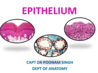
EPITHELIUM

CAPT DR POONAM SINGH
DEPT OF ANATOMY
SEQUENCE OF PPT
1.
2.
3.
4.
5.
6.
7.
8.

INTRODUCTION
DEVELOPMENT
CLASSIFICATION
HISTOLOGICAL APPEARANCE
APICAL DOMAIN & ITS MODIFICATIONS
LATERAL DOMAIN & ITS SPECIALIZATIONS
BASAL DOMAIN
GLANDS & ITS TYPES
TISSUES

4 Basic types
DEVELOPMENT
5 – 6 DAY
6th DAY

Implantation of the Blastocyst
9TH DAY
DEVELOPMENT
DEVELOPMENT OF EPITHELIUM
1. Ectoderm:- skin, hair follicles, mammary glands,
cornea, conjunctiva, some parts of mouth & anal
canal.
2. Endoderm:- GIT ( except part of mouth &anal canal),
resp tract, glands.
3. Mesoderm:-body cavities (Mesothelium),
endothelium.
EPITHELIUM
• Avascular tissue composed of cells,
1. Covers the exterior of body surfaces
2. Lines internal body cavities & body tubes
3. Forms parenchyma of glands their ducts
4. Specialized epi cells fxn as receptors for the
special senses.
•
•

Nourished by connective tissue
Regenerate & repair quickly
FUNCTIONS
1.
2.
3.
4.
5.
6.

Protection
Absorption
Secretion
lubrication
Transportation
Receptor fxn ( sensory)
Characteristics
1. Cell junctions:- specific cell-to-cell adhesion
molecules.
2. Exhibit functional & morphological polarity
a) Apical domain
b) lateral domain
c) basal domain

3. Rest on basement membrane
- anchors epithelial cells to underlying connective tissue
CLASSIFICATION
• Based on 2 factors:1. Simple / Unilaminar

2. Compound / Stratified /
Multilaminar
Based on the shape of surface cells:-

W>H

W=H

H>W
EPITHELIUM
UNILAMINAR/ SIMPLE

MULTILAMINAR/ SRTATIFIED

SQUAMOUS

STRATIFIED SQUAMOUS

CUBOIDAL
COLUMNAR

KERATINIZED
WITHOUT SPECIALIZATION

NON-KERATINIZED

WITH SPECIALIZATIONS

STRATIFIED CUBOIDAL

MICROVILLI

STRATIFIED COLUMNAR

PSEUDOSTRATIFIED

NONCILIATED

SENSORY CELLS

GLANDULAR CELLS

CILIATED

MYOEPITHELIAL CILIA

CILIA

TRANSITIONAL

STEREOCILIA
Classification of Epithelial Tissue
Simple Squamous Epithelium
• Structure
– Single Layer of flattened cells

• Function
– Diffusion and filtration
– Not effective protection – single
layer of cells.

• Location
– Endothelium
– Mesothelium
– Lung alveolus
Simple Cuboidal Epithelium
• Structure
– Single layer of cube shaped
cells

• Function
– Secretion and
transportation in glands,
filtration in kidneys

• Location
– Glands and ducts (pancreas
& salivary), kidney tubules,
germinal layer ofovaries
Simple Columnar Epithelium
• Structure
– Elongated layer of
cells with nuclei at
same level
• Function
-- Absorption, Protection
& Secretion
• Location
-- GIT
Simple Columnar Epithelium with
specializations
1. Ciliated columnar epithelium:-cell surface bears cilia.
-lines the resp tract, uterus, uterine
tubes.

2. Simple columnar epithelium with
microvilli:-visible only under EM.
-striated border:- small intestine
- brush border:- GB
- increase surface area & absorption
rate
Simple Columnar Epithelium with
specializations
3. With secretory
function: Goblet cells
- scattered in the
mucosa of stomach &
small intestines
- single cell glands,
produce protective
mucus.
Stratified Squamous Epithelium
• Structure
Many layers (usually Cuboidal/columnar at base & squamous
at surface)
 Found in those surfaces subject to friction.

• Function
– Protection
– Keratin (protein) accumulates in older cells near the
surface
• waterproofs and toughens skin

• Keratinized/ Non-keratinized
Stratified squamous
keratinized
• Loc:- skin
• Superficial cell die &
lose their nuclei
• Keratin (+)
Stratified squamous non-keratinised
• Loc:Mouth, tongue,
pharynx, esophagus,
vagina & cornea.
Stratified squamous epithelium

• Eg..
vagina
Stratified Cuboidal epithelium
• Loc:-large ducts of
sweat glands, salivary
glands, pancreas.
Stratified columnar epithelium
• Loc:- palpebral
conjunctiva
Special classification of epithelium
1. PSEUDOSTRATIFIED
EPITHELIUM
• Structure
– Irregularly shaped cells with nuclei at
different levels – appear stratified, but
aren’t.
– All cells reach basement membrane

• Function
– Absorption and Secretion
– Goblet cells produce mucus
– Cilia (larger than microvilli) sweep mucus

• Location
– Respiratory Linings & Reproductive tract
2. TRANSITIONAL
EPITHELIUM
• Structure
– Many layers / Stratified epithelium
– Very specialized
• cells at base are cuboidal or
columnar, at surface will vary.
– Change between stratified & simple
as tissue is stretched out.

• Function
– Allows stretching (change size)
– Impermeable to salts

• Location
– Urinary bladder, ureters & urethra
URINARY
BLADDER
Apical domain and its
modifications
-exhibits special structural surface
modifications to carry out specific
fxns.
1.

Microvilli
- fingerlike cytoplasmic projections
- increase surface area for absorption
- length= 5 µm
- vary
a) short, irregular, bleb-like (transepithelial transport is less).
b) tall, closely-packed, uniform ( transport fluid & absorb metabolites).
2. STEREOCILIA
• Extremely long,
immotile microvilli.
• length= 5-10 µm
Limited to:• Epididymis
• Proximal part of
ductus deferens
• Sensory cells of the
inner ear
In EM:- hairs of a paint
brush
3. Cilia
- hair like extensions of apical plasma membrane containing anoxeme.
- motile extensions
 Moves mucus, etc. over epithelial surface
 5-10 µm : length
 0.2 µm : diameter
3 Types of cilia
1. Motile:- large no. (+)nt on the apical domain of many epithelial
cells.

2. Primary / Monocilia:- solitary projections, Immotile
- single cilium per cell (+)nt
fxn:- Chemosensors mediate light sensation
Osmosensors Odorant
Mechanosensors sound perception in
multiple organs in the body
3. Nodal cilia:-Found in the embryo on the bilaminar germ disc
-Concentrated in the area that surrounds the primitive node.
Lateral domain & its
specializations
• Characterized by the presence of  CAMs
Classification of cell contacts
• Unspecialized contacts

-Cell adhesion molecule
- Each CAM is in contact with
intermediate protein.
-Force is transmitted from
cytoskeleton of one cell to
another.
-TEM:- bead-like
Specialized junctional structures
• Forms the barrier & attachment device junctional
complex responsible for joining cell together.
• Three types:1. Anchoring jxns
2. Occluding jxns/ tight
cell to cell contact:
jxns / Zonula occludens
(a). Macula adherens
(Desmosomes)
(b). Zonula adherens
3. communicating jxns/ Gap
(Adhesive belts)
jxns

Cell to extracellular matrix :
(a) Focal adhesions / Adhesive strips
(b) Hemidesmosomes / Focal spots
Desmosomes
Zonula adherens
ANCHORING
JUNCTIONS

DESMOSOME

ZONULA ADHERENS
Fascia adherens
Hemidesmosomes
Occluding junctions
Gap junctions
TYPES OF CELL JUNCTIONS
Basal domain
BASEMENT MEMBRANE
• Amorphous, dense layer of
variable thickness at the
basal surfaces of epithelia.
• Consists of:
1. Basal lamina
a). Lamina densa
b). Lamina Lucida
2. Reticular lamina
• Visible under LM
BASEMENT MEMBRANE
• Functions:
1. Adhesion
2. Act as barriers
3. Cell organization
4. Regeneration of
peripheral nerves
after injury
GLANDS
EXOCRINE GLANDS

ENDOCRINE GLANDS

-Consists of duct

- Lacks duct system

-Secretes their product into the

- Secrete their product into the CT

surface directly / thru the duct

 enter bloodstream  reach
target cells

-Secretion: Unaltered, concentrated -product called as HORMONES

- Sweat, Oil glands, Salivary glands,

- Thyroid, adrenal and pituitary

Mammary glands.

glands
Classification of Exocrine glands
1. Unicellular
- simplest, single cell
- Unbranched duct
2. Multicellular / compound
- > one cell
- Branched duct
1. Tubular:- tube like
2. Alveolar/ Acinar:- flask shaped
3. Tubuloalveolar:- tube ends in sac like dilation
** Tubular secretory portions:- straight,
branched, coiled
** Alveolar portions:- single / branched
Modes of Secretion
1. Merocrine Glands:•
secretory products del
in membrane bounded
vesicles apical surface of
cells extrude by exocytosis
•

Eg.. Pancreatic acinar cells,
sweat gland, salivary glands
Modes of Secretion
2. Apocrine Glands:• Secretory product
released in apical
portion of cell
surrounded by a thin
layer of cytoplasm
within an envelope of
plasma membrane.
•

Eg.. Mammary gland,
ceruminous gland of
ext auditory meatus
Modes of Secretion
3. Holocrine Glands:• Secretory product
accumulates within the
cell programmed cell
death
• Sec products & cell
debris discharged into
lumen
• Eg.. Sebaceous gland of
skin, meibomian glands
PARACRINE GLANDS
• Secretory material reaches
the target cells by diffusion
through the extracellular
space / subjacent CT.
ENDOCRINE GLANDS
• Eg… Pituitary gland, ovaries,
testes, pancreas
Thyroid gland, Adrenal
gland
DISCUSSION

Más contenido relacionado

La actualidad más candente

Connective tissue
Connective tissueConnective tissue
Connective tissueSiciidXanjo
 
Histology -Epithelial tissue
Histology -Epithelial tissueHistology -Epithelial tissue
Histology -Epithelial tissueDr. Devi Shankar
 
Histology of the Kidney part 1
Histology of the Kidney part 1Histology of the Kidney part 1
Histology of the Kidney part 1Maha Hammady
 
histology slides of GIT system
histology slides of GIT systemhistology slides of GIT system
histology slides of GIT systemAli Faris
 
4. smooth muscle tissue
4.  smooth muscle tissue4.  smooth muscle tissue
4. smooth muscle tissueMichael Wrock
 
Histology of the Respiratory System
Histology of the Respiratory SystemHistology of the Respiratory System
Histology of the Respiratory SystemGarry D. Lasaga
 
Nervous tissues and types of Nervous Tissue
Nervous tissues and types of Nervous TissueNervous tissues and types of Nervous Tissue
Nervous tissues and types of Nervous TissueShaista Jabeen
 
Histology of respiratory system larynx trache and lung
Histology of respiratory system larynx trache and lungHistology of respiratory system larynx trache and lung
Histology of respiratory system larynx trache and lungkohlschuetter
 
Histology of Muscle tissue
Histology of Muscle tissueHistology of Muscle tissue
Histology of Muscle tissueEneutron
 
Histology of Cartilage
Histology of CartilageHistology of Cartilage
Histology of CartilageEneutron
 
11.03.08(c): Histology of the Cardiovascular System
11.03.08(c): Histology of the Cardiovascular System11.03.08(c): Histology of the Cardiovascular System
11.03.08(c): Histology of the Cardiovascular SystemOpen.Michigan
 

La actualidad más candente (20)

Connective tissue
Connective tissueConnective tissue
Connective tissue
 
Connective Tissue PPT
Connective Tissue PPTConnective Tissue PPT
Connective Tissue PPT
 
Histology -Epithelial tissue
Histology -Epithelial tissueHistology -Epithelial tissue
Histology -Epithelial tissue
 
Connective tissue present
Connective tissue presentConnective tissue present
Connective tissue present
 
Histology of lymphatic system
Histology of lymphatic systemHistology of lymphatic system
Histology of lymphatic system
 
Histology of the Kidney part 1
Histology of the Kidney part 1Histology of the Kidney part 1
Histology of the Kidney part 1
 
Tissues
Tissues Tissues
Tissues
 
Connective tissue
Connective tissueConnective tissue
Connective tissue
 
histology slides of GIT system
histology slides of GIT systemhistology slides of GIT system
histology slides of GIT system
 
4. smooth muscle tissue
4.  smooth muscle tissue4.  smooth muscle tissue
4. smooth muscle tissue
 
Skin
SkinSkin
Skin
 
Histology of the Respiratory System
Histology of the Respiratory SystemHistology of the Respiratory System
Histology of the Respiratory System
 
Nervous tissues and types of Nervous Tissue
Nervous tissues and types of Nervous TissueNervous tissues and types of Nervous Tissue
Nervous tissues and types of Nervous Tissue
 
Histology of respiratory system larynx trache and lung
Histology of respiratory system larynx trache and lungHistology of respiratory system larynx trache and lung
Histology of respiratory system larynx trache and lung
 
Histology of Muscle tissue
Histology of Muscle tissueHistology of Muscle tissue
Histology of Muscle tissue
 
Histology of git
Histology of gitHistology of git
Histology of git
 
Histology of Cartilage
Histology of CartilageHistology of Cartilage
Histology of Cartilage
 
Tissues
TissuesTissues
Tissues
 
Muscle tissues
Muscle tissuesMuscle tissues
Muscle tissues
 
11.03.08(c): Histology of the Cardiovascular System
11.03.08(c): Histology of the Cardiovascular System11.03.08(c): Histology of the Cardiovascular System
11.03.08(c): Histology of the Cardiovascular System
 

Destacado

Chapter 6.connective tissue 2
Chapter 6.connective tissue 2Chapter 6.connective tissue 2
Chapter 6.connective tissue 2Sheryl De Villa
 
3 internal features of the heart
3 internal features of the heart3 internal features of the heart
3 internal features of the heartPoonam Singh
 
Chapter 22: Respiratory System (#1)
Chapter 22: Respiratory System (#1)Chapter 22: Respiratory System (#1)
Chapter 22: Respiratory System (#1)Caroline Tokarski
 
Epithelial cell medical images for power point
Epithelial cell medical images for power pointEpithelial cell medical images for power point
Epithelial cell medical images for power pointMedical_PPT_Images
 

Destacado (6)

Chapter 6.connective tissue 2
Chapter 6.connective tissue 2Chapter 6.connective tissue 2
Chapter 6.connective tissue 2
 
Epithelium , Dr naveen reddy
Epithelium , Dr naveen reddyEpithelium , Dr naveen reddy
Epithelium , Dr naveen reddy
 
Breast
BreastBreast
Breast
 
3 internal features of the heart
3 internal features of the heart3 internal features of the heart
3 internal features of the heart
 
Chapter 22: Respiratory System (#1)
Chapter 22: Respiratory System (#1)Chapter 22: Respiratory System (#1)
Chapter 22: Respiratory System (#1)
 
Epithelial cell medical images for power point
Epithelial cell medical images for power pointEpithelial cell medical images for power point
Epithelial cell medical images for power point
 

Similar a 18 epithelium poonam

Comparative anatomy of Integumentary System
Comparative anatomy of Integumentary SystemComparative anatomy of Integumentary System
Comparative anatomy of Integumentary Systembasimahmed15
 
Comparative Anatomy - Integumentary System
Comparative Anatomy - Integumentary SystemComparative Anatomy - Integumentary System
Comparative Anatomy - Integumentary SystemEmsi Onairpic
 
№ 6. Histo. respir. system. Skin & Their Derivatives.pptx
№ 6. Histo. respir. system. Skin & Their Derivatives.pptx№ 6. Histo. respir. system. Skin & Their Derivatives.pptx
№ 6. Histo. respir. system. Skin & Their Derivatives.pptxssuser632d38
 
LECTURE 2 TISSUES.ppt
LECTURE 2 TISSUES.pptLECTURE 2 TISSUES.ppt
LECTURE 2 TISSUES.pptBemiMac
 
Oral mucosa in health
Oral mucosa in healthOral mucosa in health
Oral mucosa in healthfathimaem
 
skin_&_fascia.ppt
skin_&_fascia.pptskin_&_fascia.ppt
skin_&_fascia.pptThuyamani M
 
Epithelium, cells,tissues & histology
Epithelium, cells,tissues & histologyEpithelium, cells,tissues & histology
Epithelium, cells,tissues & histologyDr. Waqas Nawaz
 
Anatomy of skin S.A.pptx
Anatomy of skin S.A.pptxAnatomy of skin S.A.pptx
Anatomy of skin S.A.pptxssuser3ae7cb
 
Fundamental types of tissues
Fundamental types of tissuesFundamental types of tissues
Fundamental types of tissuesBENZ BUNGGAY
 
SKIN OF VERTEBRATES (GIRJA PD.PATEL)..pptx
SKIN OF VERTEBRATES (GIRJA PD.PATEL)..pptxSKIN OF VERTEBRATES (GIRJA PD.PATEL)..pptx
SKIN OF VERTEBRATES (GIRJA PD.PATEL)..pptxGirjaPrasad
 
EPITHELIAL_TISSUE_ENG_2018.pdf
EPITHELIAL_TISSUE_ENG_2018.pdfEPITHELIAL_TISSUE_ENG_2018.pdf
EPITHELIAL_TISSUE_ENG_2018.pdfWdEaAlBoNy
 
Histology various type of epithelium
Histology   various type of epitheliumHistology   various type of epithelium
Histology various type of epitheliumMBBS IMS MSU
 
Animal Tissue Notes
Animal Tissue NotesAnimal Tissue Notes
Animal Tissue NotesManoj Dhital
 

Similar a 18 epithelium poonam (20)

5 epithelium sp
5 epithelium sp5 epithelium sp
5 epithelium sp
 
Comparative anatomy of Integumentary System
Comparative anatomy of Integumentary SystemComparative anatomy of Integumentary System
Comparative anatomy of Integumentary System
 
Comparative Anatomy - Integumentary System
Comparative Anatomy - Integumentary SystemComparative Anatomy - Integumentary System
Comparative Anatomy - Integumentary System
 
№ 6. Histo. respir. system. Skin & Their Derivatives.pptx
№ 6. Histo. respir. system. Skin & Their Derivatives.pptx№ 6. Histo. respir. system. Skin & Their Derivatives.pptx
№ 6. Histo. respir. system. Skin & Their Derivatives.pptx
 
LECTURE 2 TISSUES.ppt
LECTURE 2 TISSUES.pptLECTURE 2 TISSUES.ppt
LECTURE 2 TISSUES.ppt
 
Oral mucosa in health
Oral mucosa in healthOral mucosa in health
Oral mucosa in health
 
skin_&_fascia.ppt
skin_&_fascia.pptskin_&_fascia.ppt
skin_&_fascia.ppt
 
A and P Mod. #2 Tissues
A and P Mod. #2 TissuesA and P Mod. #2 Tissues
A and P Mod. #2 Tissues
 
Insect Cuticle or The Insect Integument.pptx
Insect Cuticle or The Insect Integument.pptxInsect Cuticle or The Insect Integument.pptx
Insect Cuticle or The Insect Integument.pptx
 
Epithelial Tissue.pdf
Epithelial Tissue.pdfEpithelial Tissue.pdf
Epithelial Tissue.pdf
 
Epithelium, cells,tissues & histology
Epithelium, cells,tissues & histologyEpithelium, cells,tissues & histology
Epithelium, cells,tissues & histology
 
TISSUE.pptx
TISSUE.pptxTISSUE.pptx
TISSUE.pptx
 
Anatomy of skin S.A.pptx
Anatomy of skin S.A.pptxAnatomy of skin S.A.pptx
Anatomy of skin S.A.pptx
 
Fundamental types of tissues
Fundamental types of tissuesFundamental types of tissues
Fundamental types of tissues
 
SKIN OF VERTEBRATES (GIRJA PD.PATEL)..pptx
SKIN OF VERTEBRATES (GIRJA PD.PATEL)..pptxSKIN OF VERTEBRATES (GIRJA PD.PATEL)..pptx
SKIN OF VERTEBRATES (GIRJA PD.PATEL)..pptx
 
EPITHELIAL_TISSUE_ENG_2018.pdf
EPITHELIAL_TISSUE_ENG_2018.pdfEPITHELIAL_TISSUE_ENG_2018.pdf
EPITHELIAL_TISSUE_ENG_2018.pdf
 
Histology various type of epithelium
Histology   various type of epitheliumHistology   various type of epithelium
Histology various type of epithelium
 
Animal Tissue Notes
Animal Tissue NotesAnimal Tissue Notes
Animal Tissue Notes
 
Integumentary System
Integumentary SystemIntegumentary System
Integumentary System
 
Integumentary System
Integumentary SystemIntegumentary System
Integumentary System
 

Más de Poonam Singh

Más de Poonam Singh (9)

6. mammary gland
6. mammary gland6. mammary gland
6. mammary gland
 
5.a.nervous tissue
5.a.nervous tissue5.a.nervous tissue
5.a.nervous tissue
 
4 blood supply of heart
4 blood supply of heart4 blood supply of heart
4 blood supply of heart
 
2 the heart
2 the heart2 the heart
2 the heart
 
1 pericardium
1 pericardium1 pericardium
1 pericardium
 
1 nerves of upper extremity
1 nerves of upper extremity1 nerves of upper extremity
1 nerves of upper extremity
 
2 popliteal fossa
2 popliteal fossa2 popliteal fossa
2 popliteal fossa
 
14 poonam bones
14 poonam bones14 poonam bones
14 poonam bones
 
joints
 joints joints
joints
 

18 epithelium poonam

  • 1. EPITHELIUM CAPT DR POONAM SINGH DEPT OF ANATOMY
  • 2. SEQUENCE OF PPT 1. 2. 3. 4. 5. 6. 7. 8. INTRODUCTION DEVELOPMENT CLASSIFICATION HISTOLOGICAL APPEARANCE APICAL DOMAIN & ITS MODIFICATIONS LATERAL DOMAIN & ITS SPECIALIZATIONS BASAL DOMAIN GLANDS & ITS TYPES
  • 5. 5 – 6 DAY
  • 6. 6th DAY Implantation of the Blastocyst
  • 8.
  • 10. DEVELOPMENT OF EPITHELIUM 1. Ectoderm:- skin, hair follicles, mammary glands, cornea, conjunctiva, some parts of mouth & anal canal. 2. Endoderm:- GIT ( except part of mouth &anal canal), resp tract, glands. 3. Mesoderm:-body cavities (Mesothelium), endothelium.
  • 11. EPITHELIUM • Avascular tissue composed of cells, 1. Covers the exterior of body surfaces 2. Lines internal body cavities & body tubes 3. Forms parenchyma of glands their ducts 4. Specialized epi cells fxn as receptors for the special senses. • • Nourished by connective tissue Regenerate & repair quickly
  • 13. Characteristics 1. Cell junctions:- specific cell-to-cell adhesion molecules. 2. Exhibit functional & morphological polarity a) Apical domain b) lateral domain c) basal domain 3. Rest on basement membrane - anchors epithelial cells to underlying connective tissue
  • 14. CLASSIFICATION • Based on 2 factors:1. Simple / Unilaminar 2. Compound / Stratified / Multilaminar
  • 15. Based on the shape of surface cells:- W>H W=H H>W
  • 16. EPITHELIUM UNILAMINAR/ SIMPLE MULTILAMINAR/ SRTATIFIED SQUAMOUS STRATIFIED SQUAMOUS CUBOIDAL COLUMNAR KERATINIZED WITHOUT SPECIALIZATION NON-KERATINIZED WITH SPECIALIZATIONS STRATIFIED CUBOIDAL MICROVILLI STRATIFIED COLUMNAR PSEUDOSTRATIFIED NONCILIATED SENSORY CELLS GLANDULAR CELLS CILIATED MYOEPITHELIAL CILIA CILIA TRANSITIONAL STEREOCILIA
  • 18. Simple Squamous Epithelium • Structure – Single Layer of flattened cells • Function – Diffusion and filtration – Not effective protection – single layer of cells. • Location – Endothelium – Mesothelium – Lung alveolus
  • 19. Simple Cuboidal Epithelium • Structure – Single layer of cube shaped cells • Function – Secretion and transportation in glands, filtration in kidneys • Location – Glands and ducts (pancreas & salivary), kidney tubules, germinal layer ofovaries
  • 20. Simple Columnar Epithelium • Structure – Elongated layer of cells with nuclei at same level • Function -- Absorption, Protection & Secretion • Location -- GIT
  • 21. Simple Columnar Epithelium with specializations 1. Ciliated columnar epithelium:-cell surface bears cilia. -lines the resp tract, uterus, uterine tubes. 2. Simple columnar epithelium with microvilli:-visible only under EM. -striated border:- small intestine - brush border:- GB - increase surface area & absorption rate
  • 22. Simple Columnar Epithelium with specializations 3. With secretory function: Goblet cells - scattered in the mucosa of stomach & small intestines - single cell glands, produce protective mucus.
  • 23. Stratified Squamous Epithelium • Structure Many layers (usually Cuboidal/columnar at base & squamous at surface)  Found in those surfaces subject to friction. • Function – Protection – Keratin (protein) accumulates in older cells near the surface • waterproofs and toughens skin • Keratinized/ Non-keratinized
  • 24. Stratified squamous keratinized • Loc:- skin • Superficial cell die & lose their nuclei • Keratin (+)
  • 25. Stratified squamous non-keratinised • Loc:Mouth, tongue, pharynx, esophagus, vagina & cornea.
  • 27. Stratified Cuboidal epithelium • Loc:-large ducts of sweat glands, salivary glands, pancreas.
  • 28. Stratified columnar epithelium • Loc:- palpebral conjunctiva
  • 29. Special classification of epithelium 1. PSEUDOSTRATIFIED EPITHELIUM • Structure – Irregularly shaped cells with nuclei at different levels – appear stratified, but aren’t. – All cells reach basement membrane • Function – Absorption and Secretion – Goblet cells produce mucus – Cilia (larger than microvilli) sweep mucus • Location – Respiratory Linings & Reproductive tract
  • 30. 2. TRANSITIONAL EPITHELIUM • Structure – Many layers / Stratified epithelium – Very specialized • cells at base are cuboidal or columnar, at surface will vary. – Change between stratified & simple as tissue is stretched out. • Function – Allows stretching (change size) – Impermeable to salts • Location – Urinary bladder, ureters & urethra
  • 32. Apical domain and its modifications -exhibits special structural surface modifications to carry out specific fxns.
  • 33. 1. Microvilli - fingerlike cytoplasmic projections - increase surface area for absorption - length= 5 µm - vary a) short, irregular, bleb-like (transepithelial transport is less). b) tall, closely-packed, uniform ( transport fluid & absorb metabolites).
  • 34. 2. STEREOCILIA • Extremely long, immotile microvilli. • length= 5-10 µm Limited to:• Epididymis • Proximal part of ductus deferens • Sensory cells of the inner ear In EM:- hairs of a paint brush
  • 35. 3. Cilia - hair like extensions of apical plasma membrane containing anoxeme. - motile extensions  Moves mucus, etc. over epithelial surface  5-10 µm : length  0.2 µm : diameter
  • 36. 3 Types of cilia 1. Motile:- large no. (+)nt on the apical domain of many epithelial cells. 2. Primary / Monocilia:- solitary projections, Immotile - single cilium per cell (+)nt fxn:- Chemosensors mediate light sensation Osmosensors Odorant Mechanosensors sound perception in multiple organs in the body 3. Nodal cilia:-Found in the embryo on the bilaminar germ disc -Concentrated in the area that surrounds the primitive node.
  • 37.
  • 38. Lateral domain & its specializations • Characterized by the presence of  CAMs
  • 39. Classification of cell contacts • Unspecialized contacts -Cell adhesion molecule - Each CAM is in contact with intermediate protein. -Force is transmitted from cytoskeleton of one cell to another. -TEM:- bead-like
  • 40. Specialized junctional structures • Forms the barrier & attachment device junctional complex responsible for joining cell together. • Three types:1. Anchoring jxns 2. Occluding jxns/ tight cell to cell contact: jxns / Zonula occludens (a). Macula adherens (Desmosomes) (b). Zonula adherens 3. communicating jxns/ Gap (Adhesive belts) jxns Cell to extracellular matrix : (a) Focal adhesions / Adhesive strips (b) Hemidesmosomes / Focal spots
  • 42.
  • 47.
  • 50.
  • 51. TYPES OF CELL JUNCTIONS
  • 52. TYPES OF CELL JUNCTIONS
  • 53. Basal domain BASEMENT MEMBRANE • Amorphous, dense layer of variable thickness at the basal surfaces of epithelia. • Consists of: 1. Basal lamina a). Lamina densa b). Lamina Lucida 2. Reticular lamina • Visible under LM
  • 54. BASEMENT MEMBRANE • Functions: 1. Adhesion 2. Act as barriers 3. Cell organization 4. Regeneration of peripheral nerves after injury
  • 55. GLANDS EXOCRINE GLANDS ENDOCRINE GLANDS -Consists of duct - Lacks duct system -Secretes their product into the - Secrete their product into the CT surface directly / thru the duct  enter bloodstream  reach target cells -Secretion: Unaltered, concentrated -product called as HORMONES - Sweat, Oil glands, Salivary glands, - Thyroid, adrenal and pituitary Mammary glands. glands
  • 56. Classification of Exocrine glands 1. Unicellular - simplest, single cell - Unbranched duct 2. Multicellular / compound - > one cell - Branched duct
  • 57. 1. Tubular:- tube like 2. Alveolar/ Acinar:- flask shaped 3. Tubuloalveolar:- tube ends in sac like dilation ** Tubular secretory portions:- straight, branched, coiled ** Alveolar portions:- single / branched
  • 58.
  • 59. Modes of Secretion 1. Merocrine Glands:• secretory products del in membrane bounded vesicles apical surface of cells extrude by exocytosis • Eg.. Pancreatic acinar cells, sweat gland, salivary glands
  • 60. Modes of Secretion 2. Apocrine Glands:• Secretory product released in apical portion of cell surrounded by a thin layer of cytoplasm within an envelope of plasma membrane. • Eg.. Mammary gland, ceruminous gland of ext auditory meatus
  • 61. Modes of Secretion 3. Holocrine Glands:• Secretory product accumulates within the cell programmed cell death • Sec products & cell debris discharged into lumen • Eg.. Sebaceous gland of skin, meibomian glands
  • 62. PARACRINE GLANDS • Secretory material reaches the target cells by diffusion through the extracellular space / subjacent CT. ENDOCRINE GLANDS • Eg… Pituitary gland, ovaries, testes, pancreas Thyroid gland, Adrenal gland