Ch23
- 1. CHAPTER 23
INFANTILE SPASMS
(WEST’S SYNDROME)
PAOLO CURATOLO, MD
Infantile spasms are a catastrophic form of epilepsy typically occurring in infancy. Because of the multiple etiologies associated
with this condition, also known as West’s syndrome, its evolution and treatment are complex. Favorable seizure and cognitive
outcomes are reported only in children whose spasms are controlled and in whom hypsarrhythmia disappears.
Infantile spasms are a catastrophic form of epileptic seizure a fraction of a second to 1 to 2 seconds. Infantile spasms
that typically occur in infancy and are characterized by sud- occur in clusters of individual jerks, with up to 60
den bilateral contractions of the muscles of the neck, trunk, seconds between spasms. Clusters typically occur upon
and extremities. Infantile spasms were first described in awakening. Infantile spasms may be flexor or extensor
1841, when Dr W.J. West published a letter in the Lancet but are most commonly mixed, although they are influ-
describing his own son’s condition and providing the first enced by position. If the trunk remains vertical, spasms
observation of the characteristic clusters of brief, tonic- will be mostly flexing, whereas if a patient is in a hori-
flexor spasms associated with developmental delay. In 1952, zontal position, spasms will be mostly extending.
Gibbs and Gibbs first described the electroencephalogram The diagnosis of spasms is easy to make when they are
(EEG) pattern commonly observed in infants with infan- typical. Sometimes motor components can be minimal.
tile spasms and named this pattern hypsarrhythmia. Polygraphic and video recordings reveal that parents miss
The estimated incidence of this condition, also known as a large number of spasms. Unusual variants have also been
West’s syndrome (WS), varies between 2 and 4 per 10,000 live described. These consist of subtle head nodding, shoulder
born, 60% being boys, and has not changed during the past shrugging, abdominal contractions, eye opening, eye
30 years. Although the onset of WS has been reported from rolling, grimacing, and yawning. Asymmetric spasms are
the newborn period to the age of 4 years, the highest inci- strongly associated with a symptomatic etiology and
dence rate is observed in infants between 4 and 7 months of should raise suspicion of an underlying cerebral lesion.
life. In 2% of patients, onset may be delayed to after the age Spasms are usually not recognized, or not reported by
of 1 year. The syndrome combines three main features: parents, at their first appearance. As a consequence, diag-
(1) sudden axial muscle contraction occurring in clusters, nosis is often delayed. Primary care physicians also may
(2) diffuse paroxysmal activity on the EEG, and (3) devel- misdiagnose spasms as colic, and consistently treat them
opmental delay or deterioration. In this chapter, we review as such, or as startle responses or even normal infant
the clinical and EEG features of the syndrome and discuss behavior. Repetitive, stereotyped characterization of any
current treatment options as well as the long-term outcome. movements in infancy should arouse the suspicion of
infantile spasms and lead to an immediate EEG recording.
Diagnosis EEG Manifestations
The term hypsarrhythmia describes a very disorganized
Ictal Manifestations interictal EEG pattern that is virtually pathognomonic of
The spasms are sudden, bilateral contractions of the WS. This pattern is characterized by high-voltage, random
muscles of the neck, trunk, and extremities, lasting from slow waves, sharp waves, and spikes in all cortical regions.
Current Management in Child Neurology, Third Edition
© 2005 Bernard L. Maria, All Rights Reserved Infantile Spasms (West’s Syndrome)
BC Decker Inc Pages 134–138
- 2. Infantile Spasms (West’s Syndrome) / 135
Spikes seem focal in one part of the cortex, but they rapidly Repeated normal EEGs, including prolonged sleep
migrate and are sometimes generalized, giving the EEG recordings, rules out the diagnosis of infantile spasms.
the typically described “chaotic” appearance. This activity Differential diagnosis of infantile spasms should take into
is continuous when the infant is awake and fragmented account both nonepileptic and epileptic seizures or syn-
during sleep. dromes with onset during the first year of life that can mimic
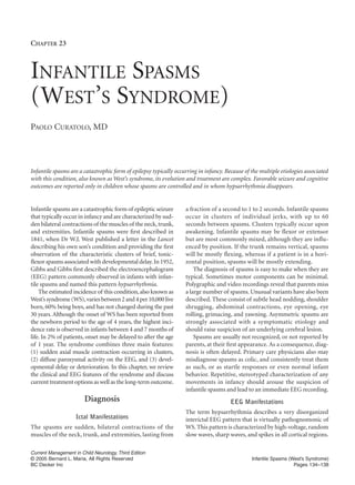
When infantile spasms occur, high-amplitude slow infantile spasms (Table 23-1). Benign myoclonus of early
waves followed by abrupt attenuation of the background infancy may closely mimic infantile spasms, but the EEG is
activity appear (Figure 23-1). Spasms need to be distin- normal. Epileptic syndromes closely related to WS include
guished from tonic seizures, in which the contraction is early myoclonic encephalopathy, early infantile epileptic
longer—lasting several seconds—and combined with low- encephalopathy, and early onset Lennox-Gastaut syndrome.
amplitude fast activity, and from myoclonic jerks, which All these conditions underlie serious brain pathology.
are shorter—lasting a fraction of a second—and combined
with a generalized spike-wave discharge. Developmental Manifestations
Although hypsarrhythmia is pathognomonic for WS, Psychomotor and behavioral regression may occur at the
other EEG patterns can occur in about one-third of onset of the spasms, but developmental delay may have
patients, most commonly represented by multifocal inde- preceded them by several months. For some infants with
pendent spike discharges. The term “modified hypsar- spasms, psychomotor retardation is present from birth. In
rhythmia” is used if there is some preservation of back- these patients, both early onset paroxysmal events and con-
ground rhythm, significant asymmetry, or synchronous tinuous nonconvulsive paroxysmal activity may account
burst of generalized spike-wave activity. Some brain mal- for the generation of developmental delay. Major cerebral
formations, such as hemimegalencephaly, lissencephaly, malformations, such as hemimegalencephaly, Aicardi’s
tuberous sclerosis, and Aicardi’s syndrome, show charac- syndrome, and lissencephaly, exhibit psychomotor delay
teristic EEG patterns. Focal features, including asymmet- before the onset of the epilepsy, which begins very early in
ric spasms and focal spikes or slow waves, indicate the life, and may consist of partial seizures before the occur-
presence of a hemispheric brain lesion, such as schizen- rence of spasms. Other patients, particularly those with
cephaly, porencephaly, polymicrogyria, or focal cortical tuberous sclerosis, usually exhibit normal development
dysplasia. In patients with focal lesions, the age of onset of until the first weeks of the disorder. Cognitive deterioration
the epilepsy has been shown to be related to the topo- in tuberous sclerosis develops slowly, owing to major
graphy, with occipital lesions producing spasms earlier epileptogenic activity in multiple areas of the cortex and to
than frontal ones. secondary generalization during sleep.
FIGURE 23-1. Ictal electroencephalogram
(EEG) showing typical spasms in a 4-month-
old child, with high-amplitude diffuse slow
waves on the EEG.
Current Management in Child Neurology, Third Edition
© 2005 Bernard L. Maria, All Rights Reserved Infantile Spasms (West’s Syndrome)
BC Decker Inc Pages 134–138
- 3. 136 / The Office Visit: Seizures and Epilepsy
TABLE 23-1. Differential Diagnosis of Infantile Spasms and to establish whether there is a need for genetic coun-
Nonepileptic syndrome mimicking infantile spasms seling. A comprehensive history, especially with regard to
Abdominal pain (colic) hereditary disorders and prenatal and perinatal events, as
Benign neonatal sleep myoclonus well as a careful clinical examination to check for signs of
Hyperexplexia tuberous sclerosis or dysmorphic features should always be
Sandifer’s syndrome
undertaken. Magnetic resonance imaging (MRI) may
Early breath-holding spells and syncopal attacks
Benign infantile dystonia reveal abnormalities in at least two-thirds of patients. If the
Increased Moro reflex and attacks of opisthotonus etiology is still unclear, a cerebrospinal fluid evaluation
Self-gratification or masturbation-like episodes and/or a screening of serum and urine for inborn error of
Benign paroxysmal tonic upward gaze metabolism, including amino acids, can be justified. A
Shuddering attacks
number of cases formerly classified as cryptogenic can now
Benign myoclonus of early infancy
be included in the symptomatic group owing to the use
Epileptic syndromes mimicking infantile spasms of more advanced imaging techniques, including high-
Early myoclonic encephalopathy
definition MRI and positron emission tomography (PET).
Early infantile epileptic encephalopathy
Benign myoclonic epilepsy of infancy In our experience, 70% of infantile spasm cases are symp-
Cryptogenic or symptomatic myoclonic-atonic-astatic epilepsies tomatic, 25% are cryptogenic, and 5% are idiopathic.
Early-onset Lennox-Gastaut syndrome In idiopathic cases, psychomotor development is normal
Partial seizures mimicking asymmetric infantile spasms at the onset of spasms, and there is no evidence of devel-
opmental arrest or regression. Approximately 40% of chil-
dren with idiopathic spasms are neurologically normal.
Cognitive disorders associated with infantile spasms are Familial occurrence of infantile spasms is rare. In recent
heterogeneous. They can consist of different combinations years, linkage to the X chromosome has been reported in two
of speech delay, visuomotor dyspraxia, autistic features, families with WS. The region of interest (Xp21.3–Xp22.1)
and mental retardation. Localized brain lesions can produce includes candidate genes involved in syndromic and non-
selective neuropsychologic disorders, depending on their syndromic mental retardation as well as radixin, a protein
topography. In this regard, tuberous sclerosis is a promis- involved in neuroaxonal processing. Familial idiopathic
ing model because the course of cognitive disorder depends spasms have also been reported, but no linkage has been
mainly on the clinical course of the epilepsy, which is related defined.
to the localization of the major cortical tubers.
Pathophysiology
Etiology
The pathophysiologic mechanisms of WS remain poorly
Many prenatal, perinatal, and postnatal abnormalities of understood. Heterogeneous pathophysiologic mecha-
the brain have been identified as presumed etiologic fac- nisms, including dysfunction of the brainstem, abnormal
tors for WS. The findings in a recent large population- cortical-subcortical interaction, disturbances of cortical
based cohort reported by Riikonen are given in Table 23- synaptogenesis, and abnormal brain-adrenal axis may be
2. Brain malformations and tuberous sclerosis may responsible for the genesis of infantile spasms.
account for up to 35% of patients. However, cerebral mal- WS could be produced by the brainstem, with pro-
formations can explain up to 45% of neuropathologic jections going both to the cortex and to the spinal cord
cases. Perinatal causes, including hypoxic-ischemic to generate hypsarrhythmia and the spasms, respec-
encephalopathy, porencephaly, multicystic encephaloma- tively. Modifications observed in the sleep-wake cycle,
lacia, and ulegyria account for about 10% of cases. Peri-
and postnatal brain lesions tend to produce later-onset
WS compared with prenatal lesions. When the cause of TABLE 23-2. Etiological Factors in a Population-Based
the spasms is identifiable (symptomatic spasms), the infant Study
is usually neurologically or developmentally abnormal Symptomatic Causes Percent
when the spasms begin. Microcephaly is more common in
Brain malformations and tuberous sclerosis 35
this group. Nonsymptomatic infantile spasms can be split Undetermined pre-/perinatal factors 19
into two groups: (1) cryptogenic cases are usually associ- Perinatal insults 9
ated with “hidden” lesions; (2) idiopathic cases are char- Symptomatic neonatal hypoglycemia 8
acterized by absence of any brain lesion. Familial or metabolic causes 9
Careful search for etiologic factors should be under- Early infections 2
Cryptogenic 18
taken in every case to determine an accurate prognosis
Current Management in Child Neurology, Third Edition
© 2005 Bernard L. Maria, All Rights Reserved Infantile Spasms (West’s Syndrome)
BC Decker Inc Pages 134–138
- 4. Infantile Spasms (West’s Syndrome) / 137
with reduced rapid eye movement sleep, are consistent VGB leads to the control of infantile spasms in up to 65%
with this hypothesis. The presence of frequent focal cor- of patients. Children with tuberous sclerosis have a 95%
tical lesions, detected by both structural and functional response rate. The efficacy of VGB can be assessed in less than
neuroimaging, has recently raised the hypothesis that a 10 days, but usually a few days’ treatment with a dose of 50
cortical lesion could trigger some subcortical structure, to 100 mg/kg/d stops infantile spasms. In contrast, steroids,
and then the basal ganglia and the cortex, resulting in benzodiazepines, or sodium valproate can take weeks to show
spasms and hypsarrhythmia. Another pathophysiologic efficacy. The cessation of spasms is associated with a marked
mechanism is inferred from the peculiar characteristics improvement in cognitive and behavioral development.
of the brain cortex in the first year of life. During this Unfortunately, in recent years, several reports have focused
period, in which there are excessive excitatory synapses, on the appearance of visual field disorders in patients treated
any triggering factor could initiate epileptic discharges with VGB. The use of VGB is associated with delayed appear-
with subsequent involvement of wide areas of the cor- ance of concentric narrowing of the visual fields in up to 40
tex. Finally, the occurrence of partial seizures during to 50% of cases. This narrowing can be severe and irre-
the cluster of infantile spasms in some children with a versible, and continuation of the drug can be associated with
symptomatic etiology suggests these two types of progressive visual-field loss. Despite this adverse effect, VGB
seizure may be generated independently at different lev- remains, in most countries, the drug of first choice for symp-
els of the brain. Because of an age-dependent peculiar tomatic infantile spasms associated with tuberous sclerosis.
cortical-subcortical interaction, infantile spasms may Currently, the minimum duration and doses of VGB treat-
be produced in these cases at a subcortical level, trig- ment that can produce side effects are unknown. The feasi-
gered by focal cortical discharges. bility of low dosages and short treatment periods (2 to
3 months) is now under investigation.
Medical Treatment Recent results of a pilot study of topiramate show that
45% of patients are seizure-free on high doses. Studies in
Seizure control is necessary to improve the natural history Japan and the United States have shown that two-thirds of
of WS. Therefore, identifying the most appropriate ther- patients become spasm-free when treated with zonisamide
apy is an urgent matter, so seizures can be controlled after other therapies have failed. Improvement of seizure
quickly, while a child still has the maximum potential for control has been observed with benzodiazepines and val-
improved development. proic acid in high doses. However, none of these drugs has
The best treatment for infantile spasms remains uncer- shown responses comparable with those of VGB or corti-
tain. There are few drugs with proven efficacy: hormonal costeroid therapy; no controlled studies have been per-
therapy (in the form of adrenocorticotropic hormone formed and their effects on EEG abnormalities are limited.
[ACTH] or prednisone), vigabatrin (VGB), topiramate, and At our institution, we recommend a 10-day course of
zonisamide. The ideal dose and duration of ACTH or pred- VGB as initial therapy. ACTH should be the first drug of
nisone have not been established. An initial dose of 20 to choice if VGB is not available. When VGB or ACTH fails to
30 IU/d for 2 to 6 weeks is just as effective as an initial dose control spasms, then topiramate can be prescribed. Clinical
of 150 IU/d tapered over 10 weeks. A 2-week course of oral experience suggests that titration has to be slow and pro-
prednisone, 2 mg/kg/d, has similar efficacy. The response to gressive. A small proportion of cases of infantile spasms
hormonal therapy is never graded; control of spasms is may represent atypical pyridoxine-dependent seizures.
either complete or nonexistent. Even when the response is High doses of pyridoxine (30 to 40 mg/kg/d) are reported
favorable, one-third of patients relapse. A second course of to be effective in 40% of patients with cryptogenic spasm
treatment is effective in 75% of cases in which the first but only in 10% of symptomatic cases.
course was successful. Failure to respond and relapse occur To conclude, it is important to underline that many
more often in symptomatic cases. ACTH, particularly in risks and adverse effects are associated with the medica-
high doses, has been shown to be more effective than tions used in the treatment of infantile spasms. As a gen-
steroids in 70 to 80% of children with WS. The enhanced eral rule, when selecting a drug regimen for a patient,
potency of ACTH compared with steroids is consistent with physicians must weigh the risk for morbidity or mortality
direct, steroid-independent actions of ACTH within the versus the benefit of increasing developmental potential if
central nervous system. Adverse events, including hyper- the seizures are controlled quickly.
tension, hypokalemia, irritability, and poor sleep are closely
related to dosage, duration of therapy, and patient’s suscep- Surgical Treatment
tibility to ACTH. Adverse events are minimized if the dosage
is quickly tapered; however, few patients with symptomatic The discovery of focal or multifocal cortical lesions has led
infantile spasms require long courses of therapy. to a surgical approach for the treatment of some patients
Current Management in Child Neurology, Third Edition
© 2005 Bernard L. Maria, All Rights Reserved Infantile Spasms (West’s Syndrome)
BC Decker Inc Pages 134–138
- 5. 138 / The Office Visit: Seizures and Epilepsy
with intractable infantile spasms. The location of these The long-term outcome is strongly influenced by the
lesions should be concordant with localization of focal structural abnormalities underlying the syndrome. The
ictal and/or interictal EEG abnormalities before proceed- outcome is worse in children with severe brain malforma-
ing with cortical resection. Interictal glucose metabolism tions and tuberous sclerosis. Risk factors for a poor prog-
PET scans show unifocal cortical hypometabolism in nosis include the presence of neurologic abnormalities
about 20% of children with intractable cryptogenic infan- before the onset of spasms, developmental regression, focal
tile spasms. When a single lesion is present on the MRI or EEG findings, failure of medication to promptly correct
PET, and there is good correlation with EEG localization, developmental arrest or regression, recurrence of spasms
surgical removal of the hypometabolic area, which usually and hypsarrhythmia after an effective response to drug,
proves to contain dysplastic tissue, often provides seizure onset before 4 months of age, presence of an underlying
control. To achieve the best seizure outcome, it is impor- structural lesion, and abnormal neuroimaging findings.
tant to resect the entire nociferous area, rather than just the Despite recent improvement in clinical and etiologic
seizure focus. However, the long-term effects of large sur- diagnosis of infantile spasms, many crucial problems
gical resection on neurologic functions have not been fully remain unresolved. Physicians should take a broad look at
evaluated, and it is not clear to what extent cognitive devel- all the therapeutic options available to achieve seizure con-
opment is affected by such radical treatment. trol and reverse the poor prognosis associated with WS.
Prognosis Suggested Readings
Curatolo P, Seri S, Verdecchia M, et al. Infantile spasms in tuber-
Most children with infantile spasms have a poor progno-
ous sclerosis complex. Brain Dev 2001;23:502–7.
sis because of intractable epilepsies, severe developmental
delay, and/or significant cognitive impairment. Epileptic Dulac O. What is West syndrome? Brain Dev 2001;23:447–52.
seizures persist in approximately 50% of patients. The nat- Shields WD. West’s syndrome. J Child Neurol 2002;17(Suppl 1):
ural history of epilepsy is characterized by the development S76–9.
of Lennox-Gastaut syndrome and temporal lobe partial
epilepsy. However, the occurrence of Lennox-Gastaut syn- Practitioner and Patient Resources
drome after WS has considerably decreased in the past
decade owing to new therapeutic options. Epilepsy Foundation
Only 5 to 10% of children with infantile spasms have 4351 Garden City Drive, Suite 406
Landover, MD 20785-2267
normal development. The vast majority of children are
Phone: (301) 459-3700 or (800) EFA-1000
left with cognitive or motor delay, and often with behav- E-mail: postmaster@efa.org
ior disorders. Psychiatric disorders, such as autism and http://www.epilepsyfoundation.org
hyperactive behavior, can occur in about 20% of children, The Epilepsy Foundation will ensure that people with seizures are
but they are significantly more frequent in children with able to participate in all life experiences and will prevent, control, and
tuberous sclerosis (about 40%). cure epilepsy through research, education, advocacy and services.
Current Management in Child Neurology, Third Edition
© 2005 Bernard L. Maria, All Rights Reserved Infantile Spasms (West’s Syndrome)
BC Decker Inc Pages 134–138