SlideShare una empresa de Scribd logo
1 de 12
Descargar para leer sin conexión
The Human Thalamus is Crucially Involved
                     in Executive Control Operations

                       Frank Marzinzik, Michael Wahl, Gerd-Helge Schneider,
                      Andreas Kupsch, Gabriel Curio, and Fabian Klostermann


Abstract
& The processing of executive control is thought to involve        prepared movement had to be performed or withheld. In fronto-
cortical as well as thalamic brain areas. However, the questions   central scalp as well as in thalamic recordings, event-related
of how thalamic structures contribute to the control of be-        potentials upon go versus no-go instructions were expressed
havior and how cortical versus thalamic processing is coor-        differentially. This task effect was unrelated to motor processes
dinated remain to be settled. We therefore aimed at specifying     and emerged significantly prior at thalamic than at scalp level.
respective activations during the performance of a go/no-go        Amplitude fluctuations of depth and scalp responses showed
task. To this end, an electroencephalogram was recorded simul-     site- and task-dependent correlations, particularly between thal-
taneously from scalp and thalamic electrodes in seven patients     amic and no-go-related activities at frontal recording sites. We
undergoing deep brain stimulation. Meanwhile, left- or right-      conclude that an early classification of go and no-go instruc-
directed precues were presented indicating with which index        tions is performed already thalamically. It further appears that
finger a button press should be putatively executed. Thereafter,   this information is subsequently utilized by cortical areas en-
2 sec elapsed until a go or no-go stimulus determined if the       gaged in the definite inhibition of the prepared action. &


INTRODUCTION
                                                                   cal regions (Behrens et al., 2003; also see Sherman &
Human behavior has to be constantly aligned with novel             Guillery, 1996 for a review). Indeed, different authors re-
environmental conditions. One of the essential abilities           ported on patients with thalamic infarctions, presenting
to exert such executive control is the inhibition of con-          with variable executive control dysfunctions (Van der
textually unsuited actions (see Smith & Jonides, 1999;             Werf et al., 2003; Mennemeier, Fennell, Valenstein, &
Norman & Shallice, 1986; Logan, 1985; Logan & Cowan,               Heilman, 1992; Eslinger, Warner, Grattan, & Easton, 1991;
1984 for reviews). Based on clinical observations in               Sandson, Daffner, Carvalho, & Mesulam, 1991). Partic-
brain-lesioned patients and on functional imaging stud-            ularly concerning inhibitory control, functional imaging
ies, the processing of such adaptive functions is mostly           studies demonstrated thalamic next to prefrontal activa-
allocated to frontal areas, in particular, the dorsolateral        tions when prepared motor responses had to be sup-
prefrontal and anterior cingulate cortices (Carter et al.,         pressed (de Jong & Paans, 2007; Garavan, Ross, Murphy,
1998; D’Esposito et al., 1995; Petrides, Alivisatos, Meyer,        Roche, & Stein, 2002). However, as functional imaging
& Evans, 1993; Shallice & Burgess, 1991; Pardo, Pardo,             hardly specifies the timing of rapidly changing activa-
Janer, & Raichle, 1990).                                           tions across different brain areas, it cannot be said how
   With the spread of neuroanatomical models on cortico-           and in which position the thalamus is involved in such
basal connectivity (see Alexander, Crutcher, & DeLong,             operations.
1990 for a review), respective research questions addi-               Exceptionally, this issue can be addressed by electro-
tionally focused on the impact of subcortical structures           encephalography (EEG), if conventional scalp recordings
on diverse higher-order operations (Rieger, Gauggel,               are combined with derivations from thalamic electrodes,
& Burmeister, 2003; Kramer, Reed, Mungas, Weiner, &                for instance, in patients with deep brain stimulation (DBS;
Chui, 2002; Rafal & Posner, 1987; also see Basso, Uhlrich,         Kuhn et al., 2004; Foffani et al., 2003; Marsden, Ashby,
& Bickford, 2005; Heyder, Suchan, & Daum, 2004; Royall             Limousin-Dowsey, Rothwell, & Brown, 2000). Intriguingly,
et al., 2002 for reviews). In this regard, the human thal-         this approach combines the high temporal resolution of
amus appears in a strategically critical position for its          EEG with otherwise inaccessible spatial information. Of
extensive reciprocal connections with almost all corti-            note in the present context, simultaneous recordings from
                                                                   the scalp and the ventral intermediate nucleus (VIM) of
                                                                   the thalamus have recently been performed in DBS pa-
Charite—University Medicine Berlin
      ´                                                            tients, who engaged in a classical oddball task. The most


D 2008 Massachusetts Institute of Technology                           Journal of Cognitive Neuroscience 20:10, pp. 1903–1914
unexpected finding of this study was that thalamic event-     in VIM. At this time, patients were without medication
related potentials (ERPs), differentially expressed upon      and almost tremor-free due to the short-lived post-
target stimuli, emerged prior to analog ERPs at scalp level   operative microthalamotomy effect (Kondziolka & Lee,
(Klostermann et al., 2006), suggestive of a central role of   2004).
thalamic processes in attentive behavior.                        The externalization phase serves to confirm the cor-
   The timing of thalamic versus cortical operations re-      rect electrode localization by MRI (which cannot be per-
lated to executive control has not been studied so far.       formed with the implanted stimulator) and to assess the
We therefore recorded EEGs simultaneously from scalp          (side) effects of DBS by a disconnectable external test
and thalamic electrodes in a number of DBS patients           stimulator, so that unfavorable electrode placements
engaging in a go/no-go task. They had to execute versus       could be corrected at this stage (no revisions necessary
inhibit previously cued finger movements. Go versus           in the present group). After this test phase, the definite
no-go instructions are known to elicit differential ERPs,     DBS stimulator is implanted in the subclavicular region
reflecting the time course of activations due to response     and subcutaneously connected with the electrode leads
inhibition in fronto-cortically assumed areas (Bokura,        (making electrode revisions thereafter more complex).
Yamaguchi, & Kobayashi, 2001; Kiefer, Marzinzik, Weisbrod,    From this programmable stimulator, high-frequency
Scherg, & Spitzer, 1998; Strik, Fallgatter, Brandeis, &       electrical impulses are continuously delivered to the
Pascual-Marqui, 1998). These go- and no-go-related scalp      electrodes, modulating the function of the target area
responses were used as a reference for the simulta-           which, in the case of tremor, is VIM (Schuurman et al.,
neously recorded thalamic ERPs. Thus, task-related am-        2000). Accordingly, ET was strongly reduced by VIM DBS
plitude and timing differences could be assessed for          in the present group (tremor scores without/with DBS:
scalp versus thalamic sites in order to analyze how pro-      3.1 ± 0.3/0.7 ± 0.7; 0 = absent; 1 = mild, intermittent;
cesses at either level related to response inhibition as a    2 = moderate; 3 = markedly abnormal, interfering with
basic function of executive control.                          many activities; 4 = severely abnormal, interfering with
                                                              most activities; scale according to Fahn et al., 1993).

METHODS
Subjects                                                      Visual Go/No-go task
Seven patients (2 women, 5 men; 36–74 years) with es-         Three hundred sixty visual trials were presented per
sential tremor (ET) participated in this study. They were     patient. Each trial was initiated by the presentation of
cognitively unimpaired (Mini-Mental State: 29 ± 1.3           a warning cue for 200 msec, either as an arrow directed
out of 30 points, range 27–30, cutoff for suspected de-       to the left or to the right. The direction of arrows indi-
mentia 23; Fillenbaum, Heyman, Williams, Prosnitz, &          cated with which index finger a motor response should
Burchett, 1990) and had normal presurgical magnetic           be prepared (Figure 1). After 1800 msec, the imperative
resonance imaging (MRI) scans. They underwent thera-          cue appeared for further 200 msec, either as a go cue
peutic DBS of the thalamic VIM, known to suppress ET          (green square), upon which the prepared movement
effectively (Schuurman et al., 2000). All patients gave       had to be executed, or as a no-go cue (red square),
informed consent to the study protocol, which was ap-         upon which it had to be withheld. Left versus right di-
proved by the local Ethics Committee.                         rected arrows and green versus red squares, respec-
   During the first days after DBS electrode placement        tively, occurred with probabilities of 50%. The resulting
(Day 4.1 ± 1), the patients engaged in a go/no-go task,       four conditions ‘‘left-go,’’ ‘‘right-go,’’ ‘‘left-no-go,’’ and
while a scalp EEG was performed together with record-         ‘‘right-no-go’’ appeared in randomized order, each com-
ings from the externalized leads of the DBS electrodes        prising 90 trials.


Figure 1. Go/no-go paradigm.
Each task trial consisted
of the presentation of a
side-instructive precue
followed by either a go or
no-go instruction. Left- versus
right-directed instructions
and go versus no-go signals
appeared equiprobably in
randomized order. The 360
trials per session appeared
at intervals of 4 sec.


1904      Journal of Cognitive Neuroscience                                                         Volume 20, Number 10
All stimuli popped up within a 5 Â 5-cm2 frame, cen-       bipolar derivations of 4.5 mm (1–3) and 7.5 mm width
tered in the middle of a 15-in. computer screen and           (0–3), respectively (cf. Figure 2). By subtraction of De-
present during the entire presentation time. The pa-          rivation 1–3 from Derivation 0–3, the upper 4.5 mm of
tients, sitting at 1.5 m distance, were instructed to look    the recording trajectory could be differentiated from
at this frame also in-between trials (intertrial interval =   the residual lower 3 mm, as the difference signal ap-
1800 msec) in order to avoid EEG artifacts from gross         proximates the physical bipolar derivation over this
eye movements.                                                caudal portion. This partitioning was done as the ori-
                                                              gins of task effects could thus be estimated. First, if an
                                                              effect was obtained only in one of the two recording
Thalamic Recordings
                                                              regions, a generation near to the sensitive derivation
Electrodes were implanted bilaterally into the VIM            could be assumed (otherwise, the effect should project
(Medtronic electrode 3387). For electrode placement,          into the upper and lower derivation, covering adjacent
standard VIM positions from the stereotactic brain atlas      areas of few millimeters along the same trajectory).
by Schaltenbrand and Wahren (1977) were referred to           Second, the lowest contact was targeted to the cau-
the individual AC–PC line (the straight sagittal connec-      dal border of VIM, which demarcates this nucleus to
tion between the anterior and posterior commissures),         nonthalamic structures, namely, the Zona incerta. Con-
exactly identified by intraoperative ventriculography.        trarily, at its posterior, anterior, cranial, and medial
Standard coordinates were adjusted for each case with         limits, VIM is surrounded by further thalamic nuclei.
respect to the individual thalamic height (15.3 ± 1 mm)       Thus, effects generated in subthalamic regions adjacent
and AC–PC length (24.8 ± 1.5 mm), determined by               to VIM should, first of all, project into the lower deriva-
matching presurgical stereotactic MRI with ventriculo-        tion, whereas processes, generated more centrally in the
graphic data. The thus-calculated coordinates for the         thalamus, will rather become discernible in the upper
lowest contact of the right/left VIM electrode (see be-       derivation.
low), expressed as (i) anteriority to PC, (ii) laterality
to AC–PC, and (iii) verticality to AC–PC, were: (i) 7.2 +
                                                              Surface Recordings
0.4 mm/7 + 0.5 mm, (ii) 14.6 + 0.5 mm/14.5 + 0.5 mm,
and (iii) À0.1 + 0.4 mm/À0.2 + 0.6 mm (minus in-              Scalp electrodes (Neuroscan system) were positioned
dicating below AC–PC). Postsurgical MRI confirmed             at F3, Fz, F4, C3, Cz, C4, P3, Pz, and P4 according to
that the planned placements were met by the implanted         the 10–20 system (impedances <5 k
) and referenced
electrodes.                                                   to linked mastoid electrodes. By this array, the typical
   DBS electrodes consist of four ring contacts (0–3 from     spatio-temporal distribution of fronto-central P300 re-
basal to cranial), longitudinally spaced at distances of      sponses upon go versus no-go stimuli could be con-
1.5 mm in VIM implants. On both recording sides, Con-         firmed per patient. It was expected that the ‘‘no-go
tacts 0 and 1 were referenced to Contact 3, resulting in      P300’’ would peak over frontal scalp sites upon the


Figure 2. Simultaneous scalp
and thalamic recordings.
Scalp EEG was derived
simultaneously with depth
recordings from the thalamic
ventral intermediate nucleus
(VIM; n = 14) in a group of
tremor patients. The MRIs
show representative DBS
electrode positions in one
patient. Two bipolar channels
covered the lower (light field)
and upper (dark field) portions
of the DBS electrodes, the
circles representing the
stereotactically defined edges
of these adjacent channels
per patient. The projection
of the recording fields on
the 13-mm paramedian slice
from the Schaltenbrand atlas
suggests that depth sampling
areas were at the lower
edge and well within VIM,
respectively.


                                                                                                Marzinzik et al.    1905
instruction to withhold a prepared movement. This ERP          peak. At scalp level, this procedure referred to the maxi-
component is a positive potential in the time window           mum component between 300 and 600 msec, a time
between 300 and 600 msec upon no-go signals and is of          domain in which ERPs upon go and no-go signals began
markedly larger magnitude than the according ERPs in           to differ and which is typical of P300 responses elicited
the go condition ( Weisbrod, Kiefer, Marzinzik, & Spitzer,     in comparable paradigms. At thalamic level, latency and
2000; Roberts, Rau, Lutzenberger, & Birbaumer, 1994;           amplitude values were assessed for the first component,
Eimer, 1993; Jodo & Inoue, 1990; Pfefferbaum & Ford,           which was differentially expressed between go and
1988; Pfefferbaum, Ford, Weller, & Kopell, 1985; Simson,       no-go conditions.
Vaughan, & Ritter, 1977). The origin of the no-go P300            The main statistical analysis primarily aimed at iden-
is allocated to prefrontal and cingulate cortical areas        tifying factors for the amplitude and latency values of
(Kiefer et al., 1998). Thus, the time course of cortical       scalp and thalamic ERPs. Therefore, three-way analyses
activations related to response inhibition can be delin-       of variance (ANOVAs), with the test factors task con-
eated by comparing the respective ERPs in the go versus        dition (two levels: go/no-go), response side (two levels:
no-go condition.                                               left/right), and electrode position, were performed, sep-
   Thalamic and scalp data were sampled continuously           arately for the scalp (nine levels: F3/Fz/F4/C3/Cz/C4/P3/
at 2 kHz with a bandpass from 0.05–500 Hz. Horizontal          Pz/P4) and the thalamus (four levels: lower left, upper
and vertical electrooculogram were registered to screen        left, lower right, upper right).
against eye-blink artifacts.                                      Further, in order to compare go/no-go-related ERPs at
                                                               scalp versus thalamic level chronometrically, the peak
                                                               latencies determined at the electrode positions with the
Analysis
                                                               largest amplitude effect for task condition (i) at scalp
Peristimulus EEG segments from À200 to +1000 msec              level and (ii) at thalamic level were utilized for a further
were averaged separately over left-go, right-go, left-no-go,   three-way ANOVA. This ANOVA comprised the test fac-
and right-no-go trials, unless they were handled incor-        tors task condition (two levels: go/no-go), response side
rectly or contained eye-blink artifacts. Further, averages     (two levels: left/right), and brain level (two levels: elec-
were calculated relative to the reaction times (button         trode positions at which the effect of task condition on
presses) in the go condition. Thus, back averages from         ERP amplitudes was strongest (i) at scalp level and (ii)
the motor response could be compared to the stimulus-          at thalamic level). As post hoc comparisons, Newman–
triggered forward averages, determining if a component         Keuls tests were performed.
reflected predominantly sensory or motor processes.               If scalp and thalamic ERPs had different peak laten-
   To assess if minor activity discernible in the back         cies, the onset of the target effect was additionally de-
average was the residual of sensory components in the          termined. Therefore, no-go ERPs were subtracted from
forward average or were proper motor potentials, an            go ERPs at either level to better display the time course
additional analysis was performed. Per patient, the re-        of differences between conditions. The task effect was
action times from the go trials were marked down to            considered to start out with the first poststimulus inter-
the no-go trials which, on this basis, could be averaged       val of at least 25 msec, in which the averaged amplitudes
in the very same way as the go trials. Such ‘‘sham aver-       exceeded the standard deviation across baseline values
ages’’ in the no-go condition (not containing any motor        by a factor > 2. Statistically, this was assessed with run-
responses) were individually compared with the back            ning t tests based on moving averages over 50 data
averages (based on reaction times). Activity in the back       points per patient.
average was considered a correlate of motor processing            If task effects were proven at both thalamic and scalp
if it exceeded according activity in the sham average,         sites, it was further tested if these effects were correlated
whereas it was viewed the residual of sensory compo-           between the different recording levels, putatively indic-
nents if it was identically identified in the sham average.    ative of thalamo-cortical networks active in executive
   Analysis of scalp and thalamic data was performed at        control. Therefore, per patient three subaverages, each
off-line filter settings from 0.5 to 20 Hz and from 20 to      over one third of the artifact-free and correct left- and
50 Hz. The former bandpass was chosen with respect to          right-hand go as well as no-go trials (each containing at
the main spectral energy of P300 components between            least 20 trials), were built, reflecting the ERP fluctuations
5 and 10 Hz in line with conventional methodology,             over subsequent time segments of the recording session
the latter was additionally applied as phase-locked tha-       in either task condition. For the resulting (sub-)ERPs,
lamic activity has hardly been described and might             the amplitude values of the monophasic thalamic com-
exceed the spectrum known from scalp ERPs, so that po-         ponent, differentially expressed between go and no-go
tentially faster components would not escape this scru-        condition (cf. Results), were then matched with the am-
tiny. Latency values were determined from the point in         plitude values of the first component in the time domain
time at which the stimulus was delivered to the peak of        of the task effect assessed at all scalp electrode sites.
a component. Amplitudes were measured from baseline            For the interindividual comparability of the results, mag-
(defined over the 200 msec prestimulus interval) to            nitudes were expressed in proportion to the mean of


1906     Journal of Cognitive Neuroscience                                                          Volume 20, Number 10
the three amplitudes determined per subject, condition,                 nitudes at scalp and thalamic levels. This difference
and recording site normalized to 1. In so doing, a                      between ‘‘forward’’ versus ‘‘back’’ averages suggests
Pearson’s coefficient of correlation could be determined                that the described components predominantly reflect
across subjects for each thalamo-cortical electrode pair                the processing of go and no-go stimuli rather than the
[per condition: 3 (subaverages) Â 2 (left/right response                motor execution of the task. This was corroborated by
side) Â 7 (subjects)]. Thus, it could be assessed if ERPs               the comparison of the ‘‘back averages’’ of the go trials
in the go and no-go condition covaried between depth                    with the ‘‘sham averages’’ of the no-go trials (replicating
and scalp recording sites or, alternatively, behaved inde-              the time criteria in the back averages; for details, cf.
pendently from each other and, secondly, how putative                   under Methods, Analysis). In the sham averages, the
thalamo-cortical relations were spatially distributed.                  residual activity from scalp as well thalamic components
                                                                        upon the no-go signal was undistinguishable from the
                                                                        activity obtained in the back average of the go trials
                                                                        (Figure 5). Thus, during the time domain of interest, the
RESULTS
                                                                        attenuation of ERPs in both conditions was due to the
Overview                                                                jitter of activity, phase-locked to go as well as no-go trials
Patients responded correctly to 88 ± 14%/98 ± 3% of                     and not to motor activity proper.
go/no-go trials (difference of error rate not significant).
The mean reaction time was slightly faster for right- than
left-hand responses (596 ± 147 msec vs. 622 ± 178 msec;                 Amplitude Analysis
difference not significant).
   Generally, ERPs were identified in the data filtered                 Scalp ERPs
from 0.5 to 20 Hz and were absent above this frequency                  An interaction Task condition  Electrode position was
band, regardless of the recording site. At scalp level,                 found [F(8, 48) = 12.85, p < .0001] according to the
positive scalp ERPs upon go as no-go stimuli peaked be-                 observation that P300 components were larger upon
tween 300 and 600 msec at all recording sites, according                no-go stimuli than upon go stimuli at rostral recording
to typical P300 responses. At parietal sites, the P300 am-              sites. Post hoc comparisons proved the difference be-
plitudes and latencies in the go condition were similar to              tween ERP magnitudes of either condition to be sig-
those in the no-go condition. At fronto-central positions,              nificant at all frontal and central positions but strongest
P300 components upon no-go stimuli were, however,                       at Fz ( p < .001). The effect was not modulated by the
larger and occurred later than upon go stimuli. In tha-                 response side.
lamic recordings, monophasic ERP components were
identified upon either stimulus class at almost identical
                                                                        Thalamic ERPs
latencies around 280 msec. This component, which pre-
ceded the fronto-central P300, was found larger upon                    According to the observation that go stimuli elicited larger
go than upon no-go stimuli (cf. Table 1 and Figure 3).                  ERPs than no-go stimuli in thalamic recordings, task con-
Further, upon go instructions, differences due to the                   dition was shown a main effect [F(1, 6) = 8.45, p < .028].
laterality of the executed response emerged only after                  The interaction Task condition  Electrode position [F(3,
the peak of this thalamic component (cf. Figure 4).                     18) = 4.12, p < .022] was due to the fact that a significant
   Averaging with respect to the motor response (go                     difference between go- and no-go-related ERPs was only
condition), resulted in a strong attenuation of ERP mag-                obtained at the upper portion of the DBS electrode, but


Table 1. Latencies and Amplitudes of ERP upon Go and No-go Stimuli
               Thalamus                                                         Scalp (P300)

Stimulus     Left       Right         F3          Fz          F4         C3          Cz          C4          P3           Pz          P4

Peak Latencies (msec)
Go         278 ± 11 280 ± 15 410 ± 19 412 ± 17 413 ± 16 413 ± 16 411 ± 16 411 ± 16 412 ± 13 412 ± 13 407 ± 14
No-go      281 ± 12 272 ± 10 465 ± 16 471 ± 20 470 ± 20 472 ± 20 459 ± 27 467 ± 18 446 ± 15 436 ± 21 426 ± 23


Amplitudes (V)
Go         6.5 ± 1.4 5.7 ± 0.9 2.9 ± 0.9 3.4 ± 1           3.8 ± 0.9 4.5 ± 1.1 4.5 ± 1.3 5.2 ± 1.2 5.6 ± 1.4 6.6 ± 1.3 6.1 ± 1.2
No-go      4.1 ± 0.9    3 ± 0.5 6.4 ± 1        7.1 ± 1.1 6.8 ± 1.1      7 ± 1.6     7 ± 1.1 7.2 ± 1.3 6.3 ± 1.2 6.8 ± 1.3            6 ± 1.1
Peak latencies (± standard error) and amplitudes (± standard error) of thalamic (upper channel) and scalp ERPs.


                                                                                                                  Marzinzik et al.     1907
Figure 3. Distribution of scalp and thalamic go and no-go ERPs. Overview of grand-averaged frontal, central, and parietal (A) as well as thalamic
(B) ERPs, averaged over ipsilateral and contralateral responses. The ERPs upon the go signal are indicated by the dashed line, the ones upon
the no-go signal by the solid line. The scalp no-go P300 peaked later than the thalamic ERPs following go and no-go signals.


not at its lower part (post hoc tests for upper/lower chan-                that P300 components upon no-go stimuli peaked signif-
nels: p  .05/p  .3). No further interactions were found.                 icantly later than upon go stimuli only at frontal and cen-
                                                                           tral recording positions, this effect being strongest at Fz
                                                                           ( p  .001).
Latency Analysis
Scalp ERPs
                                                                           Thalamic ERPs
Task condition was shown a main factor [F(1, 5) = 8.32,
p  .034], further interacting with electrode position [F(8,               The monophasic thalamic ERPs upon go versus no-go
40) = 2.29, p  .041]. The post hoc comparisons revealed                   stimuli did not differ in latency.


1908      Journal of Cognitive Neuroscience                                                                           Volume 20, Number 10
Figure 4. Thalamic ERPs in left-hand versus right-hand conditions. The amplitude of the thalamic components at peak latency was not influenced
by the side of motor execution: (A) The upper traces show the lateralization of thalamic ERP upon go instructions, compared for left-hand
(dotted line) versus right-hand (solid line) responses. Side-related differences emerged only after the peak of the monophasic thalamic ERPs.
(B) Upon no-go instructions, no lateralization was obtained.


Chronometric Comparison of the Thalamic                                  more than 2 standard deviations excess from baseline
and Scalp ERPs                                                           noise, as determined in the 200-msec interval before sig-
                                                                         nal presentation; cf. Methods, cf. Figure 6).
Brain level was a main factor of peak latencies in tha-
lamic versus scalp ERPs based on the recording positions
displaying the strongest task effects on ERP amplitudes
                                                                         Correlation between Thalamic and Scalp ERPs
of either level [left/right thalamus vs. Fz: F(1, 6) = 85.98/
76.09, p  .0001]. Further, task condition was a cofactor                Thalamic and scalp ERPs were correlated with respect to
[Brain level  Task condition for left/right thalamus vs.                the amplitude fluctuations along the recording session
Fz: F(1, 6) = 9.01/8.45, p  .024/ .027], according to                  (see Methods), but this thalamo-cortical correlation was
the fact that P300 components at Fz peaked later upon                    spatially distinct for go versus no-go ERPs. Specifically,
no-go than upon go stimuli ( p  .01), whereas thalamic                  thalamic no-go ERPs behaved similarly to accompanying
ERP latencies were similar in the go and no-go condi-                    ERPs in the time domain of the task effect at frontal sites,
tion. Thalamic ERPs, in turn, peaked prior to scalp P300                 where the no-go effect was largest. No such relation was
components both in the go and no-go condition ( p                       obtained for go ERPs which, instead, mostly correlated to
.001/p  .001).                                                          the according parietal ERPs (for summary, see Table 2).
   Accordingly, the onset of the task effect was deter-
mined earlier in thalamic than in scalp ERPs ( p  .0001).
In the subtraction curves of go versus no-go ERPs, it was
                                                                         DISCUSSION
determined to start at 196 ± 27 msec and 296 ± 57 msec
after signal presentation for thalamic ERPs and scalp                    The present study contained three major findings. First,
ERPs at Fz, respectively (criterion: during  25 msec                    as at fronto-central scalp sites, ERPs recorded from the


                                                                                                                 Marzinzik et al.       1909
Figure 5. Nonmotor character of frontal and thalamic go/no-go ERP. (A) The upper traces show scalp and thalamic ERPs to go/no-go stimuli,
contralateral to task execution/instruction, averaged to stimulus presentation (forward average). (B) The lower traces display the ERPs, averaged
with respect to the motor execution of the task for the go condition (back average; dashed line) and an according ‘‘sham ERP’’ for the no-go
condition (solid line). For the latter, the reaction times from the go trials were marked down to the no-go trials. Thus, no-go trials could be
averaged in the very same way as the go trials, although they did not contain any motor response. As activity in the back average was found
largely identical with that in the sham average, it was viewed the residual of the sensory components discernible in the forward average.


Figure 6. Difference curves
(go minus no-go condition) for
scalp and thalamic ERPs. The
task effect is shown as the
difference of ERPs in the go
versus no-go condition at scalp
and thalamic levels (grand
average). The beginning of
this effect was calculated per
patient as the first poststimulus
interval of at least 25 msec
exceeding the standard
deviation across the individual
baseline values by more
than a factor of 2. The
thus-determined onsets were
significantly earlier in thalamic
compared to scalp recordings
and are indicated by the
solid and dashed arrows,
respectively.


1910       Journal of Cognitive Neuroscience                                                                          Volume 20, Number 10
Table 2. Co-fluctuations of Thalamic and Scalp ERP Amplitudes
                                         Go Condition                                                  No-go Condition

Scalp                Left Thalamus                      Right Thalamus                Left Thalamus                  Right Thalamus

F3              r = .17 ( p  .29)                  r = .13 ( p  .42)            r = .75**(p  .001)             r = .76** (p  .001)
Fz              r = .19 ( p  .22)                  r = .19 ( p  .23)            r = .76** (p  .001)            r = .75** (p  .001)
F4              r = .13 ( p  .39)                  r = .23 ( p  .15)            r = .76** (p  .001)            r = .90** (p  .001)
C3              r = .43** (p  .005)                r = .33* (p  .05)            r = .58** (p  .001)            r = .74** (p  .001)
Cz              r = .34* (p  .03)                  r = .15 ( p  .33)            r = .24 ( p  .12)              r = .26 ( p  .11)
C4              r = .35* (p  .05)                  r = .45** (p  .01)           r = .18 ( p  .26)              r = .29 ( p  .06)
P3              r = .45** (p  .01)                 r = .45** (p  .01)           r = À.17 ( p  .28)             r = À.22 ( p  .17)
Pz              r = .52** (p  .001)                r = .55** (p  .001)          r = .07 ( p  .65)              r = .09 ( p  .58)
P4              r = .49** (p  .001)                r = .57** (p  .001)          r = À.22 ( p  .15)             r = À.06 ( p  .70)
r = Pearson’s correlation coefficient.
*Significant at .05 level.
**Significant at .01 level (two-tailed; cf. Methods).


upper intrathalamic electrode were differentially ex-                     of control processes exclusively refer to the cortical level,
pressed upon no-go versus go commands, and this dif-                      based on scalp ERPs. In this particular context, the re-
ference was not explained by motor processing. Second,                    ported depth ERPs merit a closer view.
whereas thalamic ERPs correlated to the parietal P300
component in the go condition, they corresponded to
                                                                          Origin of VIM-recorded ERPs
the task-specific frontal ERP component in the no-go
condition. Third, task-specific scalp components started                  An intrathalamic origin of VIM-recorded ERPs can be
well after thalamic go as well as no-go ERPs.                             reasonably assumed for several reasons. First, if depth
                                                                          signals were volume conducted from cortical generators,
                                                                          time courses of components at scalp and thalamic levels
Relation to Previous Studies
                                                                          should be synchronous. Accordingly, the discrepant la-
Worthwhile mentioning, the obtained spatio-temporal                       tencies of scalp versus VIM ERPs point to a closer, that is,
pattern of scalp ERPs was as described in numerous                        subcortical, origin of the latter. Second, the upper, but
studies applying comparable paradigms. Specifically, the                  not the lower, derivation from DBS electrodes caught
frontal component under scrutiny, often labeled ‘‘no-go                   significant differences between go and no-go-related
P300,’’ has been conceptualized as the reflection of ag-                  ERPs. This disparity points to an effect from near-field
gregate activity for the suppression of actions, according                activities. Far-field spread would almost equally project
to its enhancement in no-go trials (Roberts et al., 1994;                 into upper and lower derivation because of similar rela-
Eimer, 1993). The recording array used in this study does                 tive positions to both recording fields, covering few adja-
not allow to deduce the origin of this component, but                     cent millimeters along the same trajectory only. Finally,
previous studies focused on this issue. Based on source                   the absence of a the task effect in the lower thalamic
analysis of frontal P300, the provision of assigned func-                 derivations suggests that its proof in the upper record-
tions has been allocated to the anterior cingulum, the                    ings was due to processes in proximate thalamic struc-
prefrontal cortex, and the premotor cortex (Kamarajan                     tures, and not to operations in nuclei adjacent to the
et al., 2005; Bokura et al., 2001; Kiefer et al., 1998; Strik             caudal border of VIM outside the thalamus. However, an
et al., 1998). Accordingly, executive control operations                  origin in VIM proper seems unlikely, as both the upper
were proposed to be generated in a frontal lobe network,                  and lower depth recording areas were estimated to be
although a number of imaging studies and reports on                       predominantly within this nucleus.
patients with subcortical lesions point to an additional
involvement of thalamic structures (Van der Werf et al.,
                                                                          Nonmotor Nature of Thalamically Recorded ERPs
2003; Garavan et al., 2002; Mennemeier et al., 1992;
Eslinger et al., 1991; also see Van der Werf, Witter,                     Several results indicate that the thalamic activation in
Uylings,  Jolles, 2000, for a review). However, a more                   the time domain of interest is unrelated to proper motor
specific assignment of according subcortical functions is                 operations. First, thalamic ERPs in trials with left- versus
not available so far, and estimations on the chronometry                  right-sided motor responses did not differ in magnitude


                                                                                                               Marzinzik et al.    1911
at their peak latencies. Such a difference could be rea-      demands, appear aligned with more frontally located
sonably assumed if this component reflected primary           areas than throughout facilitatory processing, compati-
motor processes, as observed in later time domains of         ble with a particular involvement of dorsolateral prefron-
the ERPs. Second, thalamic ERPs were bound to stimulus        tal and anterior cingulate cortices in executive control
presentation rather than to the motor execution of the        (Carter et al., 1998; D’Esposito et al., 1995; Petrides et al.,
task, as they occurred upon go and no-go trials, al-          1993; Shallice  Burgess, 1991; Pardo et al., 1990).
though in the latter motor responses were not executed.       Such selective coupling might be a function of thalamo-
Third, a predominant binding of the ERPs to motor exe-        cortical, together with thalamic reticular neurons, whose
cution was incompatible with the finding that they were       firing properties and connectivity provide a mechanism
massively reduced in magnitude when averaged to the           for scaling and funneling information through the wide-
motor response as compared to the go instruction.             spread thalamic connections to frontal and temporo-
Fourth, the larger amplitude obtained in go versus no-        parietal cortical regions (Behrens et al., 2003; Sherman
go conditions could finally not be explained by additive       Guillery, 1996; Crick, 1984; see also Pinault, 2004;
motor operations. This was shown by the comparison of         Guillery  Sherman, 2002a, 2002b for reviews).
ERPs in go trials, averaged to the motor response, versus        Intriguingly, the chronometric comparison of thalamic
ERPs in no-go trials, averaged to the very same points        and scalp ERPs suggested that task-specific information
in time (‘‘sham average’’; cf. Methods and Results). In       was propagated in thalamo-cortical direction, that is, no
so doing, activations in the critical interval became un-     indication was found that the underlying thalamic pro-
distinguishable between conditions, delineating that the      cesses occurred under the control of related cortical
added thalamic activity was due to the go instruction         areas. This, together with the possibility of a thalamically
proper, but not to its execution.                             induced cortical coupling of go versus no-go-related
                                                              signaling, can be viewed as an experimental parallel to
                                                              ‘‘centrencephalic’’ concepts, derived from the Penfield
Conceptual Considerations
                                                              and Jasper (1954) formulation that a subcortical struc-
In this context, a number of proposed thalamic func-          ture can be ‘‘while anatomically subcortical, functionally
tions are of particular interest. During attentional and      supra-cortical,’’ further relating to the principle question
arousal processes, activation has been described for in-      if qualia can exist if subcortical structures work without
tralaminar thalamic regions (Fan, McCandliss, Fossella,       a corresponding cortex (cf. Merker, 2007; Schiff et al.,
Flombaum,  Posner, 2005; Hester, Fassbender,                2007).
Garavan, 2004; Woldorff et al., 2004; Garavan et al.,            The task-dependent expression of thalamic ERPs calls
2002; Portas et al., 1998; Coull, Frith, Dolan, Frackowiak,   for the comparison with previous results. Both go and
 Grasby, 1997; Frith  Friston, 1996), which receive         no-go instructions require specific performances with
afferent connections from the midbrain reticular forma-       respect to previously established expectancies. There-
tion and project to multiple cortical areas (McFarland       fore, their magnitudes do not appear to reflect thalamic
Haber, 2002; Macchi, Bentivoglio, Molinari,  Minciacchi,     weighing of signal relevance, given that both go and
1984; Steriade  Glenn, 1982; Bentivoglio, Macchi,           no-go signals convey behaviorally important informa-
Albanese, 1981; also see Merker, 2007; Jones, 1998 for        tion. In this sense, go and no-go signals are compara-
reviews). It has been hypothesized that midbrain signals      ble to target signals in oddball paradigms. Interestingly,
convey stimulus-related alert or orienting information        in a recent study, oddball target signals, rarely inter-
to the thalamus (Kinomura, Larsson, Gulyas,  Roland,         spersed between nontarget instructions, led to consid-
1996), which in turn communicates with specific cortical      erably larger thalamic ERPs than the present go and
regions, depending on the ongoing behavioral demands          no-go instructions (cf. Klostermann et al., 2006). There-
(see Merker, 2007; Pinault, 2004 for reviews). Similarly,     fore, our momentary working hypothesis is that the
it has been suggested that sensorily driven bottom–up         mentioned thalamic ERPs reflect distinct (rather than
signals are integrated with top–down directed informa-        unitary, but weighed) aggregate activities whose com-
tion in the center median and parafascicular nuclei of the    position imply variable thalamic contributions, specifi-
thalamus (reviewed by Sarter, Givens,  Bruno, 2001;          cally defined by the given behavioral context. However,
LaBerge, 1997).                                               the verification of this and the critical questions how and
   Of course, from the present data, any brainstem con-       by what task definitions are imposed upon the thalamus
tributions to the results remain speculative, but the cor-    remain central issues for future studies.
relations between thalamic and scalp ERPs do hint at
differential thalamo-cortical processing. According in-
                                                              Conclusions
teractions appeared task-dependent, as in the no-go
condition, thalamic ERPs co-fluctuated with the frontal       Principally, the distinct expression of thalamic ERPs upon
no-go P300, whereas in the go condition, they mainly did      go versus no-go instructions indicates thalamic involve-
so with the parietal P300. This seems to imply thalamo-       ment in executive control. Further, the fact that task-
cortical routes of information which, in case of inhibitory   specific activity emerged prior in depth recordings than


1912    Journal of Cognitive Neuroscience                                                            Volume 20, Number 10
at scalp level suggests cortical operations of response               Fan, J., McCandliss, B. D., Fossella, J., Flombaum, J. I.,
control to be subcortically prepared. Finally, the corre-                 Posner, M. I. (2005). The activation of attentional
                                                                         networks. Neuroimage, 26, 471–479.
lation of thalamic to frontal ERPs in the no-go condition             Fillenbaum, G., Heyman, A., Williams, K., Prosnitz, B.,
and of thalamic to parietal ERPs in the go condition in-                  Burchett, B. (1990). Sensitivity and specificity of
dicates that the underlying thalamo-cortical networks are                standardized screens of cognitive impairment and
flexibly activated as a function of task demands.                        dementia among elderly black and white community
                                                                         residents. Journal of Clinical Epidemiology, 43, 651–660.
                                                                      Foffani, G., Priori, A., Egidi, M., Rampini, P., Tamma, F.,
Acknowledgments                                                          Caputo, E., et al. (2003). 300-Hz subthalamic oscillations
                                                                         in Parkinson’s disease. Brain, 126, 2153–2163.
This study was supported by the Deutsche Forschungsgemein-            Frith, C. D.,  Friston, K. J. (1996). The role of the thalamus
schaft (DFG KL 1276/3-1). We thank our reviewers for their               in ‘‘top down’’ modulation of attention to sound.
constructive suggestions and comments.                                   Neuroimage, 4, 210–215.
Reprint requests should be sent to Fabian Klostermann,                Garavan, H., Ross, T. J., Murphy, K., Roche, R. A.,  Stein,
Neurology, Charite—University Medicine Berlin, Campus
                  ´                                                      E. A. (2002). Dissociable executive functions in the
Benjamin Franklin, Berlin, 12200, Germany, or via e-mail:                dynamic control of behavior: Inhibition, error detection,
fabian.klostermann@charite.de.                                           and correction. Neuroimage, 17, 1820–1829.
                                                                      Guillery, R. W.,  Sherman, S. M. (2002a). Thalamic relay
                                                                         functions and their role in corticocortical communication:
REFERENCES                                                               Generalizations from the visual system. Neuron, 33,
                                                                         163–175.
Alexander, G. E., Crutcher, M. D.,  DeLong, M. R. (1990).            Guillery, R. W.,  Sherman, S. M. (2002b). The thalamus
  Basal ganglia–thalamocortical circuits: Parallel substrates            as a monitor of motor outputs. Philosophical Transactions
  for motor, oculomotor, ‘‘prefrontal’’ and ‘‘limbic’’                   of the Royal Society of London, Series B, Biological
  functions. Progress in Brain Research, 85, 119–146.                    Sciences, 357, 1809–1821.
Basso, M. A., Uhlrich, D.,  Bickford, M. E. (2005). Cortical         Hester, R., Fassbender, C.,  Garavan, H. (2004). Individual
  function: A view from the thalamus. Neuron, 45, 485–488.               differences in error processing: A review and reanalysis
Behrens, T. E., Johansen-Berg, H., Woolrich, M. W., Smith,               of three event-related fMRI studies using the GO/NOGO
  S. M., Wheeler-Kingshott, C. A., Boulby, P. A., et al. (2003).         task. Cerebral Cortex, 14, 986–994.
  Non-invasive mapping of connections between human                   Heyder, K., Suchan, B.,  Daum, I. (2004). Cortico-subcortical
  thalamus and cortex using diffusion imaging. Nature                    contributions to executive control. Acta Psychologica
  Neuroscience, 6, 750–757.                                              (Amsterdam), 115, 271–289.
Bentivoglio, M., Macchi, G.,  Albanese, A. (1981). The               Jodo, E.,  Inoue, K. (1990). Effects of practice on the P300
  cortical projections of the thalamic intralaminar nuclei,              in a Go/NoGo task. Electroencephalography and Clinical
  as studied in cat and rat with the multiple fluorescent                Neurophysiology, 76, 249–257.
  retrograde tracing technique. Neuroscience Letters, 26,             Jones, E. G. (1998). A new view of specific and nonspecific
  5–10.                                                                  thalamocortical connections. Advances in Neurology, 77,
Bokura, H., Yamaguchi, S.,  Kobayashi, S. (2001).                       49–71 [Discussion 72–73].
  Electrophysiological correlates for response inhibition in a        Kamarajan, C., Porjesz, B., Jones, K. A., Chorlian, D. B.,
  Go/NoGo task. Clinical Neurophysiology, 112, 2224–2232.                Padmanabhapillai, A., Rangaswamy, M., et al. (2005).
Carter, C. S., Braver, T. S., Barch, D. M., Botvinick, M. M.,            Spatial–anatomical mapping of NoGo-P3 in the offspring
  Noll, D.,  Cohen, J. D. (1998). Anterior cingulate cortex,            of alcoholics: Evidence of cognitive and neural disinhibition
  error detection, and the online monitoring of performance.             as a risk for alcoholism. Clinical Neurophysiology, 116,
  Science, 280, 747–749.                                                 1049–1061.
Coull, J. T., Frith, C. D., Dolan, R. J., Frackowiak, R. S.,         Kiefer, M., Marzinzik, F., Weisbrod, M., Scherg, M., 
  Grasby, P. M. (1997). The neural correlates of the                     Spitzer, M. (1998). The time course of brain activations
  noradrenergic modulation of human attention, arousal and               during response inhibition: Evidence from event-related
  learning. European Journal of Neuroscience, 9, 589–598.                potentials in a go/no go task. NeuroReport, 9, 765–770.
Crick, F. (1984). Function of the thalamic reticular complex:         Kinomura, S., Larsson, J., Gulyas, B.,  Roland, P. E. (1996).
  The searchlight hypothesis. Proceedings of the National                Activation by attention of the human reticular formation
  Academy of Sciences, U.S.A., 81, 4586–4590.                            and thalamic intralaminar nuclei. Science, 271, 512–515.
de Jong, B. M.,  Paans, A. M. (2007). Medial versus lateral          Klostermann, F., Wahl, M., Marzinzik, F., Schneider, G. H.,
  prefrontal dissociation in movement selection and inhibitory           Kupsch, A.,  Curio, G. (2006). Mental chronometry of
  control. Brain Research, 1132, 139–147.                                target detection: Human thalamus leads cortex. Brain,
D’Esposito, M., Detre, J. A., Alsop, D. C., Shin, R. K., Atlas, S.,      129, 923–931.
   Grossman, M. (1995). The neural basis of the central              Kondziolka, D.,  Lee, J. Y. (2004). Long-lasting
  executive system of working memory. Nature, 378, 279–281.              microthalamotomy effect after temporary placement
Eimer, M. (1993). Effects of attention and stimulus probability          of a thalamic stimulating electrode. Stereotactic and
  on ERPs in a Go/Nogo task. Biological Psychology, 35,                  Functional Neurosurgery, 82, 127–130.
  123–138.                                                            Kramer, J. H., Reed, B. R., Mungas, D., Weiner, M. W., 
Eslinger, P. J., Warner, G. C., Grattan, L. M.,  Easton, J. D.          Chui, H. C. (2002). Executive dysfunction in subcortical
  (1991). ‘‘Frontal lobe’’ utilization behavior associated with          ischaemic vascular disease. Journal of Neurology,
  paramedian thalamic infarction. Neurology, 41, 450–452.                Neurosurgery and Psychiatry, 72, 217–220.
Fahn, S., Tolosa, E.,  Marin, C. (1993). Clinical rating scale for   Kuhn, A. A., Williams, D., Kupsch, A., Limousin, P., Hariz, M.,
  tremor. In J. Jankovic  E. Tolosa (Eds.), Parkinson’s                 Schneider, G. H., et al. (2004). Event-related beta
  disease and movement disorders (2nd ed., pp. 271–280).                 desynchronization in human subthalamic nucleus
  Baltimore: Williams and Wilkons.                                       correlates with motor performance. Brain, 127, 735–746.


                                                                                                           Marzinzik et al.      1913
LaBerge, D. (1997). Attention, awareness, and the triangular           discrimination. Electroencephalography and Clinical
  circuit. Consciousness and Cognition, 6, 149–181.                    Neurophysiology, 92, 44–55.
Logan, G. D. (1985). Executive control of thought and                Royall, D. R., Lauterbach, E. C., Cummings, J. L., Reeve, A.,
  action. Acta Psychologica, 60, 193–210.                              Rummans, T. A., Kaufer, D. I., et al. (2002). Executive control
Logan, G. D.,  Cowan, W. B. (1984). On the ability to                 function: A review of its promise and challenges for clinical
  inhibit thought and action: A theory of an act of control.           research. A report from the Committee on Research of
  Psychological Review, 91, 295–327.                                   the American Neuropsychiatric Association. Journal of
Macchi, G., Bentivoglio, M., Molinari, M.,  Minciacchi, D.            Neuropsychiatry and Clinical Neurosciences, 14,
  (1984). The thalamo-caudate versus thalamo-cortical                  377–405.
  projections as studied in the cat with fluorescent                 Sandson, T. A., Daffner, K. R., Carvalho, P. A.,  Mesulam,
  retrograde double labeling. Experimental Brain Research,             M. M. (1991). Frontal lobe dysfunction following infarction
  54, 225–239.                                                         of the left-sided medial thalamus. Archives of Neurology,
Marsden, J. F., Ashby, P., Limousin-Dowsey, P., Rothwell,              48, 1300–1303.
  J. C.,  Brown, P. (2000). Coherence between cerebellar            Sarter, M., Givens, B.,  Bruno, J. P. (2001). The cognitive
  thalamus, cortex and muscle in man: Cerebellar thalamus              neuroscience of sustained attention: Where top–down
  interactions. Brain, 123, 1459–1470.                                 meets bottom–up. Brain Research, Brain Research
McFarland, N. R.,  Haber, S. N. (2002). Thalamic relay nuclei         Reviews, 35, 146–160.
  of the basal ganglia form both reciprocal and nonreciprocal        Schaltenbrand, G.,  Wahren, W. (1977). Atlas for stereotaxy of
  cortical connections, linking multiple frontal cortical              the human brain. New York: Medical Publishers.
  areas. Journal of Neuroscience, 22, 8117–8132.                     Schiff, N. D., Giacino, J. T., Kalmar, K., Victor, J. D., Baker, K.,
Mennemeier, M., Fennell, E., Valenstein, E.,  Heilman, K. M.          Gerber, M., et al. (2007). Behavioural improvements with
  (1992). Contributions of the left intralaminar and medial            thalamic stimulation after severe traumatic brain injury.
  thalamic nuclei to memory. Comparisons and report of a               Nature, 448, 600–603.
  case. Archives of Neurology, 49, 1050–1058.                        Schuurman, P. R., Bosch, D. A., Bossuyt, P. M., Bonsel, G. J.,
Merker, B. (2007). Consciousness without a cerebral cortex:            van Someren, E. J., de Bie, R. M., et al. (2000). A comparison
  A challenge for neuroscience and medicine. Behavioral                of continuous thalamic stimulation and thalamotomy for
  and Brain Sciences, 30, 63–81 [Discussion 81–134].                   suppression of severe tremor. New England Journal of
Norman, D. A.,  Shallice, T. (1986). Attention to action:             Medicine, 342, 461–468.
  Willed and automatic control of behavior. In R. J. Davidson,       Shallice, T.,  Burgess, P. W. (1991). Higher-order cognitive
  G. E. Schwartz,  D. Shapiro (Eds.), Consciousness and               impairments and frontal lobe lesions in man. In S. Levin,
  self-regulation: Advances in research and theory (Vol. 4,            H. M. Eisenberg,  A. L. Benton (Eds.), Frontal lobe
  pp. 1–18). New York: Plenum.                                         function and injury (pp. 125–138). New York: Oxford
Pardo, J. V., Pardo, P. J., Janer, K. W.,  Raichle, M. E. (1990).     University Press.
  The anterior cingulate cortex mediates processing selection        Sherman, S. M.,  Guillery, R. W. (1996). Functional
  in the Stroop attentional conflict paradigm. Proceedings             organization of thalamocortical relays. Journal of
  of the National Academy of Sciences, U.S.A., 87, 256–259.            Neurophysiology, 76, 1367–1395.
Penfield, W.,  Jasper, H. H. (1954). Epilepsy and the               Simson, R., Vaughan, H. G., Jr.,  Ritter, W. (1977). The
  functional anatomy of the human brain. Boston: Little,               scalp topography of potentials in auditory and visual
  Brown  Co.                                                          Go/NoGo tasks. Electroencephalography and Clinical
Petrides, M., Alivisatos, B., Meyer, E.,  Evans, A. C. (1993).        Neurophysiology, 43, 864–875.
  Functional activation of the human frontal cortex during           Smith, E. E.,  Jonides, J. (1999). Storage and executive
  the performance of verbal working memory tasks.                      processes in the frontal lobes. Science, 283, 1657–1661.
  Proceedings of the National Academy of Sciences, U.S.A.,           Steriade, M.,  Glenn, L. L. (1982). Neocortical and caudate
  90, 878–882.                                                         projections of intralaminar thalamic neurons and their
Pfefferbaum, A.,  Ford, J. M. (1988). ERPs to stimuli requiring       synaptic excitation from midbrain reticular core. Journal
  response production and inhibition: Effects of age,                  of Neurophysiology, 48, 352–371.
  probability and visual noise. Electroencephalography               Strik, W. K., Fallgatter, A. J., Brandeis, D.,  Pascual-Marqui,
  and Clinical Neurophysiology, 71, 55–63.                             R. D. (1998). Three-dimensional tomography of event-related
Pfefferbaum, A., Ford, J. M., Weller, B. J.,  Kopell, B. S.           potentials during response inhibition: Evidence for phasic
  (1985). ERPs to response production and inhibition.                  frontal lobe activation. Electroencephalography and
  Electroencephalography and Clinical Neurophysiology,                 Clinical Neurophysiology, 108, 406–413.
  60, 423–434.                                                       Van der Werf, Y. D., Scheltens, P., Lindeboom, J., Witter,
Pinault, D. (2004). The thalamic reticular nucleus: Structure,         M. P., Uylings, H. B.,  Jolles, J. (2003). Deficits of memory,
  function and concept. Brain Research, Brain Research                 executive functioning and attention following infarction
  Reviews, 46, 1–31.                                                   in the thalamus; a study of 22 cases with localised lesions.
Portas, C. M., Rees, G., Howseman, A. M., Josephs, O., Turner,         Neuropsychologia, 41, 1330–1344.
  R.,  Frith, C. D. (1998). A specific role for the thalamus        Van der Werf, Y. D., Witter, M. P., Uylings, H. B.,  Jolles, J.
  in mediating the interaction of attention and arousal in             (2000). Neuropsychology of infarctions in the thalamus:
  humans. Journal of Neuroscience, 18, 8979–8989.                      A review. Neuropsychologia, 38, 613–627.
Rafal, R. D.,  Posner, M. I. (1987). Deficits in human visual       Weisbrod, M., Kiefer, M., Marzinzik, F.,  Spitzer, M. (2000).
  spatial attention following thalamic lesions. Proceedings of         Executive control is disturbed in schizophrenia: Evidence
  the National Academy of Sciences, U.S.A., 84, 7349–7353.             from event-related potentials in a Go/NoGo task. Biological
Rieger, M., Gauggel, S.,  Burmeister, K. (2003). Inhibition           Psychiatry, 47, 51–60.
  of ongoing responses following frontal, nonfrontal, and            Woldorff, M. G., Hazlett, C. J., Fichtenholtz, H. M., Weissman,
  basal ganglia lesions. Neuropsychology, 17, 272–282.                 D. H., Dale, A. M.,  Song, A. W. (2004). Functional
Roberts, L. E., Rau, H., Lutzenberger, W.,  Birbaumer, N.             parcellation of attentional control regions of the brain.
  (1994). Mapping P300 waves onto inhibition: Go/No-Go                 Journal of Cognitive Neuroscience, 16, 149–165.


1914     Journal of Cognitive Neuroscience                                                                     Volume 20, Number 10

Más contenido relacionado

La actualidad más candente

Tensión y Deslizamiento
Tensión y DeslizamientoTensión y Deslizamiento
Tensión y Deslizamiento
lichugojavier
 
ZMPCZM017000.11.11 Home based EMG Triggered stimulation in chronic stroke
ZMPCZM017000.11.11 Home based EMG Triggered stimulation in chronic strokeZMPCZM017000.11.11 Home based EMG Triggered stimulation in chronic stroke
ZMPCZM017000.11.11 Home based EMG Triggered stimulation in chronic stroke
painezeeman
 
ZMPCZM017000.11.03 Carey Experimentation on brain research
ZMPCZM017000.11.03 Carey Experimentation on brain researchZMPCZM017000.11.03 Carey Experimentation on brain research
ZMPCZM017000.11.03 Carey Experimentation on brain research
Painezee Specialist
 
Basic spine injections
Basic spine injectionsBasic spine injections
Basic spine injections
Tim M
 
Zmpczm0170001003 ZMPCZM017000.10.03 Neuromove clinical presentation from Pain...
Zmpczm0170001003 ZMPCZM017000.10.03 Neuromove clinical presentation from Pain...Zmpczm0170001003 ZMPCZM017000.10.03 Neuromove clinical presentation from Pain...
Zmpczm0170001003 ZMPCZM017000.10.03 Neuromove clinical presentation from Pain...
Painezee Specialist
 
19 rbeb relationship between peak and mean amplitudes v29n2
19   rbeb relationship between peak and mean amplitudes v29n219   rbeb relationship between peak and mean amplitudes v29n2
19 rbeb relationship between peak and mean amplitudes v29n2
Nathanael Amparo
 
2010.05.20 clinicians view on regeneration
2010.05.20 clinicians view on regeneration2010.05.20 clinicians view on regeneration
2010.05.20 clinicians view on regeneration
neithan
 

La actualidad más candente (18)

Tensión y Deslizamiento
Tensión y DeslizamientoTensión y Deslizamiento
Tensión y Deslizamiento
 
Fractures of the humeral shaft with radial nerve
Fractures of the humeral shaft with radial nerveFractures of the humeral shaft with radial nerve
Fractures of the humeral shaft with radial nerve
 
Spinal Injections
Spinal InjectionsSpinal Injections
Spinal Injections
 
ZMPCZM017000.11.11 Home based EMG Triggered stimulation in chronic stroke
ZMPCZM017000.11.11 Home based EMG Triggered stimulation in chronic strokeZMPCZM017000.11.11 Home based EMG Triggered stimulation in chronic stroke
ZMPCZM017000.11.11 Home based EMG Triggered stimulation in chronic stroke
 
ZMPCZM017000.11.03 Carey Experimentation on brain research
ZMPCZM017000.11.03 Carey Experimentation on brain researchZMPCZM017000.11.03 Carey Experimentation on brain research
ZMPCZM017000.11.03 Carey Experimentation on brain research
 
Basic spine injections
Basic spine injectionsBasic spine injections
Basic spine injections
 
Percutaneous Discectomy by Dr Ashok Jadon
Percutaneous Discectomy by Dr Ashok JadonPercutaneous Discectomy by Dr Ashok Jadon
Percutaneous Discectomy by Dr Ashok Jadon
 
Zmpczm0170001003 ZMPCZM017000.10.03 Neuromove clinical presentation from Pain...
Zmpczm0170001003 ZMPCZM017000.10.03 Neuromove clinical presentation from Pain...Zmpczm0170001003 ZMPCZM017000.10.03 Neuromove clinical presentation from Pain...
Zmpczm0170001003 ZMPCZM017000.10.03 Neuromove clinical presentation from Pain...
 
Percutaneous coblation disc nucleoplasty
Percutaneous coblation  disc nucleoplastyPercutaneous coblation  disc nucleoplasty
Percutaneous coblation disc nucleoplasty
 
Suzukiframepaper
SuzukiframepaperSuzukiframepaper
Suzukiframepaper
 
Interventional Pain Management in Spine - dr. Max Wirjo
Interventional Pain Management in Spine - dr. Max WirjoInterventional Pain Management in Spine - dr. Max Wirjo
Interventional Pain Management in Spine - dr. Max Wirjo
 
Spinal_Injections_v2_LinkedIN
Spinal_Injections_v2_LinkedINSpinal_Injections_v2_LinkedIN
Spinal_Injections_v2_LinkedIN
 
19 rbeb relationship between peak and mean amplitudes v29n2
19   rbeb relationship between peak and mean amplitudes v29n219   rbeb relationship between peak and mean amplitudes v29n2
19 rbeb relationship between peak and mean amplitudes v29n2
 
Spasticity in Stroke and Brain Injury Patients
Spasticity in Stroke and Brain Injury PatientsSpasticity in Stroke and Brain Injury Patients
Spasticity in Stroke and Brain Injury Patients
 
Disc regeneration
Disc regenerationDisc regeneration
Disc regeneration
 
2010.05.20 clinicians view on regeneration
2010.05.20 clinicians view on regeneration2010.05.20 clinicians view on regeneration
2010.05.20 clinicians view on regeneration
 
Ultrasound guided pulsed radiofrequency treatment of the pudendal nerve in ch...
Ultrasound guided pulsed radiofrequency treatment of the pudendal nerve in ch...Ultrasound guided pulsed radiofrequency treatment of the pudendal nerve in ch...
Ultrasound guided pulsed radiofrequency treatment of the pudendal nerve in ch...
 
Unilateral Forehead Paralysis Following Operative Repair of Facial Trauma: A ...
Unilateral Forehead Paralysis Following Operative Repair of Facial Trauma: A ...Unilateral Forehead Paralysis Following Operative Repair of Facial Trauma: A ...
Unilateral Forehead Paralysis Following Operative Repair of Facial Trauma: A ...
 

Destacado (6)

Fetal surgery for neural tube defects
Fetal surgery for neural tube defectsFetal surgery for neural tube defects
Fetal surgery for neural tube defects
 
Anesthesia for fetal surgery techniques
Anesthesia for fetal surgery techniquesAnesthesia for fetal surgery techniques
Anesthesia for fetal surgery techniques
 
Wright Testimony
Wright TestimonyWright Testimony
Wright Testimony
 
The presence of consciousness in the absence of the cerebral cortex
The presence of consciousness in the absence of the cerebral cortexThe presence of consciousness in the absence of the cerebral cortex
The presence of consciousness in the absence of the cerebral cortex
 
Anand Testimony
Anand TestimonyAnand Testimony
Anand Testimony
 
Effect of direct fetal opioid analgesia on fetal hormonal and hemodynamic str...
Effect of direct fetal opioid analgesia on fetal hormonal and hemodynamic str...Effect of direct fetal opioid analgesia on fetal hormonal and hemodynamic str...
Effect of direct fetal opioid analgesia on fetal hormonal and hemodynamic str...
 

Similar a The human thalamus is crucially involved in executive control operations

Can Illusory Deviant Stimuli Be Used As Attentional Distractors To Record V M...
Can Illusory Deviant Stimuli Be Used As Attentional Distractors To Record V M...Can Illusory Deviant Stimuli Be Used As Attentional Distractors To Record V M...
Can Illusory Deviant Stimuli Be Used As Attentional Distractors To Record V M...
guest5e4294
 
Kimpo et al_JNsci 2003
Kimpo et al_JNsci 2003Kimpo et al_JNsci 2003
Kimpo et al_JNsci 2003
Rhea Kimpo
 
Murphy_ERPfamiliarity
Murphy_ERPfamiliarityMurphy_ERPfamiliarity
Murphy_ERPfamiliarity
Tom Murphy
 
2016 RMPA POSTER_36X48_2016
2016 RMPA POSTER_36X48_20162016 RMPA POSTER_36X48_2016
2016 RMPA POSTER_36X48_2016
Jarrod Mason
 
2016 RMPA POSTER_36X48_2016
2016 RMPA POSTER_36X48_20162016 RMPA POSTER_36X48_2016
2016 RMPA POSTER_36X48_2016
Jarrod Mason
 
Mc intosh 2003
Mc intosh 2003Mc intosh 2003
Mc intosh 2003
Jacob Sheu
 
Visuomotor Learning: A Positron Emission Tomography Study by Ryuta Kawashima,...
Visuomotor Learning: A Positron Emission Tomography Study by Ryuta Kawashima,...Visuomotor Learning: A Positron Emission Tomography Study by Ryuta Kawashima,...
Visuomotor Learning: A Positron Emission Tomography Study by Ryuta Kawashima,...
Dr Brendan O'Sullivan
 
A multimodal brain imaging study of repetition suppression in the
A multimodal brain imaging study of repetition suppression in theA multimodal brain imaging study of repetition suppression in the
A multimodal brain imaging study of repetition suppression in the
fizyoloji12345
 
VEMPs in a Pediatric Population
VEMPs in a Pediatric PopulationVEMPs in a Pediatric Population
VEMPs in a Pediatric Population
Andrew Fenn
 
Final Progress Report- Farzana
Final Progress Report- FarzanaFinal Progress Report- Farzana
Final Progress Report- Farzana
Farzana Ali
 
Eva Bonda et al Amygdala & Biological Motion, Journal of Neuroscience, 1996
Eva Bonda et al Amygdala & Biological Motion, Journal of Neuroscience, 1996 Eva Bonda et al Amygdala & Biological Motion, Journal of Neuroscience, 1996
Eva Bonda et al Amygdala & Biological Motion, Journal of Neuroscience, 1996
Dr Eva Bonda, PhD
 

Similar a The human thalamus is crucially involved in executive control operations (20)

Can Illusory Deviant Stimuli Be Used As Attentional Distractors To Record V M...
Can Illusory Deviant Stimuli Be Used As Attentional Distractors To Record V M...Can Illusory Deviant Stimuli Be Used As Attentional Distractors To Record V M...
Can Illusory Deviant Stimuli Be Used As Attentional Distractors To Record V M...
 
Kimpo et al_JNsci 2003
Kimpo et al_JNsci 2003Kimpo et al_JNsci 2003
Kimpo et al_JNsci 2003
 
Journalcompneuro
JournalcompneuroJournalcompneuro
Journalcompneuro
 
Tognoli & Kelso, Society for Neuroscience 2009, diversity of 10Hz rhythms in ...
Tognoli & Kelso, Society for Neuroscience 2009, diversity of 10Hz rhythms in ...Tognoli & Kelso, Society for Neuroscience 2009, diversity of 10Hz rhythms in ...
Tognoli & Kelso, Society for Neuroscience 2009, diversity of 10Hz rhythms in ...
 
NCP_Thesis_Francesca_Bocca.pdf
NCP_Thesis_Francesca_Bocca.pdfNCP_Thesis_Francesca_Bocca.pdf
NCP_Thesis_Francesca_Bocca.pdf
 
recent investigation in epilepsy
recent investigation in epilepsyrecent investigation in epilepsy
recent investigation in epilepsy
 
Murphy_ERPfamiliarity
Murphy_ERPfamiliarityMurphy_ERPfamiliarity
Murphy_ERPfamiliarity
 
2016 RMPA POSTER_36X48_2016
2016 RMPA POSTER_36X48_20162016 RMPA POSTER_36X48_2016
2016 RMPA POSTER_36X48_2016
 
2016 RMPA POSTER_36X48_2016
2016 RMPA POSTER_36X48_20162016 RMPA POSTER_36X48_2016
2016 RMPA POSTER_36X48_2016
 
Tognoli: Neuromarkers of hapsis 2017
Tognoli: Neuromarkers of hapsis 2017Tognoli: Neuromarkers of hapsis 2017
Tognoli: Neuromarkers of hapsis 2017
 
Execitive function and fluid inteligence after frontal lobe lesions
Execitive function and fluid inteligence after frontal lobe lesionsExecitive function and fluid inteligence after frontal lobe lesions
Execitive function and fluid inteligence after frontal lobe lesions
 
Bicoherence bullock etal
Bicoherence bullock etalBicoherence bullock etal
Bicoherence bullock etal
 
Mc intosh 2003
Mc intosh 2003Mc intosh 2003
Mc intosh 2003
 
Meller et al (2012) Single Unit Firing Rates In Macaque SI (In Review)
Meller et al (2012)  Single Unit Firing Rates In Macaque SI (In Review)Meller et al (2012)  Single Unit Firing Rates In Macaque SI (In Review)
Meller et al (2012) Single Unit Firing Rates In Macaque SI (In Review)
 
Visuomotor Learning: A Positron Emission Tomography Study by Ryuta Kawashima,...
Visuomotor Learning: A Positron Emission Tomography Study by Ryuta Kawashima,...Visuomotor Learning: A Positron Emission Tomography Study by Ryuta Kawashima,...
Visuomotor Learning: A Positron Emission Tomography Study by Ryuta Kawashima,...
 
Sex differences in brain activation elicited by humor
Sex differences in brain activation elicited by humorSex differences in brain activation elicited by humor
Sex differences in brain activation elicited by humor
 
A multimodal brain imaging study of repetition suppression in the
A multimodal brain imaging study of repetition suppression in theA multimodal brain imaging study of repetition suppression in the
A multimodal brain imaging study of repetition suppression in the
 
VEMPs in a Pediatric Population
VEMPs in a Pediatric PopulationVEMPs in a Pediatric Population
VEMPs in a Pediatric Population
 
Final Progress Report- Farzana
Final Progress Report- FarzanaFinal Progress Report- Farzana
Final Progress Report- Farzana
 
Eva Bonda et al Amygdala & Biological Motion, Journal of Neuroscience, 1996
Eva Bonda et al Amygdala & Biological Motion, Journal of Neuroscience, 1996 Eva Bonda et al Amygdala & Biological Motion, Journal of Neuroscience, 1996
Eva Bonda et al Amygdala & Biological Motion, Journal of Neuroscience, 1996
 

Más de South Dakota Pain Capable Unborn Child Protection Act

Más de South Dakota Pain Capable Unborn Child Protection Act (20)

Congress on fetal pain
Congress on fetal painCongress on fetal pain
Congress on fetal pain
 
Fetal pain and implications for research and practice
Fetal pain and implications for research and practiceFetal pain and implications for research and practice
Fetal pain and implications for research and practice
 
Pain of the unborn factsheet
Pain of the unborn factsheetPain of the unborn factsheet
Pain of the unborn factsheet
 
In utero heart surgery
In utero heart surgeryIn utero heart surgery
In utero heart surgery
 
Fetal pain vulnerable period
Fetal pain vulnerable periodFetal pain vulnerable period
Fetal pain vulnerable period
 
Fetal pain
Fetal painFetal pain
Fetal pain
 
Fetal development
Fetal developmentFetal development
Fetal development
 
Fetal and embryo growth
Fetal and embryo growthFetal and embryo growth
Fetal and embryo growth
 
Dialation and evacuation abortion
Dialation and evacuation abortionDialation and evacuation abortion
Dialation and evacuation abortion
 
Critical periods in human development
Critical periods in human developmentCritical periods in human development
Critical periods in human development
 
The propositional nature of human associative learning
The propositional nature of human associative learningThe propositional nature of human associative learning
The propositional nature of human associative learning
 
Van and scheltema on fetal pain
Van and scheltema on fetal painVan and scheltema on fetal pain
Van and scheltema on fetal pain
 
Vanhatalo & niewenhuizen on fetal pain
Vanhatalo & niewenhuizen on fetal painVanhatalo & niewenhuizen on fetal pain
Vanhatalo & niewenhuizen on fetal pain
 
What is it like to be a person who knows nothing
What is it like to be a person who knows nothingWhat is it like to be a person who knows nothing
What is it like to be a person who knows nothing
 
The primary function of consciousness in the nervous system
The primary function of consciousness in the nervous systemThe primary function of consciousness in the nervous system
The primary function of consciousness in the nervous system
 
The power of the word may reside in the power to affect
The power of the word may reside in the power to affectThe power of the word may reside in the power to affect
The power of the word may reside in the power to affect
 
The importance of awareness for understanding fetal pain
The importance of awareness for understanding fetal painThe importance of awareness for understanding fetal pain
The importance of awareness for understanding fetal pain
 
The fetus may feel pain by 20 weeks
The fetus may feel pain by 20 weeksThe fetus may feel pain by 20 weeks
The fetus may feel pain by 20 weeks
 
The development of nociceptive circuits
The development of nociceptive circuitsThe development of nociceptive circuits
The development of nociceptive circuits
 
Remifentanil for fetal immobilization and maternal sedation
Remifentanil for fetal immobilization and maternal sedationRemifentanil for fetal immobilization and maternal sedation
Remifentanil for fetal immobilization and maternal sedation
 

Último

Gardella_Mateo_IntellectualProperty.pdf.
Gardella_Mateo_IntellectualProperty.pdf.Gardella_Mateo_IntellectualProperty.pdf.
Gardella_Mateo_IntellectualProperty.pdf.
MateoGardella
 
The basics of sentences session 2pptx copy.pptx
The basics of sentences session 2pptx copy.pptxThe basics of sentences session 2pptx copy.pptx
The basics of sentences session 2pptx copy.pptx
heathfieldcps1
 
An Overview of Mutual Funds Bcom Project.pdf
An Overview of Mutual Funds Bcom Project.pdfAn Overview of Mutual Funds Bcom Project.pdf
An Overview of Mutual Funds Bcom Project.pdf
SanaAli374401
 
Beyond the EU: DORA and NIS 2 Directive's Global Impact
Beyond the EU: DORA and NIS 2 Directive's Global ImpactBeyond the EU: DORA and NIS 2 Directive's Global Impact
Beyond the EU: DORA and NIS 2 Directive's Global Impact
PECB
 
1029 - Danh muc Sach Giao Khoa 10 . pdf
1029 -  Danh muc Sach Giao Khoa 10 . pdf1029 -  Danh muc Sach Giao Khoa 10 . pdf
1029 - Danh muc Sach Giao Khoa 10 . pdf
QucHHunhnh
 

Último (20)

Basic Civil Engineering first year Notes- Chapter 4 Building.pptx
Basic Civil Engineering first year Notes- Chapter 4 Building.pptxBasic Civil Engineering first year Notes- Chapter 4 Building.pptx
Basic Civil Engineering first year Notes- Chapter 4 Building.pptx
 
Mehran University Newsletter Vol-X, Issue-I, 2024
Mehran University Newsletter Vol-X, Issue-I, 2024Mehran University Newsletter Vol-X, Issue-I, 2024
Mehran University Newsletter Vol-X, Issue-I, 2024
 
Gardella_Mateo_IntellectualProperty.pdf.
Gardella_Mateo_IntellectualProperty.pdf.Gardella_Mateo_IntellectualProperty.pdf.
Gardella_Mateo_IntellectualProperty.pdf.
 
Introduction to Nonprofit Accounting: The Basics
Introduction to Nonprofit Accounting: The BasicsIntroduction to Nonprofit Accounting: The Basics
Introduction to Nonprofit Accounting: The Basics
 
INDIA QUIZ 2024 RLAC DELHI UNIVERSITY.pptx
INDIA QUIZ 2024 RLAC DELHI UNIVERSITY.pptxINDIA QUIZ 2024 RLAC DELHI UNIVERSITY.pptx
INDIA QUIZ 2024 RLAC DELHI UNIVERSITY.pptx
 
Holdier Curriculum Vitae (April 2024).pdf
Holdier Curriculum Vitae (April 2024).pdfHoldier Curriculum Vitae (April 2024).pdf
Holdier Curriculum Vitae (April 2024).pdf
 
The basics of sentences session 2pptx copy.pptx
The basics of sentences session 2pptx copy.pptxThe basics of sentences session 2pptx copy.pptx
The basics of sentences session 2pptx copy.pptx
 
Nutritional Needs Presentation - HLTH 104
Nutritional Needs Presentation - HLTH 104Nutritional Needs Presentation - HLTH 104
Nutritional Needs Presentation - HLTH 104
 
Measures of Central Tendency: Mean, Median and Mode
Measures of Central Tendency: Mean, Median and ModeMeasures of Central Tendency: Mean, Median and Mode
Measures of Central Tendency: Mean, Median and Mode
 
An Overview of Mutual Funds Bcom Project.pdf
An Overview of Mutual Funds Bcom Project.pdfAn Overview of Mutual Funds Bcom Project.pdf
An Overview of Mutual Funds Bcom Project.pdf
 
microwave assisted reaction. General introduction
microwave assisted reaction. General introductionmicrowave assisted reaction. General introduction
microwave assisted reaction. General introduction
 
Z Score,T Score, Percential Rank and Box Plot Graph
Z Score,T Score, Percential Rank and Box Plot GraphZ Score,T Score, Percential Rank and Box Plot Graph
Z Score,T Score, Percential Rank and Box Plot Graph
 
Advanced Views - Calendar View in Odoo 17
Advanced Views - Calendar View in Odoo 17Advanced Views - Calendar View in Odoo 17
Advanced Views - Calendar View in Odoo 17
 
Unit-IV- Pharma. Marketing Channels.pptx
Unit-IV- Pharma. Marketing Channels.pptxUnit-IV- Pharma. Marketing Channels.pptx
Unit-IV- Pharma. Marketing Channels.pptx
 
Web & Social Media Analytics Previous Year Question Paper.pdf
Web & Social Media Analytics Previous Year Question Paper.pdfWeb & Social Media Analytics Previous Year Question Paper.pdf
Web & Social Media Analytics Previous Year Question Paper.pdf
 
Mattingly "AI & Prompt Design: The Basics of Prompt Design"
Mattingly "AI & Prompt Design: The Basics of Prompt Design"Mattingly "AI & Prompt Design: The Basics of Prompt Design"
Mattingly "AI & Prompt Design: The Basics of Prompt Design"
 
Beyond the EU: DORA and NIS 2 Directive's Global Impact
Beyond the EU: DORA and NIS 2 Directive's Global ImpactBeyond the EU: DORA and NIS 2 Directive's Global Impact
Beyond the EU: DORA and NIS 2 Directive's Global Impact
 
Measures of Dispersion and Variability: Range, QD, AD and SD
Measures of Dispersion and Variability: Range, QD, AD and SDMeasures of Dispersion and Variability: Range, QD, AD and SD
Measures of Dispersion and Variability: Range, QD, AD and SD
 
Mixin Classes in Odoo 17 How to Extend Models Using Mixin Classes
Mixin Classes in Odoo 17  How to Extend Models Using Mixin ClassesMixin Classes in Odoo 17  How to Extend Models Using Mixin Classes
Mixin Classes in Odoo 17 How to Extend Models Using Mixin Classes
 
1029 - Danh muc Sach Giao Khoa 10 . pdf
1029 -  Danh muc Sach Giao Khoa 10 . pdf1029 -  Danh muc Sach Giao Khoa 10 . pdf
1029 - Danh muc Sach Giao Khoa 10 . pdf
 

The human thalamus is crucially involved in executive control operations

  • 1. The Human Thalamus is Crucially Involved in Executive Control Operations Frank Marzinzik, Michael Wahl, Gerd-Helge Schneider, Andreas Kupsch, Gabriel Curio, and Fabian Klostermann Abstract & The processing of executive control is thought to involve prepared movement had to be performed or withheld. In fronto- cortical as well as thalamic brain areas. However, the questions central scalp as well as in thalamic recordings, event-related of how thalamic structures contribute to the control of be- potentials upon go versus no-go instructions were expressed havior and how cortical versus thalamic processing is coor- differentially. This task effect was unrelated to motor processes dinated remain to be settled. We therefore aimed at specifying and emerged significantly prior at thalamic than at scalp level. respective activations during the performance of a go/no-go Amplitude fluctuations of depth and scalp responses showed task. To this end, an electroencephalogram was recorded simul- site- and task-dependent correlations, particularly between thal- taneously from scalp and thalamic electrodes in seven patients amic and no-go-related activities at frontal recording sites. We undergoing deep brain stimulation. Meanwhile, left- or right- conclude that an early classification of go and no-go instruc- directed precues were presented indicating with which index tions is performed already thalamically. It further appears that finger a button press should be putatively executed. Thereafter, this information is subsequently utilized by cortical areas en- 2 sec elapsed until a go or no-go stimulus determined if the gaged in the definite inhibition of the prepared action. & INTRODUCTION cal regions (Behrens et al., 2003; also see Sherman & Human behavior has to be constantly aligned with novel Guillery, 1996 for a review). Indeed, different authors re- environmental conditions. One of the essential abilities ported on patients with thalamic infarctions, presenting to exert such executive control is the inhibition of con- with variable executive control dysfunctions (Van der textually unsuited actions (see Smith & Jonides, 1999; Werf et al., 2003; Mennemeier, Fennell, Valenstein, & Norman & Shallice, 1986; Logan, 1985; Logan & Cowan, Heilman, 1992; Eslinger, Warner, Grattan, & Easton, 1991; 1984 for reviews). Based on clinical observations in Sandson, Daffner, Carvalho, & Mesulam, 1991). Partic- brain-lesioned patients and on functional imaging stud- ularly concerning inhibitory control, functional imaging ies, the processing of such adaptive functions is mostly studies demonstrated thalamic next to prefrontal activa- allocated to frontal areas, in particular, the dorsolateral tions when prepared motor responses had to be sup- prefrontal and anterior cingulate cortices (Carter et al., pressed (de Jong & Paans, 2007; Garavan, Ross, Murphy, 1998; D’Esposito et al., 1995; Petrides, Alivisatos, Meyer, Roche, & Stein, 2002). However, as functional imaging & Evans, 1993; Shallice & Burgess, 1991; Pardo, Pardo, hardly specifies the timing of rapidly changing activa- Janer, & Raichle, 1990). tions across different brain areas, it cannot be said how With the spread of neuroanatomical models on cortico- and in which position the thalamus is involved in such basal connectivity (see Alexander, Crutcher, & DeLong, operations. 1990 for a review), respective research questions addi- Exceptionally, this issue can be addressed by electro- tionally focused on the impact of subcortical structures encephalography (EEG), if conventional scalp recordings on diverse higher-order operations (Rieger, Gauggel, are combined with derivations from thalamic electrodes, & Burmeister, 2003; Kramer, Reed, Mungas, Weiner, & for instance, in patients with deep brain stimulation (DBS; Chui, 2002; Rafal & Posner, 1987; also see Basso, Uhlrich, Kuhn et al., 2004; Foffani et al., 2003; Marsden, Ashby, & Bickford, 2005; Heyder, Suchan, & Daum, 2004; Royall Limousin-Dowsey, Rothwell, & Brown, 2000). Intriguingly, et al., 2002 for reviews). In this regard, the human thal- this approach combines the high temporal resolution of amus appears in a strategically critical position for its EEG with otherwise inaccessible spatial information. Of extensive reciprocal connections with almost all corti- note in the present context, simultaneous recordings from the scalp and the ventral intermediate nucleus (VIM) of the thalamus have recently been performed in DBS pa- Charite—University Medicine Berlin ´ tients, who engaged in a classical oddball task. The most D 2008 Massachusetts Institute of Technology Journal of Cognitive Neuroscience 20:10, pp. 1903–1914
  • 2. unexpected finding of this study was that thalamic event- in VIM. At this time, patients were without medication related potentials (ERPs), differentially expressed upon and almost tremor-free due to the short-lived post- target stimuli, emerged prior to analog ERPs at scalp level operative microthalamotomy effect (Kondziolka & Lee, (Klostermann et al., 2006), suggestive of a central role of 2004). thalamic processes in attentive behavior. The externalization phase serves to confirm the cor- The timing of thalamic versus cortical operations re- rect electrode localization by MRI (which cannot be per- lated to executive control has not been studied so far. formed with the implanted stimulator) and to assess the We therefore recorded EEGs simultaneously from scalp (side) effects of DBS by a disconnectable external test and thalamic electrodes in a number of DBS patients stimulator, so that unfavorable electrode placements engaging in a go/no-go task. They had to execute versus could be corrected at this stage (no revisions necessary inhibit previously cued finger movements. Go versus in the present group). After this test phase, the definite no-go instructions are known to elicit differential ERPs, DBS stimulator is implanted in the subclavicular region reflecting the time course of activations due to response and subcutaneously connected with the electrode leads inhibition in fronto-cortically assumed areas (Bokura, (making electrode revisions thereafter more complex). Yamaguchi, & Kobayashi, 2001; Kiefer, Marzinzik, Weisbrod, From this programmable stimulator, high-frequency Scherg, & Spitzer, 1998; Strik, Fallgatter, Brandeis, & electrical impulses are continuously delivered to the Pascual-Marqui, 1998). These go- and no-go-related scalp electrodes, modulating the function of the target area responses were used as a reference for the simulta- which, in the case of tremor, is VIM (Schuurman et al., neously recorded thalamic ERPs. Thus, task-related am- 2000). Accordingly, ET was strongly reduced by VIM DBS plitude and timing differences could be assessed for in the present group (tremor scores without/with DBS: scalp versus thalamic sites in order to analyze how pro- 3.1 ± 0.3/0.7 ± 0.7; 0 = absent; 1 = mild, intermittent; cesses at either level related to response inhibition as a 2 = moderate; 3 = markedly abnormal, interfering with basic function of executive control. many activities; 4 = severely abnormal, interfering with most activities; scale according to Fahn et al., 1993). METHODS Subjects Visual Go/No-go task Seven patients (2 women, 5 men; 36–74 years) with es- Three hundred sixty visual trials were presented per sential tremor (ET) participated in this study. They were patient. Each trial was initiated by the presentation of cognitively unimpaired (Mini-Mental State: 29 ± 1.3 a warning cue for 200 msec, either as an arrow directed out of 30 points, range 27–30, cutoff for suspected de- to the left or to the right. The direction of arrows indi- mentia 23; Fillenbaum, Heyman, Williams, Prosnitz, & cated with which index finger a motor response should Burchett, 1990) and had normal presurgical magnetic be prepared (Figure 1). After 1800 msec, the imperative resonance imaging (MRI) scans. They underwent thera- cue appeared for further 200 msec, either as a go cue peutic DBS of the thalamic VIM, known to suppress ET (green square), upon which the prepared movement effectively (Schuurman et al., 2000). All patients gave had to be executed, or as a no-go cue (red square), informed consent to the study protocol, which was ap- upon which it had to be withheld. Left versus right di- proved by the local Ethics Committee. rected arrows and green versus red squares, respec- During the first days after DBS electrode placement tively, occurred with probabilities of 50%. The resulting (Day 4.1 ± 1), the patients engaged in a go/no-go task, four conditions ‘‘left-go,’’ ‘‘right-go,’’ ‘‘left-no-go,’’ and while a scalp EEG was performed together with record- ‘‘right-no-go’’ appeared in randomized order, each com- ings from the externalized leads of the DBS electrodes prising 90 trials. Figure 1. Go/no-go paradigm. Each task trial consisted of the presentation of a side-instructive precue followed by either a go or no-go instruction. Left- versus right-directed instructions and go versus no-go signals appeared equiprobably in randomized order. The 360 trials per session appeared at intervals of 4 sec. 1904 Journal of Cognitive Neuroscience Volume 20, Number 10
  • 3. All stimuli popped up within a 5 Â 5-cm2 frame, cen- bipolar derivations of 4.5 mm (1–3) and 7.5 mm width tered in the middle of a 15-in. computer screen and (0–3), respectively (cf. Figure 2). By subtraction of De- present during the entire presentation time. The pa- rivation 1–3 from Derivation 0–3, the upper 4.5 mm of tients, sitting at 1.5 m distance, were instructed to look the recording trajectory could be differentiated from at this frame also in-between trials (intertrial interval = the residual lower 3 mm, as the difference signal ap- 1800 msec) in order to avoid EEG artifacts from gross proximates the physical bipolar derivation over this eye movements. caudal portion. This partitioning was done as the ori- gins of task effects could thus be estimated. First, if an effect was obtained only in one of the two recording Thalamic Recordings regions, a generation near to the sensitive derivation Electrodes were implanted bilaterally into the VIM could be assumed (otherwise, the effect should project (Medtronic electrode 3387). For electrode placement, into the upper and lower derivation, covering adjacent standard VIM positions from the stereotactic brain atlas areas of few millimeters along the same trajectory). by Schaltenbrand and Wahren (1977) were referred to Second, the lowest contact was targeted to the cau- the individual AC–PC line (the straight sagittal connec- dal border of VIM, which demarcates this nucleus to tion between the anterior and posterior commissures), nonthalamic structures, namely, the Zona incerta. Con- exactly identified by intraoperative ventriculography. trarily, at its posterior, anterior, cranial, and medial Standard coordinates were adjusted for each case with limits, VIM is surrounded by further thalamic nuclei. respect to the individual thalamic height (15.3 ± 1 mm) Thus, effects generated in subthalamic regions adjacent and AC–PC length (24.8 ± 1.5 mm), determined by to VIM should, first of all, project into the lower deriva- matching presurgical stereotactic MRI with ventriculo- tion, whereas processes, generated more centrally in the graphic data. The thus-calculated coordinates for the thalamus, will rather become discernible in the upper lowest contact of the right/left VIM electrode (see be- derivation. low), expressed as (i) anteriority to PC, (ii) laterality to AC–PC, and (iii) verticality to AC–PC, were: (i) 7.2 + Surface Recordings 0.4 mm/7 + 0.5 mm, (ii) 14.6 + 0.5 mm/14.5 + 0.5 mm, and (iii) À0.1 + 0.4 mm/À0.2 + 0.6 mm (minus in- Scalp electrodes (Neuroscan system) were positioned dicating below AC–PC). Postsurgical MRI confirmed at F3, Fz, F4, C3, Cz, C4, P3, Pz, and P4 according to that the planned placements were met by the implanted the 10–20 system (impedances <5 k ) and referenced electrodes. to linked mastoid electrodes. By this array, the typical DBS electrodes consist of four ring contacts (0–3 from spatio-temporal distribution of fronto-central P300 re- basal to cranial), longitudinally spaced at distances of sponses upon go versus no-go stimuli could be con- 1.5 mm in VIM implants. On both recording sides, Con- firmed per patient. It was expected that the ‘‘no-go tacts 0 and 1 were referenced to Contact 3, resulting in P300’’ would peak over frontal scalp sites upon the Figure 2. Simultaneous scalp and thalamic recordings. Scalp EEG was derived simultaneously with depth recordings from the thalamic ventral intermediate nucleus (VIM; n = 14) in a group of tremor patients. The MRIs show representative DBS electrode positions in one patient. Two bipolar channels covered the lower (light field) and upper (dark field) portions of the DBS electrodes, the circles representing the stereotactically defined edges of these adjacent channels per patient. The projection of the recording fields on the 13-mm paramedian slice from the Schaltenbrand atlas suggests that depth sampling areas were at the lower edge and well within VIM, respectively. Marzinzik et al. 1905
  • 4. instruction to withhold a prepared movement. This ERP peak. At scalp level, this procedure referred to the maxi- component is a positive potential in the time window mum component between 300 and 600 msec, a time between 300 and 600 msec upon no-go signals and is of domain in which ERPs upon go and no-go signals began markedly larger magnitude than the according ERPs in to differ and which is typical of P300 responses elicited the go condition ( Weisbrod, Kiefer, Marzinzik, & Spitzer, in comparable paradigms. At thalamic level, latency and 2000; Roberts, Rau, Lutzenberger, & Birbaumer, 1994; amplitude values were assessed for the first component, Eimer, 1993; Jodo & Inoue, 1990; Pfefferbaum & Ford, which was differentially expressed between go and 1988; Pfefferbaum, Ford, Weller, & Kopell, 1985; Simson, no-go conditions. Vaughan, & Ritter, 1977). The origin of the no-go P300 The main statistical analysis primarily aimed at iden- is allocated to prefrontal and cingulate cortical areas tifying factors for the amplitude and latency values of (Kiefer et al., 1998). Thus, the time course of cortical scalp and thalamic ERPs. Therefore, three-way analyses activations related to response inhibition can be delin- of variance (ANOVAs), with the test factors task con- eated by comparing the respective ERPs in the go versus dition (two levels: go/no-go), response side (two levels: no-go condition. left/right), and electrode position, were performed, sep- Thalamic and scalp data were sampled continuously arately for the scalp (nine levels: F3/Fz/F4/C3/Cz/C4/P3/ at 2 kHz with a bandpass from 0.05–500 Hz. Horizontal Pz/P4) and the thalamus (four levels: lower left, upper and vertical electrooculogram were registered to screen left, lower right, upper right). against eye-blink artifacts. Further, in order to compare go/no-go-related ERPs at scalp versus thalamic level chronometrically, the peak latencies determined at the electrode positions with the Analysis largest amplitude effect for task condition (i) at scalp Peristimulus EEG segments from À200 to +1000 msec level and (ii) at thalamic level were utilized for a further were averaged separately over left-go, right-go, left-no-go, three-way ANOVA. This ANOVA comprised the test fac- and right-no-go trials, unless they were handled incor- tors task condition (two levels: go/no-go), response side rectly or contained eye-blink artifacts. Further, averages (two levels: left/right), and brain level (two levels: elec- were calculated relative to the reaction times (button trode positions at which the effect of task condition on presses) in the go condition. Thus, back averages from ERP amplitudes was strongest (i) at scalp level and (ii) the motor response could be compared to the stimulus- at thalamic level). As post hoc comparisons, Newman– triggered forward averages, determining if a component Keuls tests were performed. reflected predominantly sensory or motor processes. If scalp and thalamic ERPs had different peak laten- To assess if minor activity discernible in the back cies, the onset of the target effect was additionally de- average was the residual of sensory components in the termined. Therefore, no-go ERPs were subtracted from forward average or were proper motor potentials, an go ERPs at either level to better display the time course additional analysis was performed. Per patient, the re- of differences between conditions. The task effect was action times from the go trials were marked down to considered to start out with the first poststimulus inter- the no-go trials which, on this basis, could be averaged val of at least 25 msec, in which the averaged amplitudes in the very same way as the go trials. Such ‘‘sham aver- exceeded the standard deviation across baseline values ages’’ in the no-go condition (not containing any motor by a factor > 2. Statistically, this was assessed with run- responses) were individually compared with the back ning t tests based on moving averages over 50 data averages (based on reaction times). Activity in the back points per patient. average was considered a correlate of motor processing If task effects were proven at both thalamic and scalp if it exceeded according activity in the sham average, sites, it was further tested if these effects were correlated whereas it was viewed the residual of sensory compo- between the different recording levels, putatively indic- nents if it was identically identified in the sham average. ative of thalamo-cortical networks active in executive Analysis of scalp and thalamic data was performed at control. Therefore, per patient three subaverages, each off-line filter settings from 0.5 to 20 Hz and from 20 to over one third of the artifact-free and correct left- and 50 Hz. The former bandpass was chosen with respect to right-hand go as well as no-go trials (each containing at the main spectral energy of P300 components between least 20 trials), were built, reflecting the ERP fluctuations 5 and 10 Hz in line with conventional methodology, over subsequent time segments of the recording session the latter was additionally applied as phase-locked tha- in either task condition. For the resulting (sub-)ERPs, lamic activity has hardly been described and might the amplitude values of the monophasic thalamic com- exceed the spectrum known from scalp ERPs, so that po- ponent, differentially expressed between go and no-go tentially faster components would not escape this scru- condition (cf. Results), were then matched with the am- tiny. Latency values were determined from the point in plitude values of the first component in the time domain time at which the stimulus was delivered to the peak of of the task effect assessed at all scalp electrode sites. a component. Amplitudes were measured from baseline For the interindividual comparability of the results, mag- (defined over the 200 msec prestimulus interval) to nitudes were expressed in proportion to the mean of 1906 Journal of Cognitive Neuroscience Volume 20, Number 10
  • 5. the three amplitudes determined per subject, condition, nitudes at scalp and thalamic levels. This difference and recording site normalized to 1. In so doing, a between ‘‘forward’’ versus ‘‘back’’ averages suggests Pearson’s coefficient of correlation could be determined that the described components predominantly reflect across subjects for each thalamo-cortical electrode pair the processing of go and no-go stimuli rather than the [per condition: 3 (subaverages)  2 (left/right response motor execution of the task. This was corroborated by side)  7 (subjects)]. Thus, it could be assessed if ERPs the comparison of the ‘‘back averages’’ of the go trials in the go and no-go condition covaried between depth with the ‘‘sham averages’’ of the no-go trials (replicating and scalp recording sites or, alternatively, behaved inde- the time criteria in the back averages; for details, cf. pendently from each other and, secondly, how putative under Methods, Analysis). In the sham averages, the thalamo-cortical relations were spatially distributed. residual activity from scalp as well thalamic components upon the no-go signal was undistinguishable from the activity obtained in the back average of the go trials (Figure 5). Thus, during the time domain of interest, the RESULTS attenuation of ERPs in both conditions was due to the Overview jitter of activity, phase-locked to go as well as no-go trials Patients responded correctly to 88 ± 14%/98 ± 3% of and not to motor activity proper. go/no-go trials (difference of error rate not significant). The mean reaction time was slightly faster for right- than left-hand responses (596 ± 147 msec vs. 622 ± 178 msec; Amplitude Analysis difference not significant). Generally, ERPs were identified in the data filtered Scalp ERPs from 0.5 to 20 Hz and were absent above this frequency An interaction Task condition  Electrode position was band, regardless of the recording site. At scalp level, found [F(8, 48) = 12.85, p < .0001] according to the positive scalp ERPs upon go as no-go stimuli peaked be- observation that P300 components were larger upon tween 300 and 600 msec at all recording sites, according no-go stimuli than upon go stimuli at rostral recording to typical P300 responses. At parietal sites, the P300 am- sites. Post hoc comparisons proved the difference be- plitudes and latencies in the go condition were similar to tween ERP magnitudes of either condition to be sig- those in the no-go condition. At fronto-central positions, nificant at all frontal and central positions but strongest P300 components upon no-go stimuli were, however, at Fz ( p < .001). The effect was not modulated by the larger and occurred later than upon go stimuli. In tha- response side. lamic recordings, monophasic ERP components were identified upon either stimulus class at almost identical Thalamic ERPs latencies around 280 msec. This component, which pre- ceded the fronto-central P300, was found larger upon According to the observation that go stimuli elicited larger go than upon no-go stimuli (cf. Table 1 and Figure 3). ERPs than no-go stimuli in thalamic recordings, task con- Further, upon go instructions, differences due to the dition was shown a main effect [F(1, 6) = 8.45, p < .028]. laterality of the executed response emerged only after The interaction Task condition  Electrode position [F(3, the peak of this thalamic component (cf. Figure 4). 18) = 4.12, p < .022] was due to the fact that a significant Averaging with respect to the motor response (go difference between go- and no-go-related ERPs was only condition), resulted in a strong attenuation of ERP mag- obtained at the upper portion of the DBS electrode, but Table 1. Latencies and Amplitudes of ERP upon Go and No-go Stimuli Thalamus Scalp (P300) Stimulus Left Right F3 Fz F4 C3 Cz C4 P3 Pz P4 Peak Latencies (msec) Go 278 ± 11 280 ± 15 410 ± 19 412 ± 17 413 ± 16 413 ± 16 411 ± 16 411 ± 16 412 ± 13 412 ± 13 407 ± 14 No-go 281 ± 12 272 ± 10 465 ± 16 471 ± 20 470 ± 20 472 ± 20 459 ± 27 467 ± 18 446 ± 15 436 ± 21 426 ± 23 Amplitudes (V) Go 6.5 ± 1.4 5.7 ± 0.9 2.9 ± 0.9 3.4 ± 1 3.8 ± 0.9 4.5 ± 1.1 4.5 ± 1.3 5.2 ± 1.2 5.6 ± 1.4 6.6 ± 1.3 6.1 ± 1.2 No-go 4.1 ± 0.9 3 ± 0.5 6.4 ± 1 7.1 ± 1.1 6.8 ± 1.1 7 ± 1.6 7 ± 1.1 7.2 ± 1.3 6.3 ± 1.2 6.8 ± 1.3 6 ± 1.1 Peak latencies (± standard error) and amplitudes (± standard error) of thalamic (upper channel) and scalp ERPs. Marzinzik et al. 1907
  • 6. Figure 3. Distribution of scalp and thalamic go and no-go ERPs. Overview of grand-averaged frontal, central, and parietal (A) as well as thalamic (B) ERPs, averaged over ipsilateral and contralateral responses. The ERPs upon the go signal are indicated by the dashed line, the ones upon the no-go signal by the solid line. The scalp no-go P300 peaked later than the thalamic ERPs following go and no-go signals. not at its lower part (post hoc tests for upper/lower chan- that P300 components upon no-go stimuli peaked signif- nels: p .05/p .3). No further interactions were found. icantly later than upon go stimuli only at frontal and cen- tral recording positions, this effect being strongest at Fz ( p .001). Latency Analysis Scalp ERPs Thalamic ERPs Task condition was shown a main factor [F(1, 5) = 8.32, p .034], further interacting with electrode position [F(8, The monophasic thalamic ERPs upon go versus no-go 40) = 2.29, p .041]. The post hoc comparisons revealed stimuli did not differ in latency. 1908 Journal of Cognitive Neuroscience Volume 20, Number 10
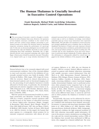
  • 7. Figure 4. Thalamic ERPs in left-hand versus right-hand conditions. The amplitude of the thalamic components at peak latency was not influenced by the side of motor execution: (A) The upper traces show the lateralization of thalamic ERP upon go instructions, compared for left-hand (dotted line) versus right-hand (solid line) responses. Side-related differences emerged only after the peak of the monophasic thalamic ERPs. (B) Upon no-go instructions, no lateralization was obtained. Chronometric Comparison of the Thalamic more than 2 standard deviations excess from baseline and Scalp ERPs noise, as determined in the 200-msec interval before sig- nal presentation; cf. Methods, cf. Figure 6). Brain level was a main factor of peak latencies in tha- lamic versus scalp ERPs based on the recording positions displaying the strongest task effects on ERP amplitudes Correlation between Thalamic and Scalp ERPs of either level [left/right thalamus vs. Fz: F(1, 6) = 85.98/ 76.09, p .0001]. Further, task condition was a cofactor Thalamic and scalp ERPs were correlated with respect to [Brain level  Task condition for left/right thalamus vs. the amplitude fluctuations along the recording session Fz: F(1, 6) = 9.01/8.45, p .024/ .027], according to (see Methods), but this thalamo-cortical correlation was the fact that P300 components at Fz peaked later upon spatially distinct for go versus no-go ERPs. Specifically, no-go than upon go stimuli ( p .01), whereas thalamic thalamic no-go ERPs behaved similarly to accompanying ERP latencies were similar in the go and no-go condi- ERPs in the time domain of the task effect at frontal sites, tion. Thalamic ERPs, in turn, peaked prior to scalp P300 where the no-go effect was largest. No such relation was components both in the go and no-go condition ( p obtained for go ERPs which, instead, mostly correlated to .001/p .001). the according parietal ERPs (for summary, see Table 2). Accordingly, the onset of the task effect was deter- mined earlier in thalamic than in scalp ERPs ( p .0001). In the subtraction curves of go versus no-go ERPs, it was DISCUSSION determined to start at 196 ± 27 msec and 296 ± 57 msec after signal presentation for thalamic ERPs and scalp The present study contained three major findings. First, ERPs at Fz, respectively (criterion: during 25 msec as at fronto-central scalp sites, ERPs recorded from the Marzinzik et al. 1909
  • 8. Figure 5. Nonmotor character of frontal and thalamic go/no-go ERP. (A) The upper traces show scalp and thalamic ERPs to go/no-go stimuli, contralateral to task execution/instruction, averaged to stimulus presentation (forward average). (B) The lower traces display the ERPs, averaged with respect to the motor execution of the task for the go condition (back average; dashed line) and an according ‘‘sham ERP’’ for the no-go condition (solid line). For the latter, the reaction times from the go trials were marked down to the no-go trials. Thus, no-go trials could be averaged in the very same way as the go trials, although they did not contain any motor response. As activity in the back average was found largely identical with that in the sham average, it was viewed the residual of the sensory components discernible in the forward average. Figure 6. Difference curves (go minus no-go condition) for scalp and thalamic ERPs. The task effect is shown as the difference of ERPs in the go versus no-go condition at scalp and thalamic levels (grand average). The beginning of this effect was calculated per patient as the first poststimulus interval of at least 25 msec exceeding the standard deviation across the individual baseline values by more than a factor of 2. The thus-determined onsets were significantly earlier in thalamic compared to scalp recordings and are indicated by the solid and dashed arrows, respectively. 1910 Journal of Cognitive Neuroscience Volume 20, Number 10
  • 9. Table 2. Co-fluctuations of Thalamic and Scalp ERP Amplitudes Go Condition No-go Condition Scalp Left Thalamus Right Thalamus Left Thalamus Right Thalamus F3 r = .17 ( p .29) r = .13 ( p .42) r = .75**(p .001) r = .76** (p .001) Fz r = .19 ( p .22) r = .19 ( p .23) r = .76** (p .001) r = .75** (p .001) F4 r = .13 ( p .39) r = .23 ( p .15) r = .76** (p .001) r = .90** (p .001) C3 r = .43** (p .005) r = .33* (p .05) r = .58** (p .001) r = .74** (p .001) Cz r = .34* (p .03) r = .15 ( p .33) r = .24 ( p .12) r = .26 ( p .11) C4 r = .35* (p .05) r = .45** (p .01) r = .18 ( p .26) r = .29 ( p .06) P3 r = .45** (p .01) r = .45** (p .01) r = À.17 ( p .28) r = À.22 ( p .17) Pz r = .52** (p .001) r = .55** (p .001) r = .07 ( p .65) r = .09 ( p .58) P4 r = .49** (p .001) r = .57** (p .001) r = À.22 ( p .15) r = À.06 ( p .70) r = Pearson’s correlation coefficient. *Significant at .05 level. **Significant at .01 level (two-tailed; cf. Methods). upper intrathalamic electrode were differentially ex- of control processes exclusively refer to the cortical level, pressed upon no-go versus go commands, and this dif- based on scalp ERPs. In this particular context, the re- ference was not explained by motor processing. Second, ported depth ERPs merit a closer view. whereas thalamic ERPs correlated to the parietal P300 component in the go condition, they corresponded to Origin of VIM-recorded ERPs the task-specific frontal ERP component in the no-go condition. Third, task-specific scalp components started An intrathalamic origin of VIM-recorded ERPs can be well after thalamic go as well as no-go ERPs. reasonably assumed for several reasons. First, if depth signals were volume conducted from cortical generators, time courses of components at scalp and thalamic levels Relation to Previous Studies should be synchronous. Accordingly, the discrepant la- Worthwhile mentioning, the obtained spatio-temporal tencies of scalp versus VIM ERPs point to a closer, that is, pattern of scalp ERPs was as described in numerous subcortical, origin of the latter. Second, the upper, but studies applying comparable paradigms. Specifically, the not the lower, derivation from DBS electrodes caught frontal component under scrutiny, often labeled ‘‘no-go significant differences between go and no-go-related P300,’’ has been conceptualized as the reflection of ag- ERPs. This disparity points to an effect from near-field gregate activity for the suppression of actions, according activities. Far-field spread would almost equally project to its enhancement in no-go trials (Roberts et al., 1994; into upper and lower derivation because of similar rela- Eimer, 1993). The recording array used in this study does tive positions to both recording fields, covering few adja- not allow to deduce the origin of this component, but cent millimeters along the same trajectory only. Finally, previous studies focused on this issue. Based on source the absence of a the task effect in the lower thalamic analysis of frontal P300, the provision of assigned func- derivations suggests that its proof in the upper record- tions has been allocated to the anterior cingulum, the ings was due to processes in proximate thalamic struc- prefrontal cortex, and the premotor cortex (Kamarajan tures, and not to operations in nuclei adjacent to the et al., 2005; Bokura et al., 2001; Kiefer et al., 1998; Strik caudal border of VIM outside the thalamus. However, an et al., 1998). Accordingly, executive control operations origin in VIM proper seems unlikely, as both the upper were proposed to be generated in a frontal lobe network, and lower depth recording areas were estimated to be although a number of imaging studies and reports on predominantly within this nucleus. patients with subcortical lesions point to an additional involvement of thalamic structures (Van der Werf et al., Nonmotor Nature of Thalamically Recorded ERPs 2003; Garavan et al., 2002; Mennemeier et al., 1992; Eslinger et al., 1991; also see Van der Werf, Witter, Several results indicate that the thalamic activation in Uylings, Jolles, 2000, for a review). However, a more the time domain of interest is unrelated to proper motor specific assignment of according subcortical functions is operations. First, thalamic ERPs in trials with left- versus not available so far, and estimations on the chronometry right-sided motor responses did not differ in magnitude Marzinzik et al. 1911
  • 10. at their peak latencies. Such a difference could be rea- demands, appear aligned with more frontally located sonably assumed if this component reflected primary areas than throughout facilitatory processing, compati- motor processes, as observed in later time domains of ble with a particular involvement of dorsolateral prefron- the ERPs. Second, thalamic ERPs were bound to stimulus tal and anterior cingulate cortices in executive control presentation rather than to the motor execution of the (Carter et al., 1998; D’Esposito et al., 1995; Petrides et al., task, as they occurred upon go and no-go trials, al- 1993; Shallice Burgess, 1991; Pardo et al., 1990). though in the latter motor responses were not executed. Such selective coupling might be a function of thalamo- Third, a predominant binding of the ERPs to motor exe- cortical, together with thalamic reticular neurons, whose cution was incompatible with the finding that they were firing properties and connectivity provide a mechanism massively reduced in magnitude when averaged to the for scaling and funneling information through the wide- motor response as compared to the go instruction. spread thalamic connections to frontal and temporo- Fourth, the larger amplitude obtained in go versus no- parietal cortical regions (Behrens et al., 2003; Sherman go conditions could finally not be explained by additive Guillery, 1996; Crick, 1984; see also Pinault, 2004; motor operations. This was shown by the comparison of Guillery Sherman, 2002a, 2002b for reviews). ERPs in go trials, averaged to the motor response, versus Intriguingly, the chronometric comparison of thalamic ERPs in no-go trials, averaged to the very same points and scalp ERPs suggested that task-specific information in time (‘‘sham average’’; cf. Methods and Results). In was propagated in thalamo-cortical direction, that is, no so doing, activations in the critical interval became un- indication was found that the underlying thalamic pro- distinguishable between conditions, delineating that the cesses occurred under the control of related cortical added thalamic activity was due to the go instruction areas. This, together with the possibility of a thalamically proper, but not to its execution. induced cortical coupling of go versus no-go-related signaling, can be viewed as an experimental parallel to ‘‘centrencephalic’’ concepts, derived from the Penfield Conceptual Considerations and Jasper (1954) formulation that a subcortical struc- In this context, a number of proposed thalamic func- ture can be ‘‘while anatomically subcortical, functionally tions are of particular interest. During attentional and supra-cortical,’’ further relating to the principle question arousal processes, activation has been described for in- if qualia can exist if subcortical structures work without tralaminar thalamic regions (Fan, McCandliss, Fossella, a corresponding cortex (cf. Merker, 2007; Schiff et al., Flombaum, Posner, 2005; Hester, Fassbender, 2007). Garavan, 2004; Woldorff et al., 2004; Garavan et al., The task-dependent expression of thalamic ERPs calls 2002; Portas et al., 1998; Coull, Frith, Dolan, Frackowiak, for the comparison with previous results. Both go and Grasby, 1997; Frith Friston, 1996), which receive no-go instructions require specific performances with afferent connections from the midbrain reticular forma- respect to previously established expectancies. There- tion and project to multiple cortical areas (McFarland fore, their magnitudes do not appear to reflect thalamic Haber, 2002; Macchi, Bentivoglio, Molinari, Minciacchi, weighing of signal relevance, given that both go and 1984; Steriade Glenn, 1982; Bentivoglio, Macchi, no-go signals convey behaviorally important informa- Albanese, 1981; also see Merker, 2007; Jones, 1998 for tion. In this sense, go and no-go signals are compara- reviews). It has been hypothesized that midbrain signals ble to target signals in oddball paradigms. Interestingly, convey stimulus-related alert or orienting information in a recent study, oddball target signals, rarely inter- to the thalamus (Kinomura, Larsson, Gulyas, Roland, spersed between nontarget instructions, led to consid- 1996), which in turn communicates with specific cortical erably larger thalamic ERPs than the present go and regions, depending on the ongoing behavioral demands no-go instructions (cf. Klostermann et al., 2006). There- (see Merker, 2007; Pinault, 2004 for reviews). Similarly, fore, our momentary working hypothesis is that the it has been suggested that sensorily driven bottom–up mentioned thalamic ERPs reflect distinct (rather than signals are integrated with top–down directed informa- unitary, but weighed) aggregate activities whose com- tion in the center median and parafascicular nuclei of the position imply variable thalamic contributions, specifi- thalamus (reviewed by Sarter, Givens, Bruno, 2001; cally defined by the given behavioral context. However, LaBerge, 1997). the verification of this and the critical questions how and Of course, from the present data, any brainstem con- by what task definitions are imposed upon the thalamus tributions to the results remain speculative, but the cor- remain central issues for future studies. relations between thalamic and scalp ERPs do hint at differential thalamo-cortical processing. According in- Conclusions teractions appeared task-dependent, as in the no-go condition, thalamic ERPs co-fluctuated with the frontal Principally, the distinct expression of thalamic ERPs upon no-go P300, whereas in the go condition, they mainly did go versus no-go instructions indicates thalamic involve- so with the parietal P300. This seems to imply thalamo- ment in executive control. Further, the fact that task- cortical routes of information which, in case of inhibitory specific activity emerged prior in depth recordings than 1912 Journal of Cognitive Neuroscience Volume 20, Number 10
  • 11. at scalp level suggests cortical operations of response Fan, J., McCandliss, B. D., Fossella, J., Flombaum, J. I., control to be subcortically prepared. Finally, the corre- Posner, M. I. (2005). The activation of attentional networks. Neuroimage, 26, 471–479. lation of thalamic to frontal ERPs in the no-go condition Fillenbaum, G., Heyman, A., Williams, K., Prosnitz, B., and of thalamic to parietal ERPs in the go condition in- Burchett, B. (1990). Sensitivity and specificity of dicates that the underlying thalamo-cortical networks are standardized screens of cognitive impairment and flexibly activated as a function of task demands. dementia among elderly black and white community residents. Journal of Clinical Epidemiology, 43, 651–660. Foffani, G., Priori, A., Egidi, M., Rampini, P., Tamma, F., Acknowledgments Caputo, E., et al. (2003). 300-Hz subthalamic oscillations in Parkinson’s disease. Brain, 126, 2153–2163. This study was supported by the Deutsche Forschungsgemein- Frith, C. D., Friston, K. J. (1996). The role of the thalamus schaft (DFG KL 1276/3-1). We thank our reviewers for their in ‘‘top down’’ modulation of attention to sound. constructive suggestions and comments. Neuroimage, 4, 210–215. Reprint requests should be sent to Fabian Klostermann, Garavan, H., Ross, T. J., Murphy, K., Roche, R. A., Stein, Neurology, Charite—University Medicine Berlin, Campus ´ E. A. (2002). Dissociable executive functions in the Benjamin Franklin, Berlin, 12200, Germany, or via e-mail: dynamic control of behavior: Inhibition, error detection, fabian.klostermann@charite.de. and correction. Neuroimage, 17, 1820–1829. Guillery, R. W., Sherman, S. M. (2002a). Thalamic relay functions and their role in corticocortical communication: REFERENCES Generalizations from the visual system. Neuron, 33, 163–175. Alexander, G. E., Crutcher, M. D., DeLong, M. R. (1990). Guillery, R. W., Sherman, S. M. (2002b). The thalamus Basal ganglia–thalamocortical circuits: Parallel substrates as a monitor of motor outputs. Philosophical Transactions for motor, oculomotor, ‘‘prefrontal’’ and ‘‘limbic’’ of the Royal Society of London, Series B, Biological functions. Progress in Brain Research, 85, 119–146. Sciences, 357, 1809–1821. Basso, M. A., Uhlrich, D., Bickford, M. E. (2005). Cortical Hester, R., Fassbender, C., Garavan, H. (2004). Individual function: A view from the thalamus. Neuron, 45, 485–488. differences in error processing: A review and reanalysis Behrens, T. E., Johansen-Berg, H., Woolrich, M. W., Smith, of three event-related fMRI studies using the GO/NOGO S. M., Wheeler-Kingshott, C. A., Boulby, P. A., et al. (2003). task. Cerebral Cortex, 14, 986–994. Non-invasive mapping of connections between human Heyder, K., Suchan, B., Daum, I. (2004). Cortico-subcortical thalamus and cortex using diffusion imaging. Nature contributions to executive control. Acta Psychologica Neuroscience, 6, 750–757. (Amsterdam), 115, 271–289. Bentivoglio, M., Macchi, G., Albanese, A. (1981). The Jodo, E., Inoue, K. (1990). Effects of practice on the P300 cortical projections of the thalamic intralaminar nuclei, in a Go/NoGo task. Electroencephalography and Clinical as studied in cat and rat with the multiple fluorescent Neurophysiology, 76, 249–257. retrograde tracing technique. Neuroscience Letters, 26, Jones, E. G. (1998). A new view of specific and nonspecific 5–10. thalamocortical connections. Advances in Neurology, 77, Bokura, H., Yamaguchi, S., Kobayashi, S. (2001). 49–71 [Discussion 72–73]. Electrophysiological correlates for response inhibition in a Kamarajan, C., Porjesz, B., Jones, K. A., Chorlian, D. B., Go/NoGo task. Clinical Neurophysiology, 112, 2224–2232. Padmanabhapillai, A., Rangaswamy, M., et al. (2005). Carter, C. S., Braver, T. S., Barch, D. M., Botvinick, M. M., Spatial–anatomical mapping of NoGo-P3 in the offspring Noll, D., Cohen, J. D. (1998). Anterior cingulate cortex, of alcoholics: Evidence of cognitive and neural disinhibition error detection, and the online monitoring of performance. as a risk for alcoholism. Clinical Neurophysiology, 116, Science, 280, 747–749. 1049–1061. Coull, J. T., Frith, C. D., Dolan, R. J., Frackowiak, R. S., Kiefer, M., Marzinzik, F., Weisbrod, M., Scherg, M., Grasby, P. M. (1997). The neural correlates of the Spitzer, M. (1998). The time course of brain activations noradrenergic modulation of human attention, arousal and during response inhibition: Evidence from event-related learning. European Journal of Neuroscience, 9, 589–598. potentials in a go/no go task. NeuroReport, 9, 765–770. Crick, F. (1984). Function of the thalamic reticular complex: Kinomura, S., Larsson, J., Gulyas, B., Roland, P. E. (1996). The searchlight hypothesis. Proceedings of the National Activation by attention of the human reticular formation Academy of Sciences, U.S.A., 81, 4586–4590. and thalamic intralaminar nuclei. Science, 271, 512–515. de Jong, B. M., Paans, A. M. (2007). Medial versus lateral Klostermann, F., Wahl, M., Marzinzik, F., Schneider, G. H., prefrontal dissociation in movement selection and inhibitory Kupsch, A., Curio, G. (2006). Mental chronometry of control. Brain Research, 1132, 139–147. target detection: Human thalamus leads cortex. Brain, D’Esposito, M., Detre, J. A., Alsop, D. C., Shin, R. K., Atlas, S., 129, 923–931. Grossman, M. (1995). The neural basis of the central Kondziolka, D., Lee, J. Y. (2004). Long-lasting executive system of working memory. Nature, 378, 279–281. microthalamotomy effect after temporary placement Eimer, M. (1993). Effects of attention and stimulus probability of a thalamic stimulating electrode. Stereotactic and on ERPs in a Go/Nogo task. Biological Psychology, 35, Functional Neurosurgery, 82, 127–130. 123–138. Kramer, J. H., Reed, B. R., Mungas, D., Weiner, M. W., Eslinger, P. J., Warner, G. C., Grattan, L. M., Easton, J. D. Chui, H. C. (2002). Executive dysfunction in subcortical (1991). ‘‘Frontal lobe’’ utilization behavior associated with ischaemic vascular disease. Journal of Neurology, paramedian thalamic infarction. Neurology, 41, 450–452. Neurosurgery and Psychiatry, 72, 217–220. Fahn, S., Tolosa, E., Marin, C. (1993). Clinical rating scale for Kuhn, A. A., Williams, D., Kupsch, A., Limousin, P., Hariz, M., tremor. In J. Jankovic E. Tolosa (Eds.), Parkinson’s Schneider, G. H., et al. (2004). Event-related beta disease and movement disorders (2nd ed., pp. 271–280). desynchronization in human subthalamic nucleus Baltimore: Williams and Wilkons. correlates with motor performance. Brain, 127, 735–746. Marzinzik et al. 1913
  • 12. LaBerge, D. (1997). Attention, awareness, and the triangular discrimination. Electroencephalography and Clinical circuit. Consciousness and Cognition, 6, 149–181. Neurophysiology, 92, 44–55. Logan, G. D. (1985). Executive control of thought and Royall, D. R., Lauterbach, E. C., Cummings, J. L., Reeve, A., action. Acta Psychologica, 60, 193–210. Rummans, T. A., Kaufer, D. I., et al. (2002). Executive control Logan, G. D., Cowan, W. B. (1984). On the ability to function: A review of its promise and challenges for clinical inhibit thought and action: A theory of an act of control. research. A report from the Committee on Research of Psychological Review, 91, 295–327. the American Neuropsychiatric Association. Journal of Macchi, G., Bentivoglio, M., Molinari, M., Minciacchi, D. Neuropsychiatry and Clinical Neurosciences, 14, (1984). The thalamo-caudate versus thalamo-cortical 377–405. projections as studied in the cat with fluorescent Sandson, T. A., Daffner, K. R., Carvalho, P. A., Mesulam, retrograde double labeling. Experimental Brain Research, M. M. (1991). Frontal lobe dysfunction following infarction 54, 225–239. of the left-sided medial thalamus. Archives of Neurology, Marsden, J. F., Ashby, P., Limousin-Dowsey, P., Rothwell, 48, 1300–1303. J. C., Brown, P. (2000). Coherence between cerebellar Sarter, M., Givens, B., Bruno, J. P. (2001). The cognitive thalamus, cortex and muscle in man: Cerebellar thalamus neuroscience of sustained attention: Where top–down interactions. Brain, 123, 1459–1470. meets bottom–up. Brain Research, Brain Research McFarland, N. R., Haber, S. N. (2002). Thalamic relay nuclei Reviews, 35, 146–160. of the basal ganglia form both reciprocal and nonreciprocal Schaltenbrand, G., Wahren, W. (1977). Atlas for stereotaxy of cortical connections, linking multiple frontal cortical the human brain. New York: Medical Publishers. areas. Journal of Neuroscience, 22, 8117–8132. Schiff, N. D., Giacino, J. T., Kalmar, K., Victor, J. D., Baker, K., Mennemeier, M., Fennell, E., Valenstein, E., Heilman, K. M. Gerber, M., et al. (2007). Behavioural improvements with (1992). Contributions of the left intralaminar and medial thalamic stimulation after severe traumatic brain injury. thalamic nuclei to memory. Comparisons and report of a Nature, 448, 600–603. case. Archives of Neurology, 49, 1050–1058. Schuurman, P. R., Bosch, D. A., Bossuyt, P. M., Bonsel, G. J., Merker, B. (2007). Consciousness without a cerebral cortex: van Someren, E. J., de Bie, R. M., et al. (2000). A comparison A challenge for neuroscience and medicine. Behavioral of continuous thalamic stimulation and thalamotomy for and Brain Sciences, 30, 63–81 [Discussion 81–134]. suppression of severe tremor. New England Journal of Norman, D. A., Shallice, T. (1986). Attention to action: Medicine, 342, 461–468. Willed and automatic control of behavior. In R. J. Davidson, Shallice, T., Burgess, P. W. (1991). Higher-order cognitive G. E. Schwartz, D. Shapiro (Eds.), Consciousness and impairments and frontal lobe lesions in man. In S. Levin, self-regulation: Advances in research and theory (Vol. 4, H. M. Eisenberg, A. L. Benton (Eds.), Frontal lobe pp. 1–18). New York: Plenum. function and injury (pp. 125–138). New York: Oxford Pardo, J. V., Pardo, P. J., Janer, K. W., Raichle, M. E. (1990). University Press. The anterior cingulate cortex mediates processing selection Sherman, S. M., Guillery, R. W. (1996). Functional in the Stroop attentional conflict paradigm. Proceedings organization of thalamocortical relays. Journal of of the National Academy of Sciences, U.S.A., 87, 256–259. Neurophysiology, 76, 1367–1395. Penfield, W., Jasper, H. H. (1954). Epilepsy and the Simson, R., Vaughan, H. G., Jr., Ritter, W. (1977). The functional anatomy of the human brain. Boston: Little, scalp topography of potentials in auditory and visual Brown Co. Go/NoGo tasks. Electroencephalography and Clinical Petrides, M., Alivisatos, B., Meyer, E., Evans, A. C. (1993). Neurophysiology, 43, 864–875. Functional activation of the human frontal cortex during Smith, E. E., Jonides, J. (1999). Storage and executive the performance of verbal working memory tasks. processes in the frontal lobes. Science, 283, 1657–1661. Proceedings of the National Academy of Sciences, U.S.A., Steriade, M., Glenn, L. L. (1982). Neocortical and caudate 90, 878–882. projections of intralaminar thalamic neurons and their Pfefferbaum, A., Ford, J. M. (1988). ERPs to stimuli requiring synaptic excitation from midbrain reticular core. Journal response production and inhibition: Effects of age, of Neurophysiology, 48, 352–371. probability and visual noise. Electroencephalography Strik, W. K., Fallgatter, A. J., Brandeis, D., Pascual-Marqui, and Clinical Neurophysiology, 71, 55–63. R. D. (1998). Three-dimensional tomography of event-related Pfefferbaum, A., Ford, J. M., Weller, B. J., Kopell, B. S. potentials during response inhibition: Evidence for phasic (1985). ERPs to response production and inhibition. frontal lobe activation. Electroencephalography and Electroencephalography and Clinical Neurophysiology, Clinical Neurophysiology, 108, 406–413. 60, 423–434. Van der Werf, Y. D., Scheltens, P., Lindeboom, J., Witter, Pinault, D. (2004). The thalamic reticular nucleus: Structure, M. P., Uylings, H. B., Jolles, J. (2003). Deficits of memory, function and concept. Brain Research, Brain Research executive functioning and attention following infarction Reviews, 46, 1–31. in the thalamus; a study of 22 cases with localised lesions. Portas, C. M., Rees, G., Howseman, A. M., Josephs, O., Turner, Neuropsychologia, 41, 1330–1344. R., Frith, C. D. (1998). A specific role for the thalamus Van der Werf, Y. D., Witter, M. P., Uylings, H. B., Jolles, J. in mediating the interaction of attention and arousal in (2000). Neuropsychology of infarctions in the thalamus: humans. Journal of Neuroscience, 18, 8979–8989. A review. Neuropsychologia, 38, 613–627. Rafal, R. D., Posner, M. I. (1987). Deficits in human visual Weisbrod, M., Kiefer, M., Marzinzik, F., Spitzer, M. (2000). spatial attention following thalamic lesions. Proceedings of Executive control is disturbed in schizophrenia: Evidence the National Academy of Sciences, U.S.A., 84, 7349–7353. from event-related potentials in a Go/NoGo task. Biological Rieger, M., Gauggel, S., Burmeister, K. (2003). Inhibition Psychiatry, 47, 51–60. of ongoing responses following frontal, nonfrontal, and Woldorff, M. G., Hazlett, C. J., Fichtenholtz, H. M., Weissman, basal ganglia lesions. Neuropsychology, 17, 272–282. D. H., Dale, A. M., Song, A. W. (2004). Functional Roberts, L. E., Rau, H., Lutzenberger, W., Birbaumer, N. parcellation of attentional control regions of the brain. (1994). Mapping P300 waves onto inhibition: Go/No-Go Journal of Cognitive Neuroscience, 16, 149–165. 1914 Journal of Cognitive Neuroscience Volume 20, Number 10