08448380779 Call Girls In Civil Lines Women Seeking Men
Handbook of the cerebellum and cerebellar disorders
1. Specification of Cerebellar and
Precerebellar Neurons 5
Mikio Hoshino, Yusuke Seto, and Mayumi Yamada
Abstract
The cerebellum is thought to participate in the regulation of movement and is
comprised of various types of neurons in the cerebellar cortex and nuclei. Each
type of neurons has morphologically, immunohistochemically, and electrophys-
iologically distinct characteristics. In addition, there are two precerebellar affer-
ent systems, the mossy fiber (MF) system and the climbing fiber (CF) system.
MF neurons are located in various nuclei throughout the brainstem and send their
axons to cerebellar granule cells, whereas CF neurons reside exclusively in the
inferior olivary nucleus (ION) and project to Purkinje cells. Recently developed
genetic lineage-tracing methods as well as gene-transfer technologies have
accelerated the studies on the molecular machinery to specify neuronal subtypes
in the cerebellum and the precerebellar systems.
Specification of Cerebellar Neurons
The cerebellum consists of three parts: cortex, white matter, and nucleus. The
cerebellar cortex contains Purkinje, Golgi, Lugaro, stellate, basket, granule, and
unipolar brush cells. The latter two cell types are glutamatergic excitatory neu-
rons, while the others are all GABAergic inhibitory neurons. The cerebellar
nucleus (CN) includes three types of neurons: large glutamatergic projection
neurons (CN-Glu neurons), mid-sized GABAergic inhibitory projection neurons
M. Hoshino (*) • M. Yamada
Department of Biochemistry and Cellular Biology, National Institute of Neuroscience, National
Center of Neurology and Psychiatry, 4-1-1 Ogawa-Higashi, Kodaira, Tokyo, 187–8502, Japan
e-mail: hoshino@ncnp.go.jp
Y. Seto
Integrative Bioscience and Biomedical Engineering, Graduate School of Science and Engineering,
Waseda University, 3-4-1 Okubo, Shinjuku-ku, Tokyo, 169–8555, Japan
M. Manto, D.L. Gruol, J.D. Schmahmann, N. Koibuchi, F. Rossi (eds.),
Handbook of the Cerebellum and Cerebellar Disorders,
DOI 10.1007/978-94-007-1333-8_5, # Springer Science+Business Media Dordrecht 2013
75
2. (CN-GABA-ION neurons), and small GABAergic interneurons (CN-GABA inter-
neurons). CN-GABA-ION neurons extend their axons to the inferior olivary
nucleus (ION) (Carletti and Rossi 2008), while CN-Glu neurons send their
axons to nuclei outside the cerebellum, including the red nucleus and the thalamus.
It is believed that all types of cerebellar neurons are generated from the
neuroepithelium of the alar plate of rhombomere 1 (r1) during development (Millet
et al. 1996; Wingate and Hatten 1999; Chizhikov and Millen 2003; Zervas et al.
2004). The dorsal-most part of the r1 neuroepithelium, that is, the roof plate, does
not produce neurons but cells of the choroid plexus (Chizhikov et al. 2006).
Neuroepithelium that produces cerebellar neurons can be divided into two regions:
the rhombic lip (RL) and the ventricular zone (VZ). These two regions can be
morphologically discriminated by a notch located on the border.
Although the history of studies on the cerebellum is very long (Ramo´n y Cajal
1911), the molecular machinery underlying cerebellar neuron development is still
unclear. In 1997, Ben-Arie et al. reported that a basic-helix-loop-helix type (bHLH)
transcription factor, Atoh1 (also called Math1), is expressed in the RL and involved
in producing cerebellar granule cells (Ben-Arie et al. 1997). However, the devel-
opment of the other types of neurons in the cerebellum remained elusive until three
breakthrough papers were published in 2005.
While generating certain transgenic lines, Hoshino et al. found a novel mutant
mouse line, cerebelless, which lacked the entire cerebellar cortex. In this mutant, all
types of GABAergic neurons are not produced in the cerebellum, which leads to the
secondary loss of glutamatergic granule cells and eventually, the entire cerebellar
cortex (Hoshino et al. 2005). The responsible gene was identified as pancreatic
transcription factor 1a (Ptf1a), which was known to participate in pancreatic
development and to encode a bHLH transcription factor. This gene is expressed
in the neuroepithelium of the VZ but not of the RL and its expression is lost in the
cerebelless mutants. Cre-loxP recombination-based lineage tracing analysis
revealed that all types of cerebellar GABAergic neurons are derived from Ptf1a-
expressing neuroepithelial cells in the VZ, but glutamatergic neurons, such as
granule cells and CN-Glu neurons, are not. Loss of Ptf1a expression in cerebelless
as well as Ptf1a-knock out mice resulted in inhibition of the production of
GABAergic neurons in the cerebellar primordium. Furthermore, ectopic introduc-
tion of Ptf1a by means of in utero electroporation resulted in the abnormal produc-
tion of neurons with GABAergic characteristics from the dorsal telencephalon that
should only produce glutamatergic neurons under normal conditions. In addition,
Pascual et al. reported that, in the Ptf1a-null mutants, the fate of neurons produced
from the VZ is changed to that of granule cells (Pascual et al. 2007). These
observations suggested that Ptf1a, expressed in the cerebellar VZ, determines
GABAergic neuronal fate in the cerebellum. PTF1A was also identified as
a causative gene for a human disease that exhibits permanent neonatal diabetes
mellitus and cerebellar agenesis (Sellick et al. 2004).
On the other hand, two other groups revealed a molecular fate map of the
derivatives of Atoh1-expressing neuroepithelial cells in the cerebellar RL (Machold
and Fishell 2005; Wang et al. 2005). They showed that not only granule cells but
76 M. Hoshino et al.
3. also, at least in part, some neurons in the CN are derived from the RL, although they
did not discriminate between neuron types in the CN. In their studies, the develop-
ment of RL-derived CN neurons was shown to be disrupted in the Atoh1-null mice.
Because Hoshino et al. reported that GABAergic but not glutamatergic CN neurons
are derived from Ptf1a-expressing neuroepithelial cells in the VZ (Hoshino et al.
2005), their findings suggest that cerebellar glutamatergic neurons such as granule
cells and CN-Glu neurons are derived from the RL. Accordingly, unipolar brush
cells, which are glutamatergic, were also shown to emerge from the RL (Englund
et al. 2006).
Together, these studies indicate the presence of two molecularly defined
neuroepithelial areas in the cerebellum, the Atoh1-expressing RL and the Ptf1a-
expressing VZ, which generate glutamatergic and GABAergic neurons, respec-
tively. Each bHLH transcription factor is involved in producing the corresponding
neuronal subtype in the cerebellum. This suggests a model in which the cerebellar
neuroepithelium is regionalized into two distinct regions, the VZ and the RL, by the
two bHLH transcription factors (Hoshino 2006). During embryonic development,
the ventral part of the cerebellar neuroepithelium expresses Ptf1a, leading to the
acquirement of cerebellar VZ characteristics to generate GABAergic neurons,
while the dorsal part of cerebellar neuroepithelium expresses Atoh1 and becomes
the cerebellar RL, producing glutamatergic neurons. In the telencephalon, similar
regionalization by bHLH transcription factors takes place. Glutamatergic neurons
emerge from dorsal neuroepithelium expressing Neurogenin 1/2 (Ngn 1/2), and
GABAergic neurons are produced from ventral neuroepithelium expressing Mash1
(Wilson and Rubenstein 2000).
How are these neuroepithelial areas formed? In general, the roof plate can affect
the dorsal structure of the neural tube (Lee et al. 2000; Millonig et al. 2000).
Chizhikov et al. revealed that the roof plate plays an important role in the formation
of the cerebellar dorsoventral domain formation by analyzing cerebellar mutants
that lack the roof plate (Chizhikov et al. 2006). Moreover, it has been suggested that
bone morphogenetic proteins (BMPs) secreted from the roof plate as well as Notch
signaling are involved in the formation of the RL and the VZ (Machold et al. 2007).
A recent study that induced Purkinje cells from ES cells suggested that loss of sonic
hedgehog signaling may give the dorsoventral spatial information of the cerebellar
VZ to the cerebellar neuroepithelium which eventually leads to the expression of
Ptf1a (Muguruma et al. 2010).
Although the machinery governing GABAergic and glutamatergic neuronal
subtype specification by transcription factors has been clarified to some extent, molec-
ular mechanisms to specify each member of GABAergic (e.g., Purkinje, Golgi, basket,
stellate cells and CN-ION, CN-interneurons) or glutamatergic (e.g., granule, unipolar
brush cells, and CN-Glu neurons) subtype remain unclear. However, birthdating
studies using 3
H-thymidine and BrdU (Chan-Palay et al. 1977; Batini et al.
1992; De Zeeuw and Berrebi 1995; Sultan et al. 2003; Leto et al. 2006) as well
as adenovirus (Hashimoto and Mikoshiba 2003) revealed that each type of
neuron is generated at distinct developmental stages. As to GABAergic neurons,
Purkinje cells are produced at an early stage (embryonic day (E) 10.5–13.5 in
5 Specification of Cerebellar and Precerebellar Neurons 77
4. mice), Golgi cells at middle stages (E14.5), and stellate/basket cells at a late
stage (Perinatal). Regarding glutamatergic neurons, in addition to the experi-
ment above, molecule-based lineage tracing analyses (Machold and Fishell
2005; Wang et al. 2005; Englund et al. 2006) have clarified that CN-Glu
neurons leave the cerebellar RL at early stages (E10.5–12.5) and granule cells
and unipolar brush cells at middle to late stages (granule cell:E12.5, ubc:
E12.5–E18.5). In addition, somatic recombination-based clonal analyses
suggested that Purkinje, Golgi, and basket/stellate cells as well as some CN
neurons (probably GABAergic) belong to the same lineage (Mathis et al. 1997;
Mathis and Nicolas 2003). These data indicate that some temporal information
in the neuroepithelium may be involved in specification of neuronal types in the
RL and VZ, respectively. However, the underlying molecular mechanisms have
not yet been clarified.
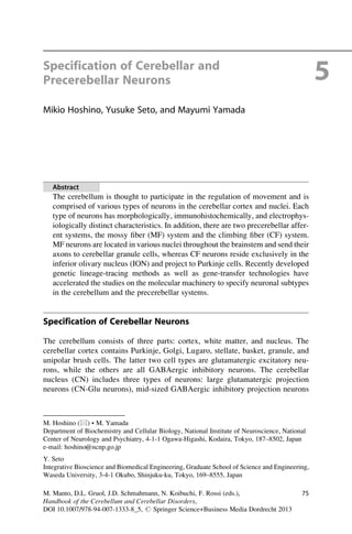
Some scientists tried to divide the structure of the cerebellar primordium into
several domains (Fig. 5.1). Chizhikov et al. defined four cellular populations
(denoted c1–c4 domains) in the cerebellar primordium by the expression of a few
transcription factors (Chizhikov et al. 2006). c1 corresponds to the Atoh1-
expressing RL and c2 is located just above the Ptf1a-expressing VZ (denoted
c1 . . .Atoh1 +
c2 . . .Lhx1/5 +
(GABAergic)
c2 (GABAergic)
c4 c3
c2v c2d
pc2dpc2v
Atoh1
pc2(Ptf1a +)
RP
c1
Pax2
Lhx1/5
Corl2
Lhx1/5
E-cadϮ
Ptf1a E-cad+++
Ptf1a
c2d . . .Corl2 +
c2v . . .Pax2 +
c3 . . .Lmx1a +
pc2 . . .Ptf1a +
pc2d . . .E-cad +++
pc2v . . .E-cadϮ
c4 . . .Lhx1/5 +
Fig. 5.1 Domain structure of the cerebellar primordium at an early developmental stage (e.g.
E12.5 in mice). The c1 domain, expressing Atoh1, corresponds to the rhombic lip that produces all
types of glutamatergic neurons in the cerebellum. The pc2 is the Ptf1a-expressing neuroepithelial
domain that generates all types of GABAergic cerebellar neurons. At early neurogenesis stages,
such as E12.5, the pc2 domain can be subdivided into pc2d and pc2v subdomains, which expresses
E-cadherin strongly and weakly, respectively. The c2 domain, expressing Lhx1/5, consists of
immature GABAergic neurons putatively generated from pc2 neuroepithelial domain. This domain
can also be subdivided into two subdomains, c2d and c2v. The c2d subdomain consists of corl2-
expressing neurons or Purkinje cells, whereas the c2v subdomain includes Pax2-positive cerebellar
GABAergic interneurons. Although c3 and c4 domains are Lmx1a- and Lhx1/5-positive, respec-
tively, cell types which consist these domains are unknown. The roof plate (RP) is located most
dorsally, and plays prominent roles in organizing this cerebellar domain structure
78 M. Hoshino et al.
5. pc2), indicating that c2 cells mainly consist of GABAergic inhibitory neurons.
Although c3 and c4 express Lmx1a and Lhx1/5 respectively, their neuronal sub-
types remain to be determined. This subdomain structure is disrupted when the roof
plate was removed (Chizhikov et al. 2006). Furthermore, at the early neurogenesis
stage (e.g., E12.5 in mice), Minaki et al. subdivided the c2 domain into dorsally
(c2d) and ventrally (c2v) located subdomains that express corl2 and Pax2, respec-
tively (Minaki et al. 2008). While corl2 is exclusively expressed in immature and
mature Purkinje cells (Minaki et al. 2008), Pax2 is expressed in GABAergic
interneurons (e.g., Golgi, stellate, basket, CN-GABA neurons) in the cerebellum
(Maricich and Herrup 1999; Weisheit et al. 2006). They also subdivided the Ptf1a-
expressing neuroepithelial domain (pc2) into pc2d and pc2v, which strongly and
weakly express E-cadherin, respectively. From the positions of the neuroepithelial
and neuronal subdomains, they suggested that the pc2d neuroepithelial subdomain
produces cells in the c2d domain which give rise to Purkinje cells and pc2v
subdomain generates cells in the c2v that become GABAergic interneurons
(Mizuhara et al. 2010). As development proceeds, pc2d and pc2v subdomains
become smaller and larger, respectively, and by E14.5 in mice, the Ptf1a-expressing
pc2 domain is comprised only by the pc2v subdomain which expresses E-cadherin
weakly. This correlates with the fact that, at E14.5 in mice, Ptf1a-expressing
neuroepithelium does not produce Purkinje cells but Pax2-positive interneurons
(Maricich and Herrup 1999; Hashimoto and Mikoshiba 2003). Expression patterns
of several other transcription factors in the cerebellar VZ during development were
also reported. For example, some groups reported the expression patterns of
proneural bHLH transcription factors, such as Ngn1, Ngn2, and Mash1 in the
cerebellar VZ although their function in cerebellar development is still unclear
(Zordan et al. 2008; Lundell et al. 2009). However, it has been recently reported
that Pax2-positive neurons, but not Purkinje cells, are reduced in the Mash1-null
cerebellum (Grimaldi et al. 2009), suggesting that these bHLH transcription factors
may play distinct roles in cerebellar development.
In addition, several transcription factors have been reported to participate in the
development of a certain type of cerebellar neurons. Double knockout of Lhx1 and
Lhx5 as well as the targeted disruption of their cofactor Ldb1 resulted in lack of
Purkinje cell production in the cerebellum although Pax2-positive interneurons did
not seem to be affected. Because Lhx1 and Lhx5 are expressed in post-mitotic cells,
this suggests that Lhx1, Lhx5, and Ldb1 are post-mitotically involved in Purkinje
cell specification (Zhao et al. 2007). In addition, targeted disruption of cyclin D2
caused loss of stellate cells in the cerebellar molecular layer, suggesting its
involvement in the development of stellate cells (Huard et al. 1999).
From the RL, several types of glutamatergic neurons, such as CN-Glu neurons,
granule cells, and unipolar brush cells, are generated. CN-Glu neurons leave the RL
at early neurogenesis stages. Some transcription factors, such as Tbr1, Irx3, Meis2,
Lhx2, and Lhx9 have been found to be expressed in post-mitotic progenitors of
CN-Glu neurons, but their roles have not been clarified (Morales and Hatten 2006).
Other molecules, such as Zic1 (Aruga et al. 1998), have been reported to play
important roles in the migration, maturation, and survival of granule cells, but the
5 Specification of Cerebellar and Precerebellar Neurons 79
6. molecular machinery underlying the specification of granule cell identity is
unknown. Although unipolar brush cells strongly express Tbr2, its function is also
elusive.
In addition to genetic analyses, heterotopic and heterochronic transplantation
studies have also provided important clues to understanding cerebellar develop-
ment (Carletti and Rossi 2008). When tissues from embryonic and postnatal
cerebella were mixed and transplanted to the fourth ventricle of an adult mouse,
the postnatal-derived cells differentiated only into interneurons such as granule,
basket, and stellate cells, but not projection neurons, such as Purkinje cells, whereas
the embryonic-derived cells were capable of becoming all types of cerebellar
neurons (Jankovski et al. 1996). In addition, it was shown that dissociated cells
taken from cerebellar primordium at early neurogenesis stages could differentiate
into all major types of cerebellar neurons, but those from postnatal cerebellum
differentiated only to Pax2-positive interneurons (Carletti et al. 2002). These
findings suggest that the differentiation competence of cerebellar progenitors
becomes restricted as development proceeds. However, the molecular mechanisms
underlying this fate restriction process have not yet been clarified. Interestingly,
Leto et al. suggested that pax2-positive interneurons, such as Golgi, stellate, basket
cells, and CN-GABA interneurons are derived from same progenitor pool (Leto
et al. 2006).
Specification of Precerebellar Neurons
There are two types of precerebellar afferent systems: mossy fiber (MF) and
climbing fiber (CF) systems. MF neurons are located in several nuclei throughout
the brain stem and extend their glutamatergic projections to granule cells conveying
peripheral and cortical information to the cerebellum. Four major nuclei containing
MF neurons are the pontine gray nucleus (PGN), the reticulotegmental nucleus
(RTN), the lateral reticular nucleus (LRN), and the external cuneate nucleus (ECN)
in the hindbrain (Altman and Bayer 1987). In addition, some MF neurons are also
located in the spinal trigeminal nucleus (Sp5) in the hindbrain and Clarke’s column
in the spinal cord. In contrast, CF neurons reside exclusively in the inferior olive
nucleus (ION), which receive input from the cerebral cortex, the red nucleus, spinal
cord, and other brain stem nuclei and send glutamatergic projections to Purkinje
cells (Ruigrok et al. 1995). Both types of precerebellar neurons also send branch
axons to the neurons in the CN. These precerebellar systems are thought to transmit
the external and internal information to the cerebellar cortex to modulate cerebellar
function, including regulation of animal movement.
Previous birthdating studies in mice revealed that CF neurons are generated at
relatively early neurogenesis stages (E9.5–11.5) and MF neurons are produced at
slightly later stages (E10.5–16.5) (Pierce 1973). Along the rostrocaudal axis, both
MF and CF neurons in the hindbrain are generated from the caudal hindbrain,
around rhombomeres 6–8 (r6–8), as suggested by avian grafting studies as well as
mammalian fate map analyses (Ambrosiani et al. 1996; Cambronero and Puelles
80 M. Hoshino et al.
7. 2000; Farago et al. 2006; Kawauchi et al. 2006). By contrast, MF neurons in the
Clarke’s nucleus are generated in the spinal cord (Bermingham et al. 2001). Classic
anatomical and immunohistochemical studies have suggested that these
precerebellar nuclei neurons in the hindbrain emerge from the dorsal part of the
hindbrain and migrate tangentially or circumferentially to their final loci (Bloch-
Gallego et al. 1999; Yee et al. 1999; Kyriakopoulou et al. 2002). However, they
take slightly different paths from each other; MF and CF neurons move extramu-
rally and intramurally, respectively. Introduction of a GFP-expressing vector into
the embryonic dorsal hindbrain allowed the dramatic visualization of migrating
precerebellar nuclei neurons during development (Kawauchi et al. 2006; Okada
et al. 2007).
Many groups have reported transcription factors that are expressed within the
dorsal neuroepithelium of the caudal (r6–8) hindbrain during embryonic develop-
ment, trying to define domains along the dorsoventral axis. The dorsal-most part
expressing Lmx1a corresponds to the roof plate which gives rise to the choroid
plexus (Chizhikov et al. 2006). Other than the roof plate, the dorsal neuroepithelium
can be divided into six domains (dP1–dP6) according to the expression pattern of
the transcription factors, such as Atoh1, Ngn1, Mash1, Ptf1a, and Olig3 (Fig. 5.2).
As to the precerebellar nuclei neurons, a series of studies have tried to clarify the
precise origins of MF and CF neurons by genetic lineage-tracing methods.
By analyzing genetically engineered mice that express lacZ or Cre recombinase
under the control of the endogenous or exogenous Atoh1 promoter, MF neurons of
PGN, RTN, LRN, and ECN were shown to emerge from the Atoh1-expressing
neuroepithelial domain (dP1, Ben-Arie et al. 2000; Rodriguez and Dymecki 2000;
RP
dP1
dP2
dP3
dP4
dP5
dP6
CF neurons
MF neurons
Wnt1
Atoh1
Ngn1
Lmx1a
Mash1
Ptf1a
Olig3
Pax6
Fig. 5.2 Neuroepithelial domain structure in the caudal hindbrain. In the caudal hindbrain (r6–8),
several transcription factors are expressed within the dorsal neuroepithelium during embryonic
development. The dorsal-most part, the roof plate (RP), expresses Lmx1a. Other than the roof
plate, the dorsal neuroepithelium can be divided into six domains (dP1–dP6) according to the
expression pattern of transcription factors such as Atoh1, Ngn1, Pax6, Mash1, Ptf1a, and Olig3.
While mossy fiber (MF) neurons are derived from the dP1 domain expressing Atoh1, climbing
fiber (CF) neurons are generated from the dP4 domain expressing Ptf1a and Olig3
5 Specification of Cerebellar and Precerebellar Neurons 81
8. Landsberg et al. 2005; Wang et al. 2005). Targeted disruption of the Atoh1 gene
resulted in loss of these MF neurons, suggesting an involvement of Atoh1 in the MF
neuron development.
Atoh1 regulates the expression of the transcription factor Barhl1 (Mbh2) that is
expressed in MF neurons. Loss of Barhl1 expression resulted in a decrease of MF
neurons, leading to a decrease in the size of MF precerebellar nuclei (Li et al. 2004).
In addition, Flora et al. reported that one of the E-proteins, Tcf4, interacts with
Atoh1 and regulates differentiation of a specific subset (PGN, RTN) of MF neurons
(Flora et al. 2007).
Landsberg et al. also performed lineage trace analysis by using two variants of
FLP (Flipperase recombinase) with different recombinase activities that were
expressed under the control of the Wnt-1 promoter whose strength is the highest
at the dorsal-most part and gradually decreases ventrally. They demonstrated that
CF neurons are derived from the neuroepithelial region where Wnt-1 is very weakly
expressed, whereas MF neurons emerge from the strongly Wnt-1-expressing region
(Landsberg et al. 2005). In addition, Nichols and Bruce generated transgenic mice
carrying a Wnt-1-enhancer/lacZ transgene and observed that MF neurons but not
CF neurons were labeled by b-gal in those mice (Nichols and Bruce 2006). These
findings suggested that CF neurons are generated from the neuroepithelial region
ventral to the Atoh1-expressing domain.
By Cre-loxP-based lineage trace analysis, Yamada et al. showed that all CF
neurons in the ION are derived from the Ptf1a-expressing neuroepithelial region
(Yamada et al. 2007). Loss of the Ptf1a gene resulted in the fate change of some CF
neurons to MF neurons, suggesting that Ptf1a plays a critical role in fate determi-
nation of CF neurons. They also showed an involvement of Ptf1a in migration,
differentiation, and survival of CF neurons. Storm et al. reported that not only MF
neurons but also CF neurons are derived from the Olig3-expressing neuroepithelial
region that broadly expands within the dorsal hindbrain (Storm et al. 2009) by Cre-
loxP-based linage tracing. Targeted disruption of the Olig3 gene caused the disor-
ganized development of MF neurons and complete loss of CF neurons (Liu et al.
2008; Storm et al. 2009). Moreover, the ectopic co-expression of Olig3 and Ptf1a
induced cells expressing a CF neuron marker in chick embryos (Storm et al. 2009).
These findings suggest that CF neurons emerge from the Ptf1a/Olig3-expressing
neuroepithelial domain (dP4) and that Ptf1a and Olig3 are cooperatively involved
in the development of CF neurons. Domain structure of the dorsal neuroepithelium
in the caudal hindbrain region is shown in Fig. 5.2.
Conclusions and Future Directions
Various types of neurons are generated from the dorsal hindbrain. As described
above, the dorsal neuroepithelium of the rostral hindbrain (r1) produces all types of
cerebellar neurons, while the dorsal regions of the caudal hindbrain (r6–8) generate
neurons that include the precerebellar system neurons, such as MF and CF neurons.
In addition, histological observations suggested that the dorsal part of the middle
82 M. Hoshino et al.
9. hindbrain produces neurons of the cochlear nucleus, where auditory information is
processed and relayed to the brain (Pierce 1967; Ivanova and Yuasa 1998). More
directly, genetic-fate-mapping studies using transgenic mice confirmed that neu-
rons of the cochlear nucleus are derived from the dorsal part of r2–5 in mice (Farago
et al. 2006), although in avians, they were shown to emerge from a broader part
(r3–8) by grafting studies (Tan and Le Douarin 1991; Cambronero and Puelles
2000; Cramer et al. 2000). As to neuronal subtypes, Fujiyama et al. identified
origins of inhibitory and excitatory neurons of the cochlear nucleus; inhibitory
(glycinergic and GABAergic) and excitatory (glutamatergic) neurons are derived
from Ptf1a- and Atoh1-expressing neuroepithelial regions, respectively (Fujiyama
et al. 2009), and their development is dependent on the corresponding bHLH
proteins.
In the hindbrain from r1 to r8, there are dorsoventral domain structures defined
by several transcription factors, which are longitudinally expressed throughout the
hindbrain. Especially, two bHLH transcription factors, Atoh1 and Ptf1a seem to
play important roles in specifying distinct neuronal subtypes. These two proteins
are expressed in different neuroepithelial regions throughout the hindbrain
Rostral Caudal
Dorsal
Atoh1
Ptf1a
r1 2 3 4 5 6 7/8
Ventral
Cerebellar
anlage
Glutamatergic
neurons
GABAergic
neurons
GABAergic/
glycinergic
neurons
Glutamatergic
neurons
Mossy fiber
neurons
Climbing fiber
neurons
cerebellum
Middle
hindbrain
Caudal
hindbrain
Cochlear nu.
Precerebellar
systems
Fig. 5.3 Basic HLH proteins and neurons produced from the dorsal hindbrain. Atoh1 and Ptf1a
are expressed in distinct neuroepithelial regions throughout the rhombomeres 1–8 (r1–8). Each
number represents the rhombomeric number. Upper side is dorsal, lower is ventral. Left side is
rostral, right side is caudal. Neuronal subtypes generated from the dorsal neuroepithelium of the
rostral, middle, and caudal hindbrain regions are shown
5 Specification of Cerebellar and Precerebellar Neurons 83
10. (Fig. 5.3). In both the rostral (r1) and middle hindbrain (r2–5 in mice), Atoh1 and
Ptf1a participate in generating excitatory and inhibitory neurons, respectively.
However, this rule is not applicable to the caudal hindbrain. The Ptf1a
neuroepithelial domain in the caudal hindbrain (r6–8 in mice) produces not only
inhibitory neurons (local circuit neurons) but also glutamatergic neurons (CF
neurons, Yamada et al. 2007), while the Atoh1 domain generates glutamatergic
MF neurons. This raises the possibility that the rostral/middle (r1–5) and caudal
(r6–8) hindbrain subregions have distinct characteristics. Overall, throughout the
hindbrain regions, transcription factors, such as Atoh1 and Ptf1a, seem to define
neuroepithelial domains along the dorsoventral axis and participate in specifying
distinct neuronal subtypes according to the rostrocaudal spatial information
(Fig. 5.3).
References
Altman J, Bayer SA (1987) Development of the precerebellar nuclei in the rat. I–IV. J Comp
Neurol 257:477–552
Ambrosiani J, Armengol JA, Martinez S, Puelles L (1996) The avian inferior olive derives from
the alar neuroepithelium of the rhombomeres 7 and 8: an analysis by using chick-quail
chimeric embryos. Neuroreport 7:1285–1288
Aruga J, Minowa O, Yaginuma H, Kuno J, Nagai T, Noda T, Mikoshiba K (1998) Mouse Zic1 is
involved in cerebellar development. J Neurosci 18:284–293
Batini C, Compoint C, Buisseret-Delmas C, Daniel H, Guegan M (1992) Cerebellar nuclei and the
nucleocortical projections in the rat: retrograde tracing coupled to GABA and glutamate
immunohistochemistry. J Comp Neurol 315:74–84
Ben-Arie N, Bellen HJ, Armstrong DL, McCall AE, Gordadze PR, Guo Q, Matzuk MM, Zoghbi
HY (1997) Math1 is essential for genesis of cerebellar granule neurons. Nature 390:169–172
Ben-Arie N, Hassan BA, Bermingham NA, Malicki DM, Armstrong D, Matzuk M, Bellen HJ,
Zoghbi HY (2000) Functional conservation of atonal and Math1 in the CNS and PNS.
Development 127:1039–1048
Bermingham NA, Hassan BA, Wang VY, Fernandez M, Banfi S, Bellen HJ, Fritzsch B, Zoghbi
HY (2001) Proprioceptor pathway development is dependent on Math1. Neuron 30:411–422
Bloch-Gallego E, Ezan F, Tessier-Lavigne M, Sotelo C (1999) Floor plate and netrin-1 are
involved in the migration and survival of inferior olivary neurons. J Neurosci 19:4407–4420
Cambronero F, Puelles L (2000) Rostrocaudal nuclear relationships in the avian medulla
oblongata: a fate map with quail chick chimeras. J Comp Neurol 427:522–545
Carletti B, Rossi F (2008) Neurogenesis in the cerebellum. Neuroscientist 14:91–100
Carletti B, Grimaldi P, Magrassi L, Rossi F (2002) Specification of cerebellar progenitors after
heterotopic-heterochronic transplantation to the embryonic CNS in vivo and in vitro.
J Neurosci 22:7132–7146
Chan-Palay V, Palay SL, Brown JT, Van Itallie C (1977) Sagittal organization of olivocerebellar
and reticulocerebellar projections: autoradiographic studies with 35 S-methionine. Exp Brain
Res 30:561–576
Chizhikov V, Millen KJ (2003) Development and malformations of the cerebellum in mice. Mol
Genet Metab 80:54–65
Chizhikov VV, Lindgren AG, Currle DS, Rose MF, Monuki ES, Millen KJ (2006) The roof plate
regulates cerebellar cell-type specification and proliferation. Development 133:2793–2804
Cramer KS, Fraser SE, Rubel EW (2000) Embryonic origins of auditory brain-stem nuclei in the
chick hindbrain. Dev Biol 224:138–151
84 M. Hoshino et al.
11. De Zeeuw CI, Berrebi AS (1995) Postsynaptic targets of Purkinje cell terminals in the cerebellar
and vestibular nuclei of the rat. Eur J Neurosci 7:2322–2333
Englund C, Kowalczyk T, Daza RA, Dagan A, Lau C, Rose MF, Hevner RF (2006) Unipolar brush
cells of the cerebellum are produced in the rhombic lip and migrate through developing white
matter. J Neurosci 26:9184–9195
Farago AF, Awatramani RB, Dymecki SM (2006) Assembly of the brainstem cochlear nuclear
complex is revealed by intersectional and subtractive genetic fate maps. Neuron 50:205–218
Flora A, Garcia JJ, Thaller C, Zoghbi HY (2007) The E-protein Tcf4 interacts with Math1 to
regulate differentiation of a specific subset of neuronal progenitors. Proc Natl Acad Sci USA
104:15382–15387
Fujiyama T, Yamada M, Terao M, Terashima T, Hioki H, Inoue YU, Inoue T, Masuyama N, Obata
K, Yanagawa Y, Kawaguchi Y, Nabeshima Y, Hoshino M (2009) Inhibitory and excitatory
subtypes of cochlear nucleus neurons are defined by distinct bHLH transcription factors, Ptf1a
and Atoh1. Development 136:2049–2058
Grimaldi P, Parras C, Guillemot F, Rossi F, Wassef M (2009) Origins and control of the
differentiation of inhibitory interneurons and glia in the cerebellum. Dev Biol 328:422–433
Hashimoto M, Mikoshiba K (2003) Mediolateral compartmentalization of the cerebellum is
determined on the “birth date” of Purkinje cells. J Neurosci 23:11342–11351
Hoshino M (2006) Molecular machinery governing GABAergic neuron specification in the
cerebellum. Cerebellum 5:193–198
Hoshino M, Nakamura S, Mori K, Kawauchi T, Terao M, Nishimura YV, Fukuda A, Fuse T,
Matsuo N, Sone M, Watanabe M, Bito H, Terashima T, Wright CV, Kawaguchi Y, Nakao K,
Nabeshima Y (2005) Ptf1a, a bHLH transcriptional gene, defines GABAergic neuronal fates in
cerebellum. Neuron 47:201–213
Huard JM, Forster CC, Carter ML, Sicinski P, Ross ME (1999) Cerebellar histogenesis is disturbed
in mice lacking cyclin D2. Development 126:1927–1935
Ivanova A, Yuasa S (1998) Neuronal migration and differentiation in the development of the
mouse dorsal cochlear nucleus. Dev Neurosci 20:495–511
Jankovski A, Rossi F, Sotelo C (1996) Neuronal precursors in the postnatal mouse cerebellum are
fully committed cells: evidence from heterochronic transplantations. Eur J Neurosci
8:2308–2319
Kawauchi D, Taniguchi H, Watanabe H, Saito T, Murakami F (2006) Direct visualization of
nucleogenesis by precerebellar neurons: involvement of ventricle-directed, radial fibre-
associated migration. Development 133:1113–1123
Kyriakopoulou K, de Diego I, Wassef M, Karagogeos D (2002) A combination of chain and
neurophilic migration involving the adhesion molecule TAG-1 in the caudal medulla. Devel-
opment 129:287–296
Landsberg RL, Awatramani RB, Hunter NL, Farago AF, DiPietrantonio HJ, Rodriguez CI,
Dymecki SM (2005) Hindbrain rhombic lip is comprised of discrete progenitor cell
populations allocated by Pax6. Neuron 48:933–947
Lee KJ, Dietrich P, Jessell TM (2000) Genetic ablation reveals that the roof plate is essential for
dorsal interneuron specification. Nature 403:734–740
Leto K, Carletti B, Williams IM, Magrassi L, Rossi F (2006) Different types of cerebellar
GABAergic interneurons originate from a common pool of multipotent progenitor cells.
J Neurosci 26:11682–11694
Li S, Qiu F, Xu A, Price SM, Xiang M (2004) Barhl1 regulates migration and survival of
cerebellar granule cells by controlling expression of the neurotrophin-3 gene. J Neurosci
24:3104–3114
Liu Z, Li H, Hu X, Yu L, Liu H, Han R, Colella R, Mower GD, Chen Y, Qiu M (2008) Control of
precerebellar neuron development by Olig3 bHLH transcription factor. J Neurosci
28:10124–10133
Lundell TG, Zhou Q, Doughty ML (2009) Neurogenin1 expression in cell lineages of the
cerebellar cortex in embryonic and postnatal mice. Dev Dyn 238:3310–3325
5 Specification of Cerebellar and Precerebellar Neurons 85
12. Machold R, Fishell G (2005) Math1 is expressed in temporally discrete pools of cerebellar
rhombic-lip neural progenitors. Neuron 48:17–24
Machold RP, Kittell DJ, Fishell GJ (2007) Antagonism between Notch and bone morphogenetic
protein receptor signaling regulates neurogenesis in the cerebellar rhombic lip. Neural Dev 2:5
Maricich SM, Herrup K (1999) Pax-2 expression defines a subset of GABAergic interneurons and
their precursors in the developing murine cerebellum. J Neurobiol 41:281–294
Mathis L, Nicolas JF (2003) Progressive restriction of cell fates in relation to neuroepithelial cell
mingling in the mouse cerebellum. Dev Biol 258:20–31
Mathis L, Bonnerot C, Puelles L, Nicolas JF (1997) Retrospective clonal analysis of the cerebel-
lum using genetic laacZ/lacZ mouse mosaics. Development 124:4089–4104
Millet S, Bloch-Gallego E, Simeone A, Alvarado-Mallart RM (1996) The caudal limit of Otx2
gene expression as a marker of the midbrain/hindbrain boundary: a study using in situ
hybridisation and chick/quail homotopic grafts. Development 122:3785–3797
Millonig JH, Millen KJ, Hatten ME (2000) The mouse Dreher gene Lmx1a controls formation of
the roof plate in the vertebrate CNS. Nature 403:764–769
Minaki Y, Nakatani T, Mizuhara E, Inoue T, Ono Y (2008) Identification of a novel transcriptional
corepressor, Corl2, as a cerebellar Purkinje cell-selective marker. Gene Expr Patterns
8:418–423
Mizuhara E, Minaki Y, Nakatani T, Kumai M, Inoue T, Muguruma K, Sasai Y, Ono Y (2010)
Purkinje cells originate from cerebellar ventricular zone progenitors positive for Neph3 and
E-cadherin. Dev Biol 338:202–214
Morales D, Hatten ME (2006) Molecular markers of neuronal progenitors in the embryonic
cerebellar anlage. J Neurosci 26:12226–12236
Muguruma K, Nishiyama A, Ono Y, Miyawaki H, Mizuhara E, Hori S, Kakizuka A, Obata K,
Yanagawa Y, Hirano T, Sasai Y (2010) Ontogeny-recapitulating generation and tissue inte-
gration of ES cell-derived Purkinje cells. Nat Neurosci 13:1171–1180
Nichols DH, Bruce LL (2006) Migratory routes and fates of cells transcribing the Wnt-1 gene in
the murine hindbrain. Dev Dyn 235:285–300
Okada T, Keino-Masu K, Masu M (2007) Migration and nucleogenesis of mouse precerebellar
neurons visualized by in utero electroporation of a green fluorescent protein gene. Neurosci
Res 57:40–49
Pascual M, Abasolo I, Mingorance-Le Meur A, Martinez A, Del Rio JA, Wright CV, Real FX,
Soriano E (2007) Cerebellar GABAergic progenitors adopt an external granule cell-like
phenotype in the absence of Ptf1a transcription factor expression. Proc Natl Acad Sci USA
104:5193–5198
Pierce ET (1967) Histogenesis of the dorsal and ventral cochlear nuclei in the mouse. An
autoradiographic study. J Comp Neurol 131:27–54
Pierce ET (1973) Time of origin of neurons in the brain stem of the mouse. Prog Brain Res
40:53–65
Ramon y Cajal S (1911) Histologie du systeme nerveux de l’homme et des vertebres (trans. L.
Azoulay). Maloine, Paris
Rodriguez CI, Dymecki SM (2000) Origin of the precerebellar system. Neuron 27:475–486
Ruigrok TJ, Cella F, Voogd J (1995) Connections of the lateral reticular nucleus to the lateral
vestibular nucleus in the rat. An anterograde tracing study with Phaseolus vulgaris
leucoagglutinin. Eur J Neurosci 7:1410–1413
Sellick GS, Barker KT, Stolte-Dijkstra I, Fleischmann C, Coleman RJ, Garrett C, Gloyn AL,
Edghill EL, Hattersley AT, Wellauer PK, Goodwin G, Houlston RS (2004) Mutations in
PTF1A cause pancreatic and cerebellar agenesis. Nat Genet 36:1301–1305
Storm R, Cholewa-Waclaw J, Reuter K, Brohl D, Sieber M, Treier M, Muller T, Birchmeier C
(2009) The bHLH transcription factor Olig3 marks the dorsal neuroepithelium of the hindbrain
and is essential for the development of brainstem nuclei. Development 136:295–305
Sultan F, Czubayko U, Thier P (2003) Morphological classification of the rat lateral cerebellar
nuclear neurons by principal component analysis. J Comp Neurol 455:139–155
86 M. Hoshino et al.
13. Tan K, Le Douarin NM (1991) Development of the nuclei and cell migration in the medulla
oblongata. Application of the quail-chick chimera system. Anat Embryol (Berl) 183:321–343
Wang VY, Rose MF, Zoghbi HY (2005) Math1 expression redefines the rhombic lip derivatives
and reveals novel lineages within the brainstem and cerebellum. Neuron 48:31–43
Weisheit G, Gliem M, Endl E, Pfeffer PL, Busslinger M, Schilling K (2006) Postnatal develop-
ment of the murine cerebellar cortex: formation and early dispersal of basket, stellate and Golgi
neurons. Eur J Neurosci 24:466–478
Wilson SW, Rubenstein JL (2000) Induction and dorsoventral patterning of the telencephalon.
Neuron 28:641–651
Wingate RJ, Hatten ME (1999) The role of the rhombic lip in avian cerebellum development.
Development 126:4395–4404
Yamada M, Terao M, Terashima T, Fujiyama T, Kawaguchi Y, Nabeshima Y, Hoshino M (2007)
Origin of climbing fiber neurons and their developmental dependence on Ptf1a. J Neurosci
27:10924–10934
Yee KT, Simon HH, Tessier-Lavigne M, O’Leary DM (1999) Extension of long leading processes
and neuronal migration in the mammalian brain directed by the chemoattractant netrin-1.
Neuron 24:607–622
Zervas M, Millet S, Ahn S, Joyner AL (2004) Cell behaviors and genetic lineages of the
mesencephalon and rhombomere 1. Neuron 43:345–357
Zhao Y, Kwan KM, Mailloux CM, Lee WK, Grinberg A, Wurst W, Behringer RR, Westphal H
(2007) LIM-homeodomain proteins Lhx1 and Lhx5, and their cofactor Ldb1, control Purkinje
cell differentiation in the developing cerebellum. Proc Natl Acad Sci USA 104:13182–13186
Zordan P, Croci L, Hawkes R, Consalez GG (2008) Comparative analysis of proneural gene
expression in the embryonic cerebellum. Dev Dyn 237:1726–1735
5 Specification of Cerebellar and Precerebellar Neurons 87