Hemodynamics
•Descargar como PPT, PDF•
54 recomendaciones•23,931 vistas

Basic Hemodynamic Monitoring.
![DEFINITION PURPOSE DEFINITION HEMODYNAMIC MONITORING ,[object Object],[object Object]](data:image/gif;base64,R0lGODlhAQABAIAAAAAAAP///yH5BAEAAAAALAAAAAABAAEAAAIBRAA7)
Recomendados
Más contenido relacionado
La actualidad más candente
La actualidad más candente (20)
Similar a Hemodynamics
Similar a Hemodynamics (20)
Más de Sherry Knowles
Más de Sherry Knowles (20)
Último
Último (20)
Hemodynamics
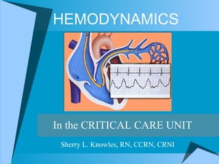
- 1. HEMODYNAMICS In the CRITICAL CARE UNIT Sherry L. Knowles, RN, CCRN, CRNI
- 19. PA CATHETER WAVEFORMS A wave - due to atrial contraction. Absent in atrial fibrillation. Enlarged in tricuspid stenosis, pulmonary stenosis and pulmonary hypertension. C wave - due to bulging of tricuspid valve into the right atrium or possibly transmitted pulsations from the carotid artery. X descent - due to atrial relaxation. V wave - due to the rise in atrial pressure before the tricuspid valve opens. Enlarged in tricuspid regurgitation. Y descent - due to atrial emptying as blood enters the ventricle. Canon waves - large waves not corresponding to a, v or c waves. Due to complete heart block or junctional arrhythmias.
- 26. RESPIRATORY VARIATION Spontaneous Breathing
- 28. END EXPIRATION
- 29. RAP WAVEFORM RAP WAVEFORM
- 30. (CVP) RA WAVEFORM & ECG
- 31. (CVP) RA WAVEFORM (CVP) RA WAVEFORM
- 33. PAWP TRACING PAWP WAVEFORM
- 34. PAWP WAVEFORM PAWP WAVEFORM
- 35. PA vs PAWP WAVEFORM PA vs PAWP WAVEFORM
- 36. PAWP WAVEFORM & ECG PAWP WAVEFORM
- 37. PAWP WAVEFORM PAWP WAVEFORM
- 38. V WAVES PAWP WAVEFORM
- 39. PAWP WITH V WAVES
- 40. SVO 2 MONITORING
- 42. POTENTIAL COMPLICATIONS POTENTIAL COMPLICATIONS Same as arterial pressure monitoring plus the following: Air emboli Cardiac tamponade Thromboembolism Dysrhythmias Catheter displacement Balloon rupture Infection Lung ischemia Inaccurate pressures Electromicroshock Equipment malfunction Pulmonary artery rupture Pneumothorax/Hemothorax Frank Hemorrhage Loss of balloon integrity Altered skin integrity Pulmonary artery extravasation PA hemorrhage or infarction Air emboli Cardiac arrest
- 46. TROUBLESHOOTING
- 48. DAMPENED PA WAVEFORM PAWP WAVEFORM
- 49. ALTERATIONS IN SVO 2
- 50. ALTERATIONS IN SVO 2
- 54. TREATMENTS
- 56. MEASUREMENTS
- 72. THE END
Notas del editor
- 1) May Be Invaluable In Diagnosing And Managing Patients. 2) Requires Critical Care Knowledge And Skill. 3) Noninvasive Hemodynamic Monitoring Is Available.
- To evaluate the hemodynamic response to fluid therapy, medication and other treatments Aspiration of air emboli
- Standard Swan-Ganz (7.0F) VIP Swan-Ganz (7.5 F) SVO 2 Monitoring Swan-Ganz (8.0F) Pacing Swan-Ganz Monitoring (Interventional) Swan-Ganz
- Components: 1. Proximal port – approximately 30 cm from tip of catheter. Also known as the CVP port (central venous pressure). It lies in the right atrium and measures CVP. It can be used for infusion of IV solutions or medications, for drawing blood and for injecting cardiac output boluses. It is usually color coded blue. 2. Distal port – opening is at the tip (end) of the catheter. A lso known as the PA port. It lies directly in the pulmonary artery and measures the pulmonary artery pressures (PAP), systolic (PAS), and diastolic (PAD). It also measures the pulmonary capillary wedge pressure (PCWP) when the balloon is inflated. The PA pressures should always be monitored continuously . NEVER USE the PA port for medication infusion. It c an be used for drawing &quot;mixed venous&quot; blood gas samples. It is u sually color coded yellow. 3. Thermistor and connector port T he thermistor connector connects the pulmonary catheter to the cardiac output computer. The connector is at the end of a separate catheter lumen outside the patient thermistor wire. Blood temperature is transmitted within the lumen (the core temperature is the most accurate reflection of the body temperature). It is used in determining cardiac output. The connector tip should always have a protective covering to protect patient from microshock. It is usually color coded yellow with a red connector. 4. Balloon port The balloon port is located < 1 cm from the tip of the catheter. When the balloon is inflated with approximately 0.8 to 1.5 cc of air, the catheter will become lodged (wedged) in the pulmonary artery and gives a wedge tracing. It r eflects the pressures that are in the left side of the heart when inflated. DO NOT INFLATE WITH LIQUID---- ALWAYS INFLATE WITH AIR. When deflated, turn stopcock to off position and leave syringe connect to the port. It is usually color coded red. 5. A 5 - lumen Swan Ganz catheter has either an infusion port or a pacing port A pacing port allows for insertion of a transvenous pacing wire. The infusion port allows for infusion of IV solutions or medications. It is usually color coded white.
- 1 ) Jugular, Subclavian, Superior Vena Cava And Right Atrial Pressures Essentially Equal Because Of No Valve Between Them. 2) Balloon Is Inflated In The Right Atria. 3) Balloon Must Be Inflated To Advance. 4) Balloon Must Be Deflated To Withdraw.
- 1) If RV Irritated… May Cause Ventricular Dysrhythmias, Including VT. 2) Must Immediately Move Catheter If Ectopy In RV. 3) Catheter May Migrate Back Into RV.
- 1) Dicrotic Notch Represents Pulmonary Valve Closure. 2) Watch For Changes In Waveforms And Pressures (PAD).
- 1) Wedging Can Cause Pulmonary Artery Rupture. 2) Prolonged Wedging Can Cause Ischemia To The Lung Tissue Distal to The Catheter. 3) Prolonged Wedging Can Cause Injury To The Pulmonary Artery. 4) Normal PA = 15-25/8-15 And Normal PAWP = 6-12. 5) Watch For Changes In Waveforms And Pressures. 6) PAWP = PACP = PCWP = PAOP = Wedge. PAWP is the most accurate reflection of left atrial pressure, therefore of left ventricular end-diastolic pressure (LVEDP), or preload.
- 1) Normal Pressures: RA = 1-7 RV = 15-25/1-7 PA = 15-25/8-15 PAD = 8-15 PAWP = 6-12
- Every one to four hours: a. Verify alarm limits are on and appropriate. b. Assess adequate fluid level in pressure bag. c. Ensure pressure bag is inflated to 300 mm Hg. Microrate infusion devices will be used on hemodynamic lines for all pediatric patients who weigh less than 20 Kg and for patients with severe fluid restrictions, i.e. ARF. d. Obtain for all hemodynamic monitoring lines: pressure readings at the phlebostatic axis level Obtain for PA catheters only: 1. cardiac output, cardiac index, systemic vascular resistance and pulmonary vascular resistance unless otherwise ordered 2. pulmonary capillary wedge pressure readings, unless otherwise ordered 3. SVO 2 readings 4. continuous cardiac output readings Every eight to twelve hours and prn: a. Zero and level all pressure transducers. b. Obtain: 1. pressure waveform tracings and readings from each line. 2. occlusion cuff pressure for arterial lines. c. For pulmonary artery catheter, ensure catheter sleeve not taped. d. For pulmonary artery catheter, check catheter length at insertion site.
- Even though there may be a small effect of PEEP on intravascular pressure measurements, it is not advisable to eliminate (turn off) PEEP temporarily while pressure measurements are being made, as this may induce hemodynamic instability due to changes in venous return and PO2. These measurements off PEEP will therefore not accurately reflect the patient's hemodynamic status when PEEP is being used.
- Even though there may be a small effect of PEEP on intravascular pressure measurements, it is not advisable to eliminate (turn off) PEEP temporarily while pressure measurements are being made, as this may induce hemodynamic instability due to changes in venous return and PO2. These measurements off PEEP will therefore not accurately reflect the patient's hemodynamic status when PEEP is being used.
- PAWP
- a wave Rise in pressure due to atrial contraction. a waves are larger in the presence of any resistance to RV filling, (tricuspid stenosis, RV failure, cardiac tamponade) because resistance will increase pressure as the atrium attempts to contract and eject blood. x descent Fall in pressure due to atrial relaxation. c wave Rise in pressure due to ventricular contraction and bulging of the closed tricuspid valve. v wave Rise in pressure during atrial filling. y descent Fall in pressure due to the opening of the tricuspid valve and the beginning of ventricular filling.
- a wave Rise in pressure due to atrial contraction. a waves are larger in the presence of any resistance to RV filling, (tricuspid stenosis, RV failure, cardiac tamponade) because resistance will increase pressure as the atrium attempts to contract and eject blood. x descent Fall in pressure due to atrial relaxation. c wave Rise in pressure due to ventricular contraction and bulging of the closed tricuspid valve. v wave Rise in pressure during atrial filling. y descent Fall in pressure due to the opening of the tricuspid valve and the beginning of ventricular filling.
- a wave = atrial contraction Large a waves will be seen with anything that increases pressure during atrial contraction, such as mitral stenosis, an ischemic LV, or an LV in failure which is not emptying completely v wave = atrial filling Large v waves will be present with any resistance to ventricular filling such as mitral regurgitation, volume overload, cardiac tamponade
- Timing of the pulmonary artery waveforms: An A wave follows the QRS wave on ECG, whereas V wave follows the T wave on ECG.
- The fall in venous pressure following opening of the A-V valve is termed the y descent .
- a waves atrial contraction large a waves will be seen with anything that increases pressure during atrial contraction, such as mitral stenosis, an ischemic LV, or an LV in failure which is not emptying completely v waves atrial filling large v waves will be present with any resistance to ventricular filling such as mitral regurgitation, volume overload, cardiac tamponade
- 1 = a wave (approx. 40mmHg) 2= v wave (approx. 52mmHg) 3= steep V descent Simultaneous recording of EKG helps identify v wave in mitral vale regurgitation, v wave corresponds to t wave of EKG. Large V waves are not always indicative of Mitral insufficiency. The size of the V wave depends on both the volume of blood entering the atrium during ventricular systole and left atrial compliance. Decreased left ventricular compliance may result in prominent V wave
- Continuous SVO2 monitoring allows the minute-to-minute assessment of total tissue oxygen balance ( i.e. , the relationship between oxygen delivery and oxygen consumption). SVO2 varies directly with cardiac output, Hb, and SaO2, and inversely with VO2 (oxygen consumption.). The normal SVO2 is 75%, which indicates that under normal conditions, tissues extract 25% of the oxygen delivered. An increase in VO2 or a decrease in arterial oxygen content (SaO2 x Hb) is compensated by increasing CO or tissue oxygen extraction. When the SVO2 is less than 30%, tissue oxygen balance is compromised, and anaerobic metabolism ensues. A normal SVO2 does not ensure a normal metabolic state but suggests that oxygen kinetics are either normal or compensated.
- Bleeding Infection Dysrhythmias Pulmonary Artery Rupture Pneumothorax Hemothorax Valvular Damage Embolization Balloon Rupture Catheter Migration
- Pulsus Paradoxus = paradoxical pulse = abnormally large inspiratory fall in arterial pressure (10mmHg or more) When left ventricular SV is decreased, blood pools in the lung and right side or the heart during inspiration, increasing intrapericardial pressure.
- Occlusion
- Measurement of the mixed venous oxygen saturation, or the saturation of blood in the pulmonary artery, can be continuous with special PA catheters. This blood is normally unoxygenated, having not yet traveled through the lungs, with a saturation of 60-80%. SvO 2 is clinically important as a function of supply and demand; how much oxygen is being extracted from the blood by the organs, before this blood is returned to the right heart. SvO 2 , therefore, serves us in evaluating the supply and demand of oxygen to the tissues. It is influenced by oxygen delivery (hemoglobin, SaO 2 , cardiac output) as well as oxygen consumption.
- 1) Beta Blockers 2) Beta Adrenergics 3) Calcium Channel Blockers 4) Epinephrine 5) Dopamine 6) Lasix 7) Bumex 8) Edecrin 9) NTG 10) Nipride 11) Neo-Synephrine 12) Levophed 13) Dobutamine 14) Dopamine 15) Primacor
- 1) Cardiac Tamponade = PA Pressures, SVR CO, CI, and BP. 2) Left Ventricular Failure = PAWP RVSW Normal 3) Right Ventricular Failure = RA/CVP, RV Pressure RVSW 4) PA Hypertension = PA Pressures, PVR May Lead To Cor Pulmonale 5) Endotoxins And Histamines Produce Vasodilation = SVR.
- 1) Normal Values: CVP = 1-7 mmHg PAWP = 6-12 mmHg CO = 4.0-8.0 L/Min. CI = 2.5-4.0 L/Min. SVR = 700=1500 dynes/sec/cm 5 PVR = 100-250 dynes/sec/cm 5 RVSW = 10-15 gm 2 /beat LVSW = 60-80 gm 2 /beat
- CVP Example
- CVP Answer
- Example 1
- Answer 1
- Example 2
- Answer 2
- Example 3
- Answer 3
- Example 4
- Answer 4
- Example 5
- Answer 5
- Example 6
- Answer 6