
Recomendados
Más contenido relacionado
La actualidad más candente
La actualidad más candente (20)
Destacado
Destacado (20)
Similar a Human Bones: Anatomy and Key Structures
Similar a Human Bones: Anatomy and Key Structures (20)
Más de abctutor
Más de abctutor (20)
Human Bones: Anatomy and Key Structures
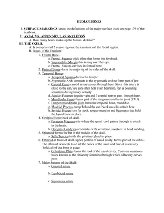
- 1. HUMAN BONES I. SURFACE MARKINGS-know the definitions of the major surface listed on page 179 of the textbook. II. AXIAL VS. APPENDICULAR SKELETON A. How many bones make-up the human skeleton? III. THE SKULL A. Is comprised of 2 major regions: the cranium and the facial region. B. Bones of the Cranium: 1. Frontal Bone- a. Frontal Squama-thick plate that forms the forehead. b. Supraorbital Margin-thickening over the eye. c. Frontal Sinuses-cavities in frontal bone. 2. Parietal Bones-form the majority of the sides of the skull. 3. Temporal Bones- a. Temporal Squama-forms the temple. b. Zygomatic Arch-connects to the zygomatic arch to form part of jaw. c. Carotid Canal-carotid artery passes through here. Since this artery is close to the ear, you can often hear your heartrate, feel a pounding sensation during heavy activity. d. Jugular Foramen-jugular vein and 3 cranial nerves pass through here. e. Mandibular Fossa-forms part of the temporomandibular joint (TMJ). f. Temporomandibular joint-between temporal bone, mandible. g. Mastoid Process-bump behind the ear. Neck muscles attach here. h. Styloid Process-site for neck, tongue muscles and ligaments that hold the hyoid bone in place. 4. Occipital Bone-back of skull. a. Foramen Magnum-site where the spinal cord passes through to attach to the brain. b. Occipital Condyles-articulates with vertebrae, involved in head nodding. 5. Sphenoid-forms the bat in the middle of the skull. a. Sella Turcica-holds the pituitary gland in place. 6. Ethmoid-at front of skull, upper portion of nasal cavity, forms part of the orbits. The ethmoid connects to all of the bones of the skull and face-it essentially holds all of the bone in place. a. Cribriform Plate-forms the roof of the nasal cavity. Contains numerous holes known as the olfactory foramina through which olfactory nerves pass. 7. Major Sutures of the Skull: a. Coronal suture b. Lambdoid suture c. Squamous suture
- 2. C. Facial Bones of the Skull 1. Nasal Bones-form the bridge of the nose, are primarily cartilage in composition. 2. Nasal Septum-separates the right and left airways in the nose. Is composed of the ethmoid bone, vomer bone and cartilage. There are three bony processes in the nasal cavity known as concha (Middle, Inferior, Superior).The concha primarily form the walls of the nasal cavity. 3. Maxillae-form the upper jaw. These paired bones hold the upper teeth in place and they form the boundaries of three cavities: the roof of the mouth, the floor of the nose and the floor of the orbits. a. Cleft palate-condition in which the maxillary bones are not completely joined. This often leads to a cleft lip. This condition is often repaired via surgery. 4. Zygomatic Bones-cheek bones. These articulate with the maxilla, the temporal bones, and the sphenoid bone. 5. The Mandible-largest and strongest bone of the face. It forms the lower jaw bone. It holds the lower teeth in place. 6. The Vomer-unpaired facial bone that forms part of the nasal septum. a. Deviated nasal septum-physical disorder in which the vomer is pushed to one side or another. Is often caused by trauma to the face. 7. The Lacrimal Bones-smallest and most fragile bones of the face. These bones contain the lacrimal duct which produces tears. 8. The Orbit-is the cavity or socket that the eye is located in. 9. The Hyoid Bone-where is this bone located? a. What attaches to this bone? IV. THE VERTEBRAL COLUMN A. Region of the Vertebral Column-How many vertebrae are in each region? 1. Cervical Region- 2. Thoracic Region- 3. Lumbar Region- 4. Sacral Region- 5. Coccygeal Region- B. Normal Curves of the Vertebral Column 1. Cervical Curve and Lumbar Curve-posteriorly concave. 2. Thoracic Curve and Sacral Curve-posteriorly convex. 3. What is the significance of these curves? C. Verebrae-bones that make up the spinal column. The major parts of a vertebra include: 1. The Intervertebral Disc-cartilage pad between the vertebrae. 2. Body(Centrum)-major weight-bearing structure. 3. Vertebral Arch-also bears weight. Composed of: a. The Pedicle-forms the arches. b. The Lamina-what is a Laminectomy? 4. Vertebral Foramen-what passes through here?
- 3. 5. Intervertebral Foramina-nerves pass through these. 6. Processes on Vertebrae: Where are each of these located? a. Spinous Process b. 2 Transverse Processes c. Articular Processes 7. The Atlas-where is this located? 8. The Axis-where is this located? a. The Dens (Odontoid Process)-pivot point on the axis for skull rotation. D. The Sacrum-triangular bone, composed of 5 fused vertebrae. 1. Sacral Foramina-holes through which nerves and blood vessels pass through. 2. Sacral Canal-extension of the vertebral foramen on the sacrum. E. The Coccyx-what is this structure? V. THE STERNUM A. Major Regions of the Sternum: 1. The Manubrium-superior portion, clavicle attaches here at the clavicular notch. a. Jugular notch-superior portion of the manubrium. 2. The Body-attaches to cartilages from ribs 2-7 3. Xiphoid Process-primarily cartilage, becomes hardened (ossified) as we age. a. Abdominal muscles attach here. b. Landmark for CPR. VI. RIBS-everyone has 12 pairs of ribs. A. True Ribs-their cartilage attaches directly to the sternum. False Ribs-their cartilage does not attach directly to the sternum. Floating Ribs-do not attach to the sternum at all. B. Parts of a Rib 1. The Head-attaches to the vertebrae, the neck is near the head of a rib. 2. The Shaft-forms the length of a rib. Has a costal groove where nerves and blood vessels are located. 3. The Tubercle-attaches to the thoracic vertebrae. VII. THE CLAVICLE A. Sternal Extremity-site where the clavicle articulates with the sternum. B. Acromial Extremity-site where the clavicle articulates with the acromion. C. Conoid Tubercle-site of muscle attachments. VIII. THE SCAPULA A. Acromion-upper portion of scapula, forms the Acromioclavicular joint. B. Glenoid Cavity-fossa where the humerus attaches to form the shoulder joint. C. Suprascapular Notch-notch through which major nerves pass. D. Coracoid Process-posterior structure, site of muscle attachment. E. Supraspinous Fossa-depression where the supraspinatus muscle sits. F. Infraspinous Fossa-depression where the infraspinatus muscle sits. G. Subscapular Fossa-depression where the subscapularis muscle sits. IX. THE HUMERUS A. The Head-articulates with the glenoid cavity to form the shoulder joint. B. The Anatomical Neck-site of the epiphyseal plate. C. The Greater Tubercle-knob, where major muscles attach. D. The Lesser Tubercle-smaller knob, major muscle also attach here. E. The Surgical Neck-areas that is easily broken, requires surgery to repair.
- 4. F. The Intertubercular Sulcus-groove between the tubercles, nerves run through here G. The Deltoid Tuberosity-site where the deltoid muscle attaches. H. The Capitulum-allows the humerus to articulate with the ulna. I. The Trochlea-also allows for articulation of the humerus at the elbow joint. J. The Coronoid Fossa-forms part of the elbow joint. K. The Olecranon Fossa-forms part of the elbow joint. L. The Medial and Lateral Epidcondyles-bumps, sites of muscle attachment. X. THE ULNA A. The Olecranon Process- B. The Coronoid Process- C. The Radial Notch-site of radial articulation with the ulna. XI. THE RADIUS A. The Head-nail-shaped structure that articulates with the humerus. B. The Radial Tuberosity-site for muscle attachment. C. Ulnar notch-low on the radius, articulates with the ulna. XII. THE HAND A. Carpals-8 bones that form the wrist. B. Metacarpals-5 bones that from the palm of the hand. C. Phalanges-14 of these in each hand, these form the fingers. D. Disorders of the Forearm/Hand: 1. Carpal Tunnel Syndrome-produced by a pinched nerve in the wrist. The nerve carries feeling to the thumb and controls hand movements. Caused by repetitive movement, strain, vibration. 2. Lyme Disease-caused by a tick, can lead to rheumatoid arthritis (an autoimmune disease). 3. Osteoarthritis-degeneration of articular cartilage. Very common in the hand, wrist. Occurs as individuals age. XIII. BONES OF THE PELVIC GIRDLE A. THE ILLIUM 1. Sacroiliac Joint-site where the sacrum and ilium attach. 2. Greater Sciatic Notch-allows passage of sciatic nerve to leg. 3. The Iliac Fossa-flat surface of the ilium. B. THE ISCHIUM 1. The Obturator Foramen-large hole, blood vessels and nerves pass through here. It is nearly closed by a fibrous membrane. 2. The Ischial Tuberosity-inferior surface of the ischial body, is rough and thickened. This support our weight when we sit. This tuberosity is the strongest part of the hip bone. 3. The Acetabulum-deep socket that receives the head of the femur or thigh bone. C. THE PUBIS BONE 1. The Pubic Symphysis-formed by the rami of the pubic bones. This symphysis is held together by fibrocartilage. The pubic symphysis forms the pubic arch. 2. The female pubis is wider in females to allow for childbirth. XIV. THE FEMUR A. Head of the Femur-forms the pelvic girdle B. Greater and Lesser Trochanters-knobs, for major muscle attachment, including the
- 5. gluteal muscles. C. The Intertrochanteric Line-lines between the trochanters, anterior side. D. The Intertrochanteric Crest-between the trochanters, posterior side. E. The Medial and Lateral Condyles-articulate with the same structures of the tibia to form the knee joint. F. The Gluteal Tuberosity-rough area on femur, where gluteus maximus attaches. XV. THE PATELLA-what is this? A. Increases leverage of the leg. Held in place by the patellar ligament. XVI. THE TIBIA-second largest bone of the body. A. The Medial and Lateral Condyles-articulate with femur to form knee joint. B. The Medial Malleolus-forms the medial knob of the ankle. C. The Tibial Tuberosity-site for patellar ligament attachment. D. The Fibular Notch-site where the fibula attaches to the tibia. XVII. THE FIBULA A. The Lateral Malleolus-forms the lateral side of the ankle. XVIII. THE FOOT A. Tarsus-with the calcaneus, talus. B. Metatarsus-composed of metatarsal bones. C. Phalanges-toes D. Arches of the foot-for support XIX. MALE VS. FEMALE SKELETON-major differences are in the pelvic girdle. XX. CLINICAL TERMS/DISORDERS