SlideShare una empresa de Scribd logo
1 de 208
MRI OF THE KNEE JOINT.
Dr/ ABD ALLAH NAZEER. MD.
Clinical Indication
Acute pain secondary to trauma in the presence of an effusion
without fracture on x-ray.
Suspected unstable knee with history of knee locking or positive
McMurray test on examination.
Suspected stable meniscus tear following treatment with
analgesia and physiotherapy and activity modification for at least
4 weeks in the presence of persistent joint effusion or history of
locking
Suspected cruciate ligament injury with a history of knee giving
way and laxity of between 5 and 11mm) on examination
grade II-III instability (medial laxity between 5 and 11mm on
examination.
Suspected multi-ligamentous or lateral collateral ligament injury
when there is grade II-III instability
Suspected medial collateral ligament injury with grade II-III
instability despite treatment with brace and activity modification
for at least 6 weeks
Other knee conditions of unknown etiology when there are both
symptoms and signs that suggest a significant underlying injury
and when knee x-ray is non-diagnostic for the etiology of the
underlying condition
Knee - AP View
 Lateral View
Skyline View
Intercondylar
View
Protocol of examination
 Axial T1 localizer
 Sagittal T1, PD, T2
 Coronal gradient echo,
STIR
 If contrast is injected
[ Axial, Sagittal ,coronal T1 WIs ]
How to know the pulse sequence?!
T1 T2 Gradient STIR
Upper EgyptLower Egypt
Items to be evaluated Where?!
 
 Menisci (medial & lateral)
 Ligaments
- Cruciate (ACL, PCL)
- Collateral
- Retinacular
 Tendons ( Quadriceps, Patellar)
 Bones
 Synovial effusion
      
Sagittal PD
Coronal.
Sagittal,coronal
and axial.
Coronal
Axial
Sagittal PD
Sagittal T1& T2
Sagittal T2
Anatomy of the meniscus.
Meniscus
Medial meniscus
 Semilunar - shaped
Posterior horn wider,
longer, taller than
anterior horn
Posterior horn tightly
attached to the capsule
Lateral meniscus
 C- shape
 Posterior and anterior horns are symmetric
 Anterior horn may be hypo plastic, extremely
thin
 Discoid meniscus and meniscal cysts more
common
AC
B
A B C
Lateral meniscus
Medial meniscus
Medial and lateral meniscus.
Medial and lateral meniscusMedial and lateral meniscus..
Patellar retinacular ligaments
Collateral
Ligaments
Proton density coronal image shows the normal medial collateral
ligament as a thin, taut, well-defined, low-signal structure extending
from the medial femoral epicondyle to the medial tibial metaphysis
Coronal and sagittal proton density image demonstrating the
normal lateral collateral ligament in its entirety, from the
femoral condyle origin to the fibular head insertion.
MeniscalMeniscal
LesionsLesions
Degeneration
 Tear
 Cyst
Discoid
Simple Tear Complex Tear
 Type II
 Type I  Normal
Types of Meniscal degenerationTypes of Meniscal degeneration
 Grade IGrade I
 Grade IIGrade II
 Meniscal frayingMeniscal fraying
Tear
 Grade 1 degeneration. Grade 11 degeneration
Meniscal degeneration with free edge fraying.
Meniscal degeneration with free edge fraying
Types of Meniscal TearsTypes of Meniscal Tears
 SimpleSimple
 ComplexComplex
 Special typesSpecial types
Simple Meniscal TearSimple Meniscal Tear
 HorizontalHorizontal
 VerticalVertical
 RadialRadial
Horizontal tear
Horizontal tear
Horizontal tear
Vertical tear
Occurs typically in the outer 1/3 of
the posterior horn or body of the
meniscus
]rare in the anterior horn[
Vertical
tear
Vertical tear
Body radial tear.
Full thickness body radial tear
Marching cleft sign in discoid lateral
meniscus indicates radial meniscal tear
Meniscal radial tear (arrow).
Vertical radial meniscal tear in the mid-body lateral
meniscus. (A) A sagittal fat-suppressed proton-density image
shows a vertical tear (arrow) of the central edge of the lateral
meniscus, oriented perpendicular to the curvature of the meniscus.
Note the superimposed horizontal tear of the anterior horn. (B) A
coronal fat-suppressed T2-weighted image shows the same tear
with truncation of the central meniscal margin (arrow).
Radial tear of lateral meniscus.
Vertical tear of the free edge of the meniscus [Root tear[
Ghost meniscus
Ghost meniscus
If there is no history of
Meniscal surgery and
the posterior horn is
absent near the
intercondylar notch
Meniscus Extrusion
Meniscal extrusion
Special Meniscal TearsSpecial Meniscal Tears
 FlapFlap
 Bucket handleBucket handle
 MC separationMC separation
Flap tear:displaced horizontal
or longitudinal tears
Flap tear [Oblique[
Should have tow components , horizontal and
Vertical common in the medial meniscus
Flap tear.
Bucket handle tearBucket handle tear
Small sized
posterior horn
[ sagittal[
Medially displaced
fragment[ coronal[
Double PCL sign
]sagittal[
Bucket Handel tearBucket Handel tear
Flipped meniscus : Double Delta Sign
Bucket Handel tear , Lateral meniscus
Flipped meniscus : Double Delta Sign
Parrot beak tear (displaced radial oblique tear(.
MENISCOCAPSULAR SEPARATIONMENISCOCAPSULAR SEPARATION..
Post-traumatic contusion
of the lateral femoral and
tibial condyles
Discoid meniscus
Dysplastic meniscus with loss of normal semi lunar shape.
50%or more coverage of the tibial plateau.
Meniscal body segment seen in 3 or more sagittal images
Discoid meniscusDiscoid meniscus
Meniscal cyst
A Cyst extending from a meniscal tear
Common sites : Anterior horn LM , Posterior horn MM
Meniscal cyst
Meniscal cyst
Magic angle phenomenon
5555
Closed MRI
Open MRI
Open MRIClosed MRI
In closed MRI scanner,In closed MRI scanner,
magic angle artifact canmagic angle artifact can
be found in one locationbe found in one location::
The up-sloping of theThe up-sloping of the
posterior horn lateralposterior horn lateral
meniscusmeniscus..
In open, vertical field magnets,
magic angle artifact will be
found in two locations:
1)1(junction of anterior horn
and body of meniscus and
2)2(junction of posterior horn
and body of meniscus
Normal intrameniscal signal evolution in the site of surgery, with very good final result.
Normal healing sequence. Typical worsening in the first scans - intrameniscal
signal after surgery is worse then before surgery. No healing disturbance.
Abnormal healing of degenerated meniscus.
Ligamentous
Lesions
 ACL
 PCL
 Collateral
 Retinacular
Anterior cruciate ligament
MRI shows the normalMRI shows the normal
linear low signal intensitylinear low signal intensity
ACL adjacent to theACL adjacent to the
lateral bony wall of thelateral bony wall of the
upper intercondylar notchupper intercondylar notch
(arrow). The normal ACL(arrow). The normal ACL
moves away from the wallmoves away from the wall
and diverges into multipleand diverges into multiple
fascicles on more distalfascicles on more distal
imagesimages.
Anterior cruciate ligament Posterior cruciate ligament
Primary signs [ In the ligament ]
Total discontinuity
Abnormal signal
Abnormal configuration
Abrupt angulation
Wavy appearance
Abnormal axis
Anterior cruciate ligament injury
Intercondylar
roof
Anterior cruciate ligament injury
Anterior cruciate ligament injury.
Normal ACL
Normal ACL
Non visualization of the ACL with a cloud of edema and hemorrhage
Secondary signs [ Outside the
ligament ]
• Blumensaat angle sign.
• Bone contusions [Pivot- shift bruises ]
• Anterior translocation of the tibia
• Uncovered meniscus sign
• Avulsion fracture of the tibial insertion
• Segond fracture 70-100% with ACL tear
• PCL buckling
• PCL line sign
Anterior cruciate ligament injury
Hyperextension ACL tear withHyperextension ACL tear with
"kissing bone bruises"kissing bone bruises."."
Negative Blumensaat angleNegative Blumensaat angle..
Positive Blumensaat anglePositive Blumensaat angle..
ACL Graft with negative Blumensaat angleACL Graft with negative Blumensaat angle..
The probability of anThe probability of an
ACL tear is very highACL tear is very high
if both such boneif both such bone
bruises are present,bruises are present,
only slightly lower ifonly slightly lower if
the tibial bone bruisethe tibial bone bruise
is present in isolation,is present in isolation,
and still slightlyand still slightly
lower with anlower with an
isolated femoral boneisolated femoral bone
bruise of thisbruise of this
appearance.appearance.
Anterior tibial translocation
Anterior tibial translocation with” uncovered meniscus sign”
Segond fractureSegond fracture..
An elliptical verticallyAn elliptical vertically
3x10mm bone3x10mm bone
fragment parallel to thefragment parallel to the
lateral tibial cortex,lateral tibial cortex,
about 4mm distal toabout 4mm distal to
the plateau. Best seenthe plateau. Best seen
on AP or tunnelon AP or tunnel
radiographic viewsradiographic views
75-100%association with ACL tear
Segond fracture inSegond fracture in
patient with ACLpatient with ACL
tear. T1- weightedtear. T1- weighted
coronal MRI shows acoronal MRI shows a
small, low-signalsmall, low-signal
elongated fractureelongated fracture
fragment that isfragment that is
parallel to the lateralparallel to the lateral
tibia. The associationtibia. The association
of Segond fracturesof Segond fractures
with ACL tearswith ACL tears
approaches 100%.approaches 100%.
ACL injury
+ve
PCL LINE SIGN
PCL redundancy as a secondary sign of ACL tear. This is a relativelyPCL redundancy as a secondary sign of ACL tear. This is a relatively
unreliable secondary sign of ACL tear.unreliable secondary sign of ACL tear.
Partial ACL tearPartial ACL tear
 Common about 10-43% of ACL tearsCommon about 10-43% of ACL tears
 Suboptimal accuracy of MRISuboptimal accuracy of MRI
 Subtle 1ry and 2ry signsSubtle 1ry and 2ry signs
 Focal angulationsFocal angulations
 Focal increase T2 signalFocal increase T2 signal
[[non specificnon specific ]]
 Single bundle signSingle bundle sign
Sagittal MRI shows an abruptlySagittal MRI shows an abruptly
angulated mid-ACL (arrow) .A wavy orangulated mid-ACL (arrow) .A wavy or
sharply angulated appearance issharply angulated appearance is
abnormal.abnormal.
T1-weighted sagittalT1-weighted sagittal
MRI shows a normal-MRI shows a normal-
appearing ACL.appearing ACL.
Partial ACL tear
T1-weighted sagittal MRI imageT1-weighted sagittal MRI image
immediately adjacent to theimmediately adjacent to the
previous image shows a partiallyprevious image shows a partially
disrupted ACLdisrupted ACL
Partial ACL tear
Partial ACL tear with thickening, angulations and abnormal brightPartial ACL tear with thickening, angulations and abnormal bright
signal inside. The tibial and femoral attachment is preservedsignal inside. The tibial and femoral attachment is preserved..
Partial ACL tear with thickening and abnormal bright signal insidePartial ACL tear with thickening and abnormal bright signal inside..
Partial ACL tear with diffuse thickening and abnormal signal insidePartial ACL tear with diffuse thickening and abnormal signal inside..
Chronic ACL TearChronic ACL Tear
 Fragmented ACL [ common finding ]Fragmented ACL [ common finding ]
 Absent bone edema and contusionsAbsent bone edema and contusions
 Empty notch signEmpty notch sign
 ACL attached to PCLACL attached to PCL
ACL tear on axial image showing non-visualization of the anteriorACL tear on axial image showing non-visualization of the anterior
cruciate ligament (ACL) in the upper inter-condylar notchcruciate ligament (ACL) in the upper inter-condylar notch
A large knee effusion and a Baker cyst are noted incidentally.A large knee effusion and a Baker cyst are noted incidentally.
Empty notch sign
Chronic ACL tear, empty notch sign. T1-weighted coronal MRI shows fat in theChronic ACL tear, empty notch sign. T1-weighted coronal MRI shows fat in the
lateral intercondylar notch, ACL is absent. This is a frequent MRI appearance of alateral intercondylar notch, ACL is absent. This is a frequent MRI appearance of a
chronic ACL tear after resolution of acute edema and hemorrhage.chronic ACL tear after resolution of acute edema and hemorrhage.
ACL DegenerationACL Degeneration
 Intra ligamentous cystIntra ligamentous cyst
 May be mistaken for a tearMay be mistaken for a tear
 Arthroscopic decompressionArthroscopic decompression
Intercondylar notch cystIntercondylar notch cyst
 1% of knee MRIs1% of knee MRIs
 Usually an incidental findingUsually an incidental finding
 Painful if erodes the bonePainful if erodes the bone
 Post-traumatic chronic partialPost-traumatic chronic partial
cruciate ligament tear with internalcruciate ligament tear with internal
degenerationdegeneration
 More common in the ACLMore common in the ACL
 Oval , rounded may be multilocularOval , rounded may be multilocular
 Rim enhancement if inflamedRim enhancement if inflamed
 Arthroscopic drainageArthroscopic drainage
Cruciate ligament cyst
Intraligamentous ganglion cyst
A 3-D cutaway at the
intercondylar notch in
the sagittal plane
reveals normal
positioning for a
patellar tendon ACL
autograft. The tibial
tunnel should lie
posterior to the line
drawn parallel to the
intercondylar notch
(red) and the femoral
attachment should lie
posterior to a line
drawn parallel to the
cortex of the distal
femoral diaphysis
(blue(.
Femoral tunnel is normally positioned
at the junction of the physeal scar and
posterior intercondylar roof (asterisk(.
Abnormal placement of the femoral
tunnel (asterisk), which lies significantly
anterior to a line drawn along the
posterior cortex of the femoral diaphysis
with graft degeneration.
Tibial tunnel cyst after ACL graft reconstruction.
Cyclops lesion (arrowheads) attached to the ACL (arrow) with a head-like
appearance, showing a focal area of discoloration resembling an eye (curved arrow(
Hypointense to isointense nodule (arrowhead) attached to the anterior
surface of the anterior cruciate ligament (ACL) graft (arrow). Sagittal
T1W MRI image (C) shows a hypointense nodule (arrowhead) in the
anterior intercondylar notch, related to Cyclops.
ACL reconstruction are provided. Diffuse abnormal fluid signal
intensity is seen along the course of the graft (arrows) on the sagittal
image, compatible with extensive graft ganglion formation.
ACL Surgery Failure with stem cells injection.
Posterior cruciate ligament.Posterior cruciate ligament.
 The major stabilizer of the kneeThe major stabilizer of the knee
 Uniform low signal , no striationsUniform low signal , no striations
 Twice strong as the ACLTwice strong as the ACL
 The menisco-femoral ligaments are intimately related to PCL.The menisco-femoral ligaments are intimately related to PCL.
They connect the posterior horn of the lateral meniscus to theThey connect the posterior horn of the lateral meniscus to the
medial femoral condylemedial femoral condyle
Ligament of Humphrey anterior to PCLLigament of Humphrey anterior to PCL
Ligament of Weisberg posterior to PCLLigament of Weisberg posterior to PCL
Proton-dense sagittal image demonstrates the normal
tibial insertion of the PCL. The insertion site is a
vertically inclined posterior to the articular surface.
 PCL injuries represent about 12% of knee injuriesPCL injuries represent about 12% of knee injuries
 Combined PCL injuries represent 97%Combined PCL injuries represent 97%
With ACL 65%With ACL 65%
With MCL 50%With MCL 50%
With MM 30%With MM 30%
Posterior cruciate ligamentPosterior cruciate ligament
TYPES OF PCL INJURES
Complete tear 40%
Partial tear 55%
Avulsion tear 7%
NORMAL PCL
TORN PCL
MR FINDINGS
Increased signal due to hemorrhage and edema
Diffuse enlargement of PCL
COMPLETE PCL TEAR
An enlarged, intermediate signal (obviously torn) PCL.
NORMAL PCL
AVULSION TEAR
• Involves the tibial insertion
• Retracted bone fragment
• Bone marrow edema at avulsion site
• The actual PCL may be normal
AVULSION PCL TEAR
AVULSION PCL TEAR
PARTIAL PCL TEAR
PD sagittal image
shows partial tear
of the mid-substance
of the PCL. The
normal ligament of
Humphrey (small
arrow) is visualized
better because it is
adjacent to the high
signal intensity edema
of the torn PCL.
PARTIAL PCL TEAR
Collateral ligaments.
MCL is about 8-11 cm
LCL is about 5-7 cm
Isolated injuries are rare,
usually with ACL and MM
Collateral ligaments
Grade I : microscopic tear
Grade II :partial tear
Grade III : complete tear
GRADING SYSTEM
Grade I,II and isolated grade III are treated conservatively, while grade
III tears associated with ACL tears are treated by repairing ACL only
Proton density coronal image shows the normal medial collateral
ligament as a thin, taut, well-defined, low-signal structure extending
from the medial femoral epicondyle to the medial tibial metaphysis
Coronal and sagittal proton density image demonstrating
the normal lateral collateral ligament in its entirety, from
the femoral condyle origin to the fibular head insertion.
Grade I medial collateral ligament tear with surrounding
edema (straight arrows) on a T2WI. Note the normal
thickness and signal of the medial collateral ligament and
continued close apposition to the femoral and tibial cortices.
Grade 1 sprain of the medial collateral ligamentGrade 1 sprain of the medial collateral ligament..
Grade 11 sprain of the medial collateral ligamentGrade 11 sprain of the medial collateral ligament..
Grade II medial collateral ligament tear seen on a coronal proton
density image shows slight thickening of the medial collateral
ligament and separation from the underlying cortices. Bone marrow
edema of the lateral tibial plateau is seen due to valgus stress
7months after
conservative treatment
Grade II medial collateral ligament tear seen on a coronal T1
and STIR images showing slight thickening of the medial
collateral ligament and separation from the underlying cortices.
Grade 111 tear of the MCLGrade 111 tear of the MCL..
Grade III medial
collateral ligament
tear on a coronal
fast spin-echo T2-
weighted image
demonstrates a
disrupted ligament
that is thickened and
retracted with
surrounding edema
(black arrow).
Acute grade III tear with a folded ligament (arrow) and
surrounding edema on a coronal proton density image.
Acute tear of
the proximal
portion of the
lateral collateral
ligament is seen
on this coronal
proton density
image (white
arrow). Note the
associated grade
II medial
collateral ligament
tear.
Grade III MCL tear with retraction
Grade III MCL tear with
abnormal signal and edema
Grade 111 tear of the LCLGrade 111 tear of the LCL..
Ilio-tibial band syndrome. Distal tendon of IT fascia and insert at gerdy,sIlio-tibial band syndrome. Distal tendon of IT fascia and insert at gerdy,s
tubercle of the tibia. It occur in runner, cyclists, football players and weight liftertubercle of the tibia. It occur in runner, cyclists, football players and weight lifter..
Pre-patellar bursitis.(housemaid bursitisPre-patellar bursitis.(housemaid bursitis((
Backer and Pes anserine cystBacker and Pes anserine cyst..
Patellar and quadriceps tendons
Patellar
tendons
Complete tear of the patellar tendon with ACL teaComplete tear of the patellar tendon with ACL tearr
Complete tear of the patellar tendon with ACL tear
Partial tear of the patellar tendon
Lateral pressure syndrome
 Thickening of the lateral retinaculum
 Lateral knee pain
 Obese, athletic patients
 May be associated with
chondromalacia
Patella alta
 Sequlae of
patellofemoral
dysplasia
 Lengthening of the
infrapatellar tendon
 May be associated with
chondromalacia
 Length of patellar
tendon/ length of
patella > 1.3
Patella Baja
 Poliomyelitis
 Achondroplasia
 JRA
Hyaline cartilage
Hyaline cartilage
Hyaline cartilage
T2* MT
Articular cartilage
MT STIR
Chondromalacia patellae
Degeneration of the hyaline cartilage
Anterior knee pain in young adults
Four stages
Signal abnormalities
Ulceration [ fraying , partial or
full thickness defects ]
Reactive bone changes [ edema ,
cyst formation , sclerosis ]
Osteoartheritic changes
Chondromalacia patella
Chondromalacia patella.
Loose bodies
• Read with plain films
• Low signal fragments
Synovial osteochondromatosis.
Loose bodies
Synovial osteochondromatosisSynovial osteochondromatosis
Metaplasia of subsynovial soft tissues cartilage formation
Affects any joint [ knee , hip , elbow[
Age incidence 40 years M : F = 2 : 1
FINDINGSFINDINGS
Widening of the joint space
Bone erosions
Intra articular loose bodies
Secondary osteoarthritic changes
Synovial chondromatosisSynovial chondromatosis
Synovial chondromatosis.
Lipoma arborescenceLipoma arborescence
RareRare
IdiopathicIdiopathic
Fatty synovial infiltrations formingFatty synovial infiltrations forming
variable sized villous projectionsvariable sized villous projections
withinwithin
the joint capsule commonly in thethe joint capsule commonly in the
supra- patellar pouchsupra- patellar pouch
Associated with joint effusionAssociated with joint effusion
Painless swellingPainless swelling
Treatment by synovectomyTreatment by synovectomy
Lipoma arborescence
Lipoma
arborescens
Pigmented villo-nodular synovitisPigmented villo-nodular synovitis
 IdiopathicIdiopathic
 Monoarticular disease 1% incidenceMonoarticular disease 1% incidence
 Hypertrophic synovial masses with hemosiderinHypertrophic synovial masses with hemosiderin
laden macrophages bone erosionsladen macrophages bone erosions
 Intermediate signal in T1 andIntermediate signal in T1 and low signal in T2low signal in T2 withwith
enhancement after contrast injectionenhancement after contrast injection
 Typical location posterior to HoffaTypical location posterior to Hoffa’’s fat pads fat pad
 Painless swelling , pain with progressive diseasePainless swelling , pain with progressive disease
 Treatment by synovectomyTreatment by synovectomy
PIGMENTED VELLONODULAR
SYNOVITIS
PIGMENTED VILLONODULAR SYNOVITIS
PIGMENTED VILLONODULAR SYNOVITIS VERSUS
LIPOMA ARBORESCENS
Pigmented
villo-
nodular
synovitis
POPLITEAL CYST
Fluid in the bursa which is usually
communicating with the joint space
Other names
Baker’s cyst
Gastrocnemius/semimembranosus
bursa
Medial plica syndromeMedial plica syndrome
 Inflamed synovial plica causing pain , crepitusInflamed synovial plica causing pain , crepitus
and pseudolockingand pseudolocking
 Often in adolescents and athleticsOften in adolescents and athletics
 No measurement for plica thicknessNo measurement for plica thickness
Four types of plicaFour types of plica
Suprapatellar 90%Suprapatellar 90%
Medial 15 -30%Medial 15 -30%
InfrapatellarInfrapatellar
Lateral [ rare]Lateral [ rare]
PLICA SYNDROME
Medial plica
Osteochondritis dissecansOsteochondritis dissecans
 Osteochondral fragmentOsteochondral fragment in a typical locationin a typical location
 Young maleYoung male
 Lateral aspect of the medial femoral condoyleLateral aspect of the medial femoral condoyle
 Variable sized fragment attached or detachedVariable sized fragment attached or detached
 Criteria of unstable fragmentCriteria of unstable fragment
Large size more than 1cmLarge size more than 1cm
Fluid between the fragment and donor boneFluid between the fragment and donor bone
Cystic changes at the donor siteCystic changes at the donor site
Enhancement of the separation lineEnhancement of the separation line
Osteochondritis
Dissecans
Osteochondritis Dissecans along the medial femoral condyleOsteochondritis Dissecans along the medial femoral condyle
OSTEOCHONDRITIS DISSECANS
Red marrow recon version / marrowRed marrow recon version / marrow
lesionlesion
Bone marrow contusion
Migratory osteoporosis
Bone infarctsBone infarcts
 Serpigenous lesions in the bone marrowSerpigenous lesions in the bone marrow
 Variable in size [ Chinese figures ]Variable in size [ Chinese figures ]
 Double line sign is diagnostic [peripheralDouble line sign is diagnostic [peripheral
hyperintense with hypointense inner border on T2hyperintense with hypointense inner border on T2
 CAUSESCAUSES
POSTTRAUMATICPOSTTRAUMATIC
STEROIDSSTEROIDS
COLLAGEN DISEASESCOLLAGEN DISEASES
ALCOHOLISMALCOHOLISM
PANCREATITISPANCREATITIS
SPONTANEOUSSPONTANEOUS
BONE INFARCTS.
BONE INFARCTS
BONE INFARCTS
EnchondromaEnchondroma..
Thank You.

Más contenido relacionado

La actualidad más candente

Presentation1, radiological film reading of knee joint.
Presentation1, radiological film reading of knee joint.Presentation1, radiological film reading of knee joint.
Presentation1, radiological film reading of knee joint.Abdellah Nazeer
 
26. acetabular fractures anatomy, evaluation and classification - muhammad...
26. acetabular fractures   anatomy, evaluation and classification  - muhammad...26. acetabular fractures   anatomy, evaluation and classification  - muhammad...
26. acetabular fractures anatomy, evaluation and classification - muhammad...Muhammad Abdelghani
 
Presentation1.pptx ankle joint..
Presentation1.pptx ankle joint..Presentation1.pptx ankle joint..
Presentation1.pptx ankle joint..Abdellah Nazeer
 
Elbow MRI
Elbow MRIElbow MRI
Elbow MRIfagr s
 
HOW TO READ SHOULDER MRI
HOW TO READ SHOULDER MRI HOW TO READ SHOULDER MRI
HOW TO READ SHOULDER MRI Arjun Kouloth
 
Presentation1.pptx, radiological anatomy of the shoulder joint.
Presentation1.pptx, radiological anatomy of the shoulder joint.Presentation1.pptx, radiological anatomy of the shoulder joint.
Presentation1.pptx, radiological anatomy of the shoulder joint.Abdellah Nazeer
 
MRI KNEE OF ORTHOPEDIC IMPORTANCE
MRI KNEE OF ORTHOPEDIC IMPORTANCEMRI KNEE OF ORTHOPEDIC IMPORTANCE
MRI KNEE OF ORTHOPEDIC IMPORTANCEBenthungo Tungoe
 
Presentation1.pptx. shoulder joint.
Presentation1.pptx. shoulder joint.Presentation1.pptx. shoulder joint.
Presentation1.pptx. shoulder joint.Abdellah Nazeer
 
Presentation1 radiological film reading of wrist joint.
Presentation1 radiological film reading of wrist joint.Presentation1 radiological film reading of wrist joint.
Presentation1 radiological film reading of wrist joint.Abdellah Nazeer
 
Presentation1, radiological imaging of anterior knee pain.
Presentation1, radiological imaging of anterior knee pain.Presentation1, radiological imaging of anterior knee pain.
Presentation1, radiological imaging of anterior knee pain.Abdellah Nazeer
 
Presentation1.pptx, radiological imaging of keinbok,s disease
Presentation1.pptx, radiological imaging of keinbok,s diseasePresentation1.pptx, radiological imaging of keinbok,s disease
Presentation1.pptx, radiological imaging of keinbok,s diseaseAbdellah Nazeer
 
Imaging of spinal trauma
Imaging of spinal traumaImaging of spinal trauma
Imaging of spinal traumaVishal Sankpal
 
Presentation1, radiological imaging of popliteal fossa masses.
Presentation1, radiological imaging of popliteal fossa masses.Presentation1, radiological imaging of popliteal fossa masses.
Presentation1, radiological imaging of popliteal fossa masses.Abdellah Nazeer
 
Systemic approach to bone tumor radiology
Systemic approach to bone tumor radiologySystemic approach to bone tumor radiology
Systemic approach to bone tumor radiologyRamin Sadeghi
 
Presentation1, radiological imaging of lateral hindfoot impingement.
Presentation1, radiological imaging of lateral hindfoot impingement.Presentation1, radiological imaging of lateral hindfoot impingement.
Presentation1, radiological imaging of lateral hindfoot impingement.Abdellah Nazeer
 

La actualidad más candente (20)

Presentation1, radiological film reading of knee joint.
Presentation1, radiological film reading of knee joint.Presentation1, radiological film reading of knee joint.
Presentation1, radiological film reading of knee joint.
 
MRI of Shoulder anatomy
MRI of Shoulder anatomyMRI of Shoulder anatomy
MRI of Shoulder anatomy
 
26. acetabular fractures anatomy, evaluation and classification - muhammad...
26. acetabular fractures   anatomy, evaluation and classification  - muhammad...26. acetabular fractures   anatomy, evaluation and classification  - muhammad...
26. acetabular fractures anatomy, evaluation and classification - muhammad...
 
Mri of knee
Mri of kneeMri of knee
Mri of knee
 
Presentation1.pptx ankle joint..
Presentation1.pptx ankle joint..Presentation1.pptx ankle joint..
Presentation1.pptx ankle joint..
 
Elbow MRI
Elbow MRIElbow MRI
Elbow MRI
 
HOW TO READ SHOULDER MRI
HOW TO READ SHOULDER MRI HOW TO READ SHOULDER MRI
HOW TO READ SHOULDER MRI
 
Presentation1.pptx, radiological anatomy of the shoulder joint.
Presentation1.pptx, radiological anatomy of the shoulder joint.Presentation1.pptx, radiological anatomy of the shoulder joint.
Presentation1.pptx, radiological anatomy of the shoulder joint.
 
MRI KNEE OF ORTHOPEDIC IMPORTANCE
MRI KNEE OF ORTHOPEDIC IMPORTANCEMRI KNEE OF ORTHOPEDIC IMPORTANCE
MRI KNEE OF ORTHOPEDIC IMPORTANCE
 
Presentation1.pptx. shoulder joint.
Presentation1.pptx. shoulder joint.Presentation1.pptx. shoulder joint.
Presentation1.pptx. shoulder joint.
 
Presentation1 radiological film reading of wrist joint.
Presentation1 radiological film reading of wrist joint.Presentation1 radiological film reading of wrist joint.
Presentation1 radiological film reading of wrist joint.
 
Presentation1, radiological imaging of anterior knee pain.
Presentation1, radiological imaging of anterior knee pain.Presentation1, radiological imaging of anterior knee pain.
Presentation1, radiological imaging of anterior knee pain.
 
Knee mri
Knee mriKnee mri
Knee mri
 
Hip joint
Hip jointHip joint
Hip joint
 
Presentation1.pptx, radiological imaging of keinbok,s disease
Presentation1.pptx, radiological imaging of keinbok,s diseasePresentation1.pptx, radiological imaging of keinbok,s disease
Presentation1.pptx, radiological imaging of keinbok,s disease
 
Imaging of spinal trauma
Imaging of spinal traumaImaging of spinal trauma
Imaging of spinal trauma
 
Presentation1, radiological imaging of popliteal fossa masses.
Presentation1, radiological imaging of popliteal fossa masses.Presentation1, radiological imaging of popliteal fossa masses.
Presentation1, radiological imaging of popliteal fossa masses.
 
Systemic approach to bone tumor radiology
Systemic approach to bone tumor radiologySystemic approach to bone tumor radiology
Systemic approach to bone tumor radiology
 
Mri anatomy of ankle
Mri anatomy of ankleMri anatomy of ankle
Mri anatomy of ankle
 
Presentation1, radiological imaging of lateral hindfoot impingement.
Presentation1, radiological imaging of lateral hindfoot impingement.Presentation1, radiological imaging of lateral hindfoot impingement.
Presentation1, radiological imaging of lateral hindfoot impingement.
 

Destacado

Periprosthetic fractures of knee
Periprosthetic fractures of kneePeriprosthetic fractures of knee
Periprosthetic fractures of kneeRukmangada Naidu
 
G20 nonunions with defects
G20 nonunions with defectsG20 nonunions with defects
G20 nonunions with defectsClaudiu Cucu
 
External fixation in open tibial fractures best evidence
External fixation in open tibial fractures best evidenceExternal fixation in open tibial fractures best evidence
External fixation in open tibial fractures best evidenceorthoprinciples
 
Post-Traumatic Tibial Defects Treated by the Ilizarov Method
Post-Traumatic Tibial Defects Treated by the Ilizarov MethodPost-Traumatic Tibial Defects Treated by the Ilizarov Method
Post-Traumatic Tibial Defects Treated by the Ilizarov MethodHospital for Special Surgery
 
Lower limb fractures
Lower limb fracturesLower limb fractures
Lower limb fracturesairwave12
 
Bone graft substitutes
Bone graft substitutesBone graft substitutes
Bone graft substitutesPaudel Sushil
 
39. tibial plafond (pilon) fractures
39. tibial plafond (pilon) fractures39. tibial plafond (pilon) fractures
39. tibial plafond (pilon) fracturesMuhammad Abdelghani
 

Destacado (10)

Periprosthetic fractures of knee
Periprosthetic fractures of kneePeriprosthetic fractures of knee
Periprosthetic fractures of knee
 
G20 nonunions with defects
G20 nonunions with defectsG20 nonunions with defects
G20 nonunions with defects
 
External fixation in open tibial fractures best evidence
External fixation in open tibial fractures best evidenceExternal fixation in open tibial fractures best evidence
External fixation in open tibial fractures best evidence
 
Mri knee bone
Mri knee boneMri knee bone
Mri knee bone
 
Post-Traumatic Tibial Defects Treated by the Ilizarov Method
Post-Traumatic Tibial Defects Treated by the Ilizarov MethodPost-Traumatic Tibial Defects Treated by the Ilizarov Method
Post-Traumatic Tibial Defects Treated by the Ilizarov Method
 
Lower limb fractures
Lower limb fracturesLower limb fractures
Lower limb fractures
 
Bone graft substitutes
Bone graft substitutesBone graft substitutes
Bone graft substitutes
 
Non Union
Non UnionNon Union
Non Union
 
39. tibial plafond (pilon) fractures
39. tibial plafond (pilon) fractures39. tibial plafond (pilon) fractures
39. tibial plafond (pilon) fractures
 
Compound Fracture Tibia
Compound Fracture TibiaCompound Fracture Tibia
Compound Fracture Tibia
 

Similar a Knee 2

MRI Knee Interpretation - Some Insights.pptx
MRI Knee Interpretation - Some Insights.pptxMRI Knee Interpretation - Some Insights.pptx
MRI Knee Interpretation - Some Insights.pptxssuser80b0e8
 
Final final madhu sir
Final final  madhu sirFinal final  madhu sir
Final final madhu sirvaruntandra
 
Ankle joint pathology imaging
Ankle joint pathology imagingAnkle joint pathology imaging
Ankle joint pathology imagingmacshrestha
 
fractures of proximal tibia.pptx
fractures of proximal tibia.pptxfractures of proximal tibia.pptx
fractures of proximal tibia.pptxSaurabh Agrawal
 
Lateral condyle of humerus fracture in children
Lateral condyle of humerus fracture in childrenLateral condyle of humerus fracture in children
Lateral condyle of humerus fracture in childrenAnilKC5
 
Calcaneal fractures
Calcaneal fracturesCalcaneal fractures
Calcaneal fracturesRohit Vikas
 
Presentation1.pptx mri of elbow joint
Presentation1.pptx mri of elbow jointPresentation1.pptx mri of elbow joint
Presentation1.pptx mri of elbow jointAbdellah Nazeer
 
Meniscal pathology
Meniscal pathologyMeniscal pathology
Meniscal pathologykishtwara
 
Craniovertebral juction 1 by dr mohammad mushtaq
Craniovertebral juction 1 by dr mohammad mushtaqCraniovertebral juction 1 by dr mohammad mushtaq
Craniovertebral juction 1 by dr mohammad mushtaqdrmushtaq22
 
Mandibular fracture
Mandibular fracture Mandibular fracture
Mandibular fracture Abhishek PT
 
Mandibular fractures
Mandibular fracturesMandibular fractures
Mandibular fracturesArjun Shenoy
 
PELVIC FRACTURES – ANATOMY, TYPES, MANAGEMENT &OUTCOME - EVIDENCE BASED ANALYSIS
PELVIC FRACTURES – ANATOMY, TYPES, MANAGEMENT &OUTCOME - EVIDENCE BASED ANALYSISPELVIC FRACTURES – ANATOMY, TYPES, MANAGEMENT &OUTCOME - EVIDENCE BASED ANALYSIS
PELVIC FRACTURES – ANATOMY, TYPES, MANAGEMENT &OUTCOME - EVIDENCE BASED ANALYSISanytumour
 

Similar a Knee 2 (20)

MRI Knee Interpretation - Some Insights.pptx
MRI Knee Interpretation - Some Insights.pptxMRI Knee Interpretation - Some Insights.pptx
MRI Knee Interpretation - Some Insights.pptx
 
Final final madhu sir
Final final  madhu sirFinal final  madhu sir
Final final madhu sir
 
Meniscal injuries
Meniscal injuriesMeniscal injuries
Meniscal injuries
 
Ankle joint pathology imaging
Ankle joint pathology imagingAnkle joint pathology imaging
Ankle joint pathology imaging
 
MRI Knee trauma
MRI Knee traumaMRI Knee trauma
MRI Knee trauma
 
Mri knee trauma
Mri knee traumaMri knee trauma
Mri knee trauma
 
Meniscal injury
Meniscal injuryMeniscal injury
Meniscal injury
 
fractures of proximal tibia.pptx
fractures of proximal tibia.pptxfractures of proximal tibia.pptx
fractures of proximal tibia.pptx
 
Msk signs edited
Msk signs   editedMsk signs   edited
Msk signs edited
 
Ankle fracture
Ankle fractureAnkle fracture
Ankle fracture
 
Lateral condyle of humerus fracture in children
Lateral condyle of humerus fracture in childrenLateral condyle of humerus fracture in children
Lateral condyle of humerus fracture in children
 
Meniscal injury
Meniscal injury Meniscal injury
Meniscal injury
 
Calcaneal fractures
Calcaneal fracturesCalcaneal fractures
Calcaneal fractures
 
Presentation1.pptx mri of elbow joint
Presentation1.pptx mri of elbow jointPresentation1.pptx mri of elbow joint
Presentation1.pptx mri of elbow joint
 
Meniscal pathology
Meniscal pathologyMeniscal pathology
Meniscal pathology
 
Craniovertebral juction 1 by dr mohammad mushtaq
Craniovertebral juction 1 by dr mohammad mushtaqCraniovertebral juction 1 by dr mohammad mushtaq
Craniovertebral juction 1 by dr mohammad mushtaq
 
Mandibular fracture
Mandibular fracture Mandibular fracture
Mandibular fracture
 
Fracture talus
Fracture talusFracture talus
Fracture talus
 
Mandibular fractures
Mandibular fracturesMandibular fractures
Mandibular fractures
 
PELVIC FRACTURES – ANATOMY, TYPES, MANAGEMENT &OUTCOME - EVIDENCE BASED ANALYSIS
PELVIC FRACTURES – ANATOMY, TYPES, MANAGEMENT &OUTCOME - EVIDENCE BASED ANALYSISPELVIC FRACTURES – ANATOMY, TYPES, MANAGEMENT &OUTCOME - EVIDENCE BASED ANALYSIS
PELVIC FRACTURES – ANATOMY, TYPES, MANAGEMENT &OUTCOME - EVIDENCE BASED ANALYSIS
 

Más de Abdellah Nazeer

Muculoskeletal Pediatic Imaging..pptx
Muculoskeletal Pediatic Imaging..pptxMuculoskeletal Pediatic Imaging..pptx
Muculoskeletal Pediatic Imaging..pptxAbdellah Nazeer
 
Presentation1, Ultrasound of the bowel loops and the lymph nodes..pptx
Presentation1, Ultrasound of the bowel loops and the lymph nodes..pptxPresentation1, Ultrasound of the bowel loops and the lymph nodes..pptx
Presentation1, Ultrasound of the bowel loops and the lymph nodes..pptxAbdellah Nazeer
 
Presentation1 Short cases MD..pptx
Presentation1 Short cases MD..pptxPresentation1 Short cases MD..pptx
Presentation1 Short cases MD..pptxAbdellah Nazeer
 
Presentation1, MD MCQ Cases..pptx
Presentation1, MD MCQ Cases..pptxPresentation1, MD MCQ Cases..pptx
Presentation1, MD MCQ Cases..pptxAbdellah Nazeer
 
Presentation1, Short Cases Quiz..pptx
Presentation1, Short Cases Quiz..pptxPresentation1, Short Cases Quiz..pptx
Presentation1, Short Cases Quiz..pptxAbdellah Nazeer
 
Presentation2, radiological anatomy of the liver and spleen.
Presentation2, radiological anatomy of the liver and spleen.Presentation2, radiological anatomy of the liver and spleen.
Presentation2, radiological anatomy of the liver and spleen.Abdellah Nazeer
 
Presentation1, artifacts and pitfalls of the wrist and elbow joints.
Presentation1, artifacts and pitfalls of the wrist and elbow joints.Presentation1, artifacts and pitfalls of the wrist and elbow joints.
Presentation1, artifacts and pitfalls of the wrist and elbow joints.Abdellah Nazeer
 
Presentation1, artifact and pitfalls of the knee, hip and ankle joints.
Presentation1, artifact and pitfalls of the knee, hip and ankle joints.Presentation1, artifact and pitfalls of the knee, hip and ankle joints.
Presentation1, artifact and pitfalls of the knee, hip and ankle joints.Abdellah Nazeer
 
Presentation1, radiological imaging of artifact and pitfalls in shoulder join...
Presentation1, radiological imaging of artifact and pitfalls in shoulder join...Presentation1, radiological imaging of artifact and pitfalls in shoulder join...
Presentation1, radiological imaging of artifact and pitfalls in shoulder join...Abdellah Nazeer
 
Presentation1, radiological imaging of internal abdominal hernia.
Presentation1, radiological imaging of internal abdominal hernia.Presentation1, radiological imaging of internal abdominal hernia.
Presentation1, radiological imaging of internal abdominal hernia.Abdellah Nazeer
 
Presentation11, radiological imaging of ovarian torsion.
Presentation11, radiological imaging of ovarian torsion.Presentation11, radiological imaging of ovarian torsion.
Presentation11, radiological imaging of ovarian torsion.Abdellah Nazeer
 
Presentation1, musculoskeletal anatomy.
Presentation1, musculoskeletal anatomy.Presentation1, musculoskeletal anatomy.
Presentation1, musculoskeletal anatomy.Abdellah Nazeer
 
Presentation1, new mri techniques in the diagnosis and monitoring of multiple...
Presentation1, new mri techniques in the diagnosis and monitoring of multiple...Presentation1, new mri techniques in the diagnosis and monitoring of multiple...
Presentation1, new mri techniques in the diagnosis and monitoring of multiple...Abdellah Nazeer
 
Presentation1, radiological application of diffusion weighted mri in neck mas...
Presentation1, radiological application of diffusion weighted mri in neck mas...Presentation1, radiological application of diffusion weighted mri in neck mas...
Presentation1, radiological application of diffusion weighted mri in neck mas...Abdellah Nazeer
 
Presentation1, radiological application of diffusion weighted images in breas...
Presentation1, radiological application of diffusion weighted images in breas...Presentation1, radiological application of diffusion weighted images in breas...
Presentation1, radiological application of diffusion weighted images in breas...Abdellah Nazeer
 
Presentation1, radiological application of diffusion weighted images in abdom...
Presentation1, radiological application of diffusion weighted images in abdom...Presentation1, radiological application of diffusion weighted images in abdom...
Presentation1, radiological application of diffusion weighted images in abdom...Abdellah Nazeer
 
Presentation1, radiological application of diffusion weighted imges in neuror...
Presentation1, radiological application of diffusion weighted imges in neuror...Presentation1, radiological application of diffusion weighted imges in neuror...
Presentation1, radiological application of diffusion weighted imges in neuror...Abdellah Nazeer
 
Presentation1, mr physics.
Presentation1, mr physics.Presentation1, mr physics.
Presentation1, mr physics.Abdellah Nazeer
 
Presentation1. ct physics.
Presentation1. ct physics.Presentation1. ct physics.
Presentation1. ct physics.Abdellah Nazeer
 
Presentation1, ultrasound physics.
Presentation1, ultrasound physics.Presentation1, ultrasound physics.
Presentation1, ultrasound physics.Abdellah Nazeer
 

Más de Abdellah Nazeer (20)

Muculoskeletal Pediatic Imaging..pptx
Muculoskeletal Pediatic Imaging..pptxMuculoskeletal Pediatic Imaging..pptx
Muculoskeletal Pediatic Imaging..pptx
 
Presentation1, Ultrasound of the bowel loops and the lymph nodes..pptx
Presentation1, Ultrasound of the bowel loops and the lymph nodes..pptxPresentation1, Ultrasound of the bowel loops and the lymph nodes..pptx
Presentation1, Ultrasound of the bowel loops and the lymph nodes..pptx
 
Presentation1 Short cases MD..pptx
Presentation1 Short cases MD..pptxPresentation1 Short cases MD..pptx
Presentation1 Short cases MD..pptx
 
Presentation1, MD MCQ Cases..pptx
Presentation1, MD MCQ Cases..pptxPresentation1, MD MCQ Cases..pptx
Presentation1, MD MCQ Cases..pptx
 
Presentation1, Short Cases Quiz..pptx
Presentation1, Short Cases Quiz..pptxPresentation1, Short Cases Quiz..pptx
Presentation1, Short Cases Quiz..pptx
 
Presentation2, radiological anatomy of the liver and spleen.
Presentation2, radiological anatomy of the liver and spleen.Presentation2, radiological anatomy of the liver and spleen.
Presentation2, radiological anatomy of the liver and spleen.
 
Presentation1, artifacts and pitfalls of the wrist and elbow joints.
Presentation1, artifacts and pitfalls of the wrist and elbow joints.Presentation1, artifacts and pitfalls of the wrist and elbow joints.
Presentation1, artifacts and pitfalls of the wrist and elbow joints.
 
Presentation1, artifact and pitfalls of the knee, hip and ankle joints.
Presentation1, artifact and pitfalls of the knee, hip and ankle joints.Presentation1, artifact and pitfalls of the knee, hip and ankle joints.
Presentation1, artifact and pitfalls of the knee, hip and ankle joints.
 
Presentation1, radiological imaging of artifact and pitfalls in shoulder join...
Presentation1, radiological imaging of artifact and pitfalls in shoulder join...Presentation1, radiological imaging of artifact and pitfalls in shoulder join...
Presentation1, radiological imaging of artifact and pitfalls in shoulder join...
 
Presentation1, radiological imaging of internal abdominal hernia.
Presentation1, radiological imaging of internal abdominal hernia.Presentation1, radiological imaging of internal abdominal hernia.
Presentation1, radiological imaging of internal abdominal hernia.
 
Presentation11, radiological imaging of ovarian torsion.
Presentation11, radiological imaging of ovarian torsion.Presentation11, radiological imaging of ovarian torsion.
Presentation11, radiological imaging of ovarian torsion.
 
Presentation1, musculoskeletal anatomy.
Presentation1, musculoskeletal anatomy.Presentation1, musculoskeletal anatomy.
Presentation1, musculoskeletal anatomy.
 
Presentation1, new mri techniques in the diagnosis and monitoring of multiple...
Presentation1, new mri techniques in the diagnosis and monitoring of multiple...Presentation1, new mri techniques in the diagnosis and monitoring of multiple...
Presentation1, new mri techniques in the diagnosis and monitoring of multiple...
 
Presentation1, radiological application of diffusion weighted mri in neck mas...
Presentation1, radiological application of diffusion weighted mri in neck mas...Presentation1, radiological application of diffusion weighted mri in neck mas...
Presentation1, radiological application of diffusion weighted mri in neck mas...
 
Presentation1, radiological application of diffusion weighted images in breas...
Presentation1, radiological application of diffusion weighted images in breas...Presentation1, radiological application of diffusion weighted images in breas...
Presentation1, radiological application of diffusion weighted images in breas...
 
Presentation1, radiological application of diffusion weighted images in abdom...
Presentation1, radiological application of diffusion weighted images in abdom...Presentation1, radiological application of diffusion weighted images in abdom...
Presentation1, radiological application of diffusion weighted images in abdom...
 
Presentation1, radiological application of diffusion weighted imges in neuror...
Presentation1, radiological application of diffusion weighted imges in neuror...Presentation1, radiological application of diffusion weighted imges in neuror...
Presentation1, radiological application of diffusion weighted imges in neuror...
 
Presentation1, mr physics.
Presentation1, mr physics.Presentation1, mr physics.
Presentation1, mr physics.
 
Presentation1. ct physics.
Presentation1. ct physics.Presentation1. ct physics.
Presentation1. ct physics.
 
Presentation1, ultrasound physics.
Presentation1, ultrasound physics.Presentation1, ultrasound physics.
Presentation1, ultrasound physics.
 

Knee 2

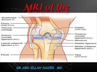
  • 1. MRI OF THE KNEE JOINT. Dr/ ABD ALLAH NAZEER. MD.
  • 2. Clinical Indication Acute pain secondary to trauma in the presence of an effusion without fracture on x-ray. Suspected unstable knee with history of knee locking or positive McMurray test on examination. Suspected stable meniscus tear following treatment with analgesia and physiotherapy and activity modification for at least 4 weeks in the presence of persistent joint effusion or history of locking Suspected cruciate ligament injury with a history of knee giving way and laxity of between 5 and 11mm) on examination grade II-III instability (medial laxity between 5 and 11mm on examination. Suspected multi-ligamentous or lateral collateral ligament injury when there is grade II-III instability Suspected medial collateral ligament injury with grade II-III instability despite treatment with brace and activity modification for at least 6 weeks Other knee conditions of unknown etiology when there are both symptoms and signs that suggest a significant underlying injury and when knee x-ray is non-diagnostic for the etiology of the underlying condition
  • 3. Knee - AP View
  • 6. Protocol of examination  Axial T1 localizer  Sagittal T1, PD, T2  Coronal gradient echo, STIR  If contrast is injected [ Axial, Sagittal ,coronal T1 WIs ]
  • 7. How to know the pulse sequence?! T1 T2 Gradient STIR Upper EgyptLower Egypt
  • 8. Items to be evaluated Where?!    Menisci (medial & lateral)  Ligaments - Cruciate (ACL, PCL) - Collateral - Retinacular  Tendons ( Quadriceps, Patellar)  Bones  Synovial effusion        Sagittal PD Coronal. Sagittal,coronal and axial. Coronal Axial Sagittal PD Sagittal T1& T2 Sagittal T2
  • 9. Anatomy of the meniscus.
  • 10. Meniscus Medial meniscus  Semilunar - shaped Posterior horn wider, longer, taller than anterior horn Posterior horn tightly attached to the capsule
  • 11. Lateral meniscus  C- shape  Posterior and anterior horns are symmetric  Anterior horn may be hypo plastic, extremely thin  Discoid meniscus and meniscal cysts more common
  • 15. Medial and lateral meniscus.
  • 16.
  • 17. Medial and lateral meniscusMedial and lateral meniscus..
  • 18.
  • 19.
  • 20.
  • 23. Proton density coronal image shows the normal medial collateral ligament as a thin, taut, well-defined, low-signal structure extending from the medial femoral epicondyle to the medial tibial metaphysis
  • 24. Coronal and sagittal proton density image demonstrating the normal lateral collateral ligament in its entirety, from the femoral condyle origin to the fibular head insertion.
  • 26. Simple Tear Complex Tear  Type II  Type I  Normal
  • 27. Types of Meniscal degenerationTypes of Meniscal degeneration  Grade IGrade I  Grade IIGrade II  Meniscal frayingMeniscal fraying
  • 28. Tear
  • 29.  Grade 1 degeneration. Grade 11 degeneration
  • 30. Meniscal degeneration with free edge fraying.
  • 31. Meniscal degeneration with free edge fraying
  • 32.
  • 33. Types of Meniscal TearsTypes of Meniscal Tears  SimpleSimple  ComplexComplex  Special typesSpecial types
  • 34. Simple Meniscal TearSimple Meniscal Tear  HorizontalHorizontal  VerticalVertical  RadialRadial
  • 35.
  • 39.
  • 40. Vertical tear Occurs typically in the outer 1/3 of the posterior horn or body of the meniscus ]rare in the anterior horn[
  • 43.
  • 44.
  • 45.
  • 47. Full thickness body radial tear
  • 48. Marching cleft sign in discoid lateral meniscus indicates radial meniscal tear
  • 50. Vertical radial meniscal tear in the mid-body lateral meniscus. (A) A sagittal fat-suppressed proton-density image shows a vertical tear (arrow) of the central edge of the lateral meniscus, oriented perpendicular to the curvature of the meniscus. Note the superimposed horizontal tear of the anterior horn. (B) A coronal fat-suppressed T2-weighted image shows the same tear with truncation of the central meniscal margin (arrow).
  • 51. Radial tear of lateral meniscus.
  • 52.
  • 53. Vertical tear of the free edge of the meniscus [Root tear[ Ghost meniscus
  • 54. Ghost meniscus If there is no history of Meniscal surgery and the posterior horn is absent near the intercondylar notch
  • 57. Special Meniscal TearsSpecial Meniscal Tears  FlapFlap  Bucket handleBucket handle  MC separationMC separation
  • 58. Flap tear:displaced horizontal or longitudinal tears
  • 59. Flap tear [Oblique[ Should have tow components , horizontal and Vertical common in the medial meniscus
  • 62. Small sized posterior horn [ sagittal[ Medially displaced fragment[ coronal[ Double PCL sign ]sagittal[
  • 63.
  • 66. Flipped meniscus : Double Delta Sign Bucket Handel tear , Lateral meniscus
  • 67. Flipped meniscus : Double Delta Sign
  • 68. Flipped meniscus : Double Delta Sign
  • 69. Flipped meniscus : Double Delta Sign
  • 70. Parrot beak tear (displaced radial oblique tear(.
  • 72. Post-traumatic contusion of the lateral femoral and tibial condyles
  • 73.
  • 74. Discoid meniscus Dysplastic meniscus with loss of normal semi lunar shape. 50%or more coverage of the tibial plateau. Meniscal body segment seen in 3 or more sagittal images
  • 77. Meniscal cyst A Cyst extending from a meniscal tear Common sites : Anterior horn LM , Posterior horn MM
  • 80.
  • 81.
  • 85. In closed MRI scanner,In closed MRI scanner, magic angle artifact canmagic angle artifact can be found in one locationbe found in one location:: The up-sloping of theThe up-sloping of the posterior horn lateralposterior horn lateral meniscusmeniscus.. In open, vertical field magnets, magic angle artifact will be found in two locations: 1)1(junction of anterior horn and body of meniscus and 2)2(junction of posterior horn and body of meniscus
  • 86. Normal intrameniscal signal evolution in the site of surgery, with very good final result.
  • 87. Normal healing sequence. Typical worsening in the first scans - intrameniscal signal after surgery is worse then before surgery. No healing disturbance.
  • 88. Abnormal healing of degenerated meniscus.
  • 89. Ligamentous Lesions  ACL  PCL  Collateral  Retinacular
  • 92. MRI shows the normalMRI shows the normal linear low signal intensitylinear low signal intensity ACL adjacent to theACL adjacent to the lateral bony wall of thelateral bony wall of the upper intercondylar notchupper intercondylar notch (arrow). The normal ACL(arrow). The normal ACL moves away from the wallmoves away from the wall and diverges into multipleand diverges into multiple fascicles on more distalfascicles on more distal imagesimages.
  • 93. Anterior cruciate ligament Posterior cruciate ligament
  • 94. Primary signs [ In the ligament ] Total discontinuity Abnormal signal Abnormal configuration Abrupt angulation Wavy appearance Abnormal axis Anterior cruciate ligament injury Intercondylar roof
  • 98. Normal ACL Non visualization of the ACL with a cloud of edema and hemorrhage
  • 99. Secondary signs [ Outside the ligament ] • Blumensaat angle sign. • Bone contusions [Pivot- shift bruises ] • Anterior translocation of the tibia • Uncovered meniscus sign • Avulsion fracture of the tibial insertion • Segond fracture 70-100% with ACL tear • PCL buckling • PCL line sign Anterior cruciate ligament injury Hyperextension ACL tear withHyperextension ACL tear with "kissing bone bruises"kissing bone bruises."."
  • 100. Negative Blumensaat angleNegative Blumensaat angle..
  • 101. Positive Blumensaat anglePositive Blumensaat angle..
  • 102. ACL Graft with negative Blumensaat angleACL Graft with negative Blumensaat angle..
  • 103. The probability of anThe probability of an ACL tear is very highACL tear is very high if both such boneif both such bone bruises are present,bruises are present, only slightly lower ifonly slightly lower if the tibial bone bruisethe tibial bone bruise is present in isolation,is present in isolation, and still slightlyand still slightly lower with anlower with an isolated femoral boneisolated femoral bone bruise of thisbruise of this appearance.appearance.
  • 105. Anterior tibial translocation with” uncovered meniscus sign”
  • 106. Segond fractureSegond fracture.. An elliptical verticallyAn elliptical vertically 3x10mm bone3x10mm bone fragment parallel to thefragment parallel to the lateral tibial cortex,lateral tibial cortex, about 4mm distal toabout 4mm distal to the plateau. Best seenthe plateau. Best seen on AP or tunnelon AP or tunnel radiographic viewsradiographic views 75-100%association with ACL tear
  • 107. Segond fracture inSegond fracture in patient with ACLpatient with ACL tear. T1- weightedtear. T1- weighted coronal MRI shows acoronal MRI shows a small, low-signalsmall, low-signal elongated fractureelongated fracture fragment that isfragment that is parallel to the lateralparallel to the lateral tibia. The associationtibia. The association of Segond fracturesof Segond fractures with ACL tearswith ACL tears approaches 100%.approaches 100%.
  • 110. PCL redundancy as a secondary sign of ACL tear. This is a relativelyPCL redundancy as a secondary sign of ACL tear. This is a relatively unreliable secondary sign of ACL tear.unreliable secondary sign of ACL tear.
  • 111. Partial ACL tearPartial ACL tear  Common about 10-43% of ACL tearsCommon about 10-43% of ACL tears  Suboptimal accuracy of MRISuboptimal accuracy of MRI  Subtle 1ry and 2ry signsSubtle 1ry and 2ry signs  Focal angulationsFocal angulations  Focal increase T2 signalFocal increase T2 signal [[non specificnon specific ]]  Single bundle signSingle bundle sign Sagittal MRI shows an abruptlySagittal MRI shows an abruptly angulated mid-ACL (arrow) .A wavy orangulated mid-ACL (arrow) .A wavy or sharply angulated appearance issharply angulated appearance is abnormal.abnormal.
  • 112. T1-weighted sagittalT1-weighted sagittal MRI shows a normal-MRI shows a normal- appearing ACL.appearing ACL. Partial ACL tear T1-weighted sagittal MRI imageT1-weighted sagittal MRI image immediately adjacent to theimmediately adjacent to the previous image shows a partiallyprevious image shows a partially disrupted ACLdisrupted ACL
  • 114. Partial ACL tear with thickening, angulations and abnormal brightPartial ACL tear with thickening, angulations and abnormal bright signal inside. The tibial and femoral attachment is preservedsignal inside. The tibial and femoral attachment is preserved..
  • 115. Partial ACL tear with thickening and abnormal bright signal insidePartial ACL tear with thickening and abnormal bright signal inside..
  • 116. Partial ACL tear with diffuse thickening and abnormal signal insidePartial ACL tear with diffuse thickening and abnormal signal inside..
  • 117. Chronic ACL TearChronic ACL Tear  Fragmented ACL [ common finding ]Fragmented ACL [ common finding ]  Absent bone edema and contusionsAbsent bone edema and contusions  Empty notch signEmpty notch sign  ACL attached to PCLACL attached to PCL
  • 118. ACL tear on axial image showing non-visualization of the anteriorACL tear on axial image showing non-visualization of the anterior cruciate ligament (ACL) in the upper inter-condylar notchcruciate ligament (ACL) in the upper inter-condylar notch A large knee effusion and a Baker cyst are noted incidentally.A large knee effusion and a Baker cyst are noted incidentally. Empty notch sign
  • 119. Chronic ACL tear, empty notch sign. T1-weighted coronal MRI shows fat in theChronic ACL tear, empty notch sign. T1-weighted coronal MRI shows fat in the lateral intercondylar notch, ACL is absent. This is a frequent MRI appearance of alateral intercondylar notch, ACL is absent. This is a frequent MRI appearance of a chronic ACL tear after resolution of acute edema and hemorrhage.chronic ACL tear after resolution of acute edema and hemorrhage.
  • 120. ACL DegenerationACL Degeneration  Intra ligamentous cystIntra ligamentous cyst  May be mistaken for a tearMay be mistaken for a tear  Arthroscopic decompressionArthroscopic decompression
  • 121. Intercondylar notch cystIntercondylar notch cyst  1% of knee MRIs1% of knee MRIs  Usually an incidental findingUsually an incidental finding  Painful if erodes the bonePainful if erodes the bone  Post-traumatic chronic partialPost-traumatic chronic partial cruciate ligament tear with internalcruciate ligament tear with internal degenerationdegeneration  More common in the ACLMore common in the ACL  Oval , rounded may be multilocularOval , rounded may be multilocular  Rim enhancement if inflamedRim enhancement if inflamed  Arthroscopic drainageArthroscopic drainage Cruciate ligament cyst
  • 123. A 3-D cutaway at the intercondylar notch in the sagittal plane reveals normal positioning for a patellar tendon ACL autograft. The tibial tunnel should lie posterior to the line drawn parallel to the intercondylar notch (red) and the femoral attachment should lie posterior to a line drawn parallel to the cortex of the distal femoral diaphysis (blue(.
  • 124. Femoral tunnel is normally positioned at the junction of the physeal scar and posterior intercondylar roof (asterisk(. Abnormal placement of the femoral tunnel (asterisk), which lies significantly anterior to a line drawn along the posterior cortex of the femoral diaphysis with graft degeneration.
  • 125. Tibial tunnel cyst after ACL graft reconstruction.
  • 126. Cyclops lesion (arrowheads) attached to the ACL (arrow) with a head-like appearance, showing a focal area of discoloration resembling an eye (curved arrow(
  • 127. Hypointense to isointense nodule (arrowhead) attached to the anterior surface of the anterior cruciate ligament (ACL) graft (arrow). Sagittal T1W MRI image (C) shows a hypointense nodule (arrowhead) in the anterior intercondylar notch, related to Cyclops.
  • 128. ACL reconstruction are provided. Diffuse abnormal fluid signal intensity is seen along the course of the graft (arrows) on the sagittal image, compatible with extensive graft ganglion formation.
  • 129. ACL Surgery Failure with stem cells injection.
  • 130. Posterior cruciate ligament.Posterior cruciate ligament.  The major stabilizer of the kneeThe major stabilizer of the knee  Uniform low signal , no striationsUniform low signal , no striations  Twice strong as the ACLTwice strong as the ACL  The menisco-femoral ligaments are intimately related to PCL.The menisco-femoral ligaments are intimately related to PCL. They connect the posterior horn of the lateral meniscus to theThey connect the posterior horn of the lateral meniscus to the medial femoral condylemedial femoral condyle Ligament of Humphrey anterior to PCLLigament of Humphrey anterior to PCL Ligament of Weisberg posterior to PCLLigament of Weisberg posterior to PCL
  • 131. Proton-dense sagittal image demonstrates the normal tibial insertion of the PCL. The insertion site is a vertically inclined posterior to the articular surface.
  • 132.  PCL injuries represent about 12% of knee injuriesPCL injuries represent about 12% of knee injuries  Combined PCL injuries represent 97%Combined PCL injuries represent 97% With ACL 65%With ACL 65% With MCL 50%With MCL 50% With MM 30%With MM 30% Posterior cruciate ligamentPosterior cruciate ligament TYPES OF PCL INJURES Complete tear 40% Partial tear 55% Avulsion tear 7%
  • 133. NORMAL PCL TORN PCL MR FINDINGS Increased signal due to hemorrhage and edema Diffuse enlargement of PCL
  • 135. An enlarged, intermediate signal (obviously torn) PCL.
  • 136. NORMAL PCL AVULSION TEAR • Involves the tibial insertion • Retracted bone fragment • Bone marrow edema at avulsion site • The actual PCL may be normal
  • 140. PD sagittal image shows partial tear of the mid-substance of the PCL. The normal ligament of Humphrey (small arrow) is visualized better because it is adjacent to the high signal intensity edema of the torn PCL. PARTIAL PCL TEAR
  • 141. Collateral ligaments. MCL is about 8-11 cm LCL is about 5-7 cm Isolated injuries are rare, usually with ACL and MM
  • 142. Collateral ligaments Grade I : microscopic tear Grade II :partial tear Grade III : complete tear GRADING SYSTEM Grade I,II and isolated grade III are treated conservatively, while grade III tears associated with ACL tears are treated by repairing ACL only
  • 143. Proton density coronal image shows the normal medial collateral ligament as a thin, taut, well-defined, low-signal structure extending from the medial femoral epicondyle to the medial tibial metaphysis
  • 144. Coronal and sagittal proton density image demonstrating the normal lateral collateral ligament in its entirety, from the femoral condyle origin to the fibular head insertion.
  • 145. Grade I medial collateral ligament tear with surrounding edema (straight arrows) on a T2WI. Note the normal thickness and signal of the medial collateral ligament and continued close apposition to the femoral and tibial cortices.
  • 146. Grade 1 sprain of the medial collateral ligamentGrade 1 sprain of the medial collateral ligament..
  • 147. Grade 11 sprain of the medial collateral ligamentGrade 11 sprain of the medial collateral ligament..
  • 148. Grade II medial collateral ligament tear seen on a coronal proton density image shows slight thickening of the medial collateral ligament and separation from the underlying cortices. Bone marrow edema of the lateral tibial plateau is seen due to valgus stress 7months after conservative treatment
  • 149. Grade II medial collateral ligament tear seen on a coronal T1 and STIR images showing slight thickening of the medial collateral ligament and separation from the underlying cortices.
  • 150. Grade 111 tear of the MCLGrade 111 tear of the MCL..
  • 151. Grade III medial collateral ligament tear on a coronal fast spin-echo T2- weighted image demonstrates a disrupted ligament that is thickened and retracted with surrounding edema (black arrow).
  • 152. Acute grade III tear with a folded ligament (arrow) and surrounding edema on a coronal proton density image.
  • 153. Acute tear of the proximal portion of the lateral collateral ligament is seen on this coronal proton density image (white arrow). Note the associated grade II medial collateral ligament tear.
  • 154. Grade III MCL tear with retraction
  • 155. Grade III MCL tear with abnormal signal and edema
  • 156. Grade 111 tear of the LCLGrade 111 tear of the LCL..
  • 157. Ilio-tibial band syndrome. Distal tendon of IT fascia and insert at gerdy,sIlio-tibial band syndrome. Distal tendon of IT fascia and insert at gerdy,s tubercle of the tibia. It occur in runner, cyclists, football players and weight liftertubercle of the tibia. It occur in runner, cyclists, football players and weight lifter..
  • 158. Pre-patellar bursitis.(housemaid bursitisPre-patellar bursitis.(housemaid bursitis((
  • 159.
  • 160. Backer and Pes anserine cystBacker and Pes anserine cyst..
  • 162.
  • 163.
  • 165. Complete tear of the patellar tendon with ACL teaComplete tear of the patellar tendon with ACL tearr
  • 166. Complete tear of the patellar tendon with ACL tear
  • 167. Partial tear of the patellar tendon
  • 168. Lateral pressure syndrome  Thickening of the lateral retinaculum  Lateral knee pain  Obese, athletic patients  May be associated with chondromalacia
  • 169. Patella alta  Sequlae of patellofemoral dysplasia  Lengthening of the infrapatellar tendon  May be associated with chondromalacia  Length of patellar tendon/ length of patella > 1.3
  • 170. Patella Baja  Poliomyelitis  Achondroplasia  JRA
  • 176. Chondromalacia patellae Degeneration of the hyaline cartilage Anterior knee pain in young adults Four stages Signal abnormalities Ulceration [ fraying , partial or full thickness defects ] Reactive bone changes [ edema , cyst formation , sclerosis ] Osteoartheritic changes
  • 180. Loose bodies • Read with plain films • Low signal fragments Synovial osteochondromatosis.
  • 182. Synovial osteochondromatosisSynovial osteochondromatosis Metaplasia of subsynovial soft tissues cartilage formation Affects any joint [ knee , hip , elbow[ Age incidence 40 years M : F = 2 : 1 FINDINGSFINDINGS Widening of the joint space Bone erosions Intra articular loose bodies Secondary osteoarthritic changes
  • 185. Lipoma arborescenceLipoma arborescence RareRare IdiopathicIdiopathic Fatty synovial infiltrations formingFatty synovial infiltrations forming variable sized villous projectionsvariable sized villous projections withinwithin the joint capsule commonly in thethe joint capsule commonly in the supra- patellar pouchsupra- patellar pouch Associated with joint effusionAssociated with joint effusion Painless swellingPainless swelling Treatment by synovectomyTreatment by synovectomy
  • 187. Pigmented villo-nodular synovitisPigmented villo-nodular synovitis  IdiopathicIdiopathic  Monoarticular disease 1% incidenceMonoarticular disease 1% incidence  Hypertrophic synovial masses with hemosiderinHypertrophic synovial masses with hemosiderin laden macrophages bone erosionsladen macrophages bone erosions  Intermediate signal in T1 andIntermediate signal in T1 and low signal in T2low signal in T2 withwith enhancement after contrast injectionenhancement after contrast injection  Typical location posterior to HoffaTypical location posterior to Hoffa’’s fat pads fat pad  Painless swelling , pain with progressive diseasePainless swelling , pain with progressive disease  Treatment by synovectomyTreatment by synovectomy
  • 190. PIGMENTED VILLONODULAR SYNOVITIS VERSUS LIPOMA ARBORESCENS
  • 192. POPLITEAL CYST Fluid in the bursa which is usually communicating with the joint space Other names Baker’s cyst Gastrocnemius/semimembranosus bursa
  • 193. Medial plica syndromeMedial plica syndrome  Inflamed synovial plica causing pain , crepitusInflamed synovial plica causing pain , crepitus and pseudolockingand pseudolocking  Often in adolescents and athleticsOften in adolescents and athletics  No measurement for plica thicknessNo measurement for plica thickness Four types of plicaFour types of plica Suprapatellar 90%Suprapatellar 90% Medial 15 -30%Medial 15 -30% InfrapatellarInfrapatellar Lateral [ rare]Lateral [ rare]
  • 196. Osteochondritis dissecansOsteochondritis dissecans  Osteochondral fragmentOsteochondral fragment in a typical locationin a typical location  Young maleYoung male  Lateral aspect of the medial femoral condoyleLateral aspect of the medial femoral condoyle  Variable sized fragment attached or detachedVariable sized fragment attached or detached  Criteria of unstable fragmentCriteria of unstable fragment Large size more than 1cmLarge size more than 1cm Fluid between the fragment and donor boneFluid between the fragment and donor bone Cystic changes at the donor siteCystic changes at the donor site Enhancement of the separation lineEnhancement of the separation line
  • 198. Osteochondritis Dissecans along the medial femoral condyleOsteochondritis Dissecans along the medial femoral condyle
  • 200. Red marrow recon version / marrowRed marrow recon version / marrow lesionlesion
  • 203. Bone infarctsBone infarcts  Serpigenous lesions in the bone marrowSerpigenous lesions in the bone marrow  Variable in size [ Chinese figures ]Variable in size [ Chinese figures ]  Double line sign is diagnostic [peripheralDouble line sign is diagnostic [peripheral hyperintense with hypointense inner border on T2hyperintense with hypointense inner border on T2  CAUSESCAUSES POSTTRAUMATICPOSTTRAUMATIC STEROIDSSTEROIDS COLLAGEN DISEASESCOLLAGEN DISEASES ALCOHOLISMALCOHOLISM PANCREATITISPANCREATITIS SPONTANEOUSSPONTANEOUS