![[object Object],[object Object],[object Object],[object Object]](data:image/gif;base64,R0lGODlhAQABAIAAAAAAAP///yH5BAEAAAAALAAAAAABAAEAAAIBRAA7)
Recomendados
Más contenido relacionado
La actualidad más candente
La actualidad más candente (20)
Destacado
Similar a Myocardial infarction
Similar a Myocardial infarction (20)
Más de adolescent4u
Último
Último (20)
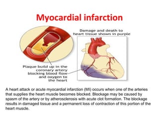
Myocardial infarction
- 1. Myocardial infarction A heart attack or acute myocardial infarction (MI) occurs when one of the arteries that supplies the heart muscle becomes blocked. Blockage may be caused by spasm of the artery or by atherosclerosis with acute clot formation. The blockage results in damaged tissue and a permanent loss of contraction of this portion of the heart muscle.
- 8. These serial sections of a coronary artery demonstrate grossly the appearance of lumenal narrowing with atherosclerosis.
- 10. myocardial infarction (2) of the tip of the anterior wall of the heart (an apical infarct ) after occlusion (1) of a branch of the left coronary artery(LCA, right coronar
- 11. Ohr 2hr 24hr
- 12. This is an acute myocardial infarction in the septum. After several days, there is a yellowish center with necrosis and inflammation surrounded by a hyperemic border.
- 13. This is an acute myocardial infarction of the anterior left ventricular free wall and septum in cross section. Note that the infarction is nearly transmural. There is a yellowish center with necrosis and inflammation surrounded by a hyperemic borde
- 14. When the infarction is 3 to 5 days old, the necrosis and inflammation are most extensive, and the myocardium is the softest, so that transmural infarctions may be complicated by rupture. A papillary muscle may rupture as well to produce sudden valvular insufficiency. Rupture through the septum results in a left-to-right shunt and right heart failure.
- 15. Remote myocardial infarction (weeks to years)
- 16. Gross morphologic changes evolve over time as follows : Time from Onset Gross Morphologic Finding 18 - 24 Hours Pallor of myocardium 24 - 72 Hours Pallor with some hyperemia 3 - 7 Days Hyperemic border with central yellowing 10 - 21 Days Maximally yellow and soft with vascular margins 7 weeks White fibrosis
- 17. This is normal myocardium. There are cross striations and central nuclei. Pale pink intercalated disks are also present.
- 20. This is an early acute myocardial infarction. (<iday) Note the prominent pink contraction bands.
- 21. 1-2 daysThis is an early acute myocardial infarction. There is increasing loss of cross striations, and some contraction bands are also seen, and the nuclei are undergoing karyolysis. Some neutrophils are beginning to infiltrate the myocardium.
- 22. 1-2days This is an acute myocardial infarction. There is loss of cross striations, and the nuclei are not present. There is extensive hemorrhage here at the border of the infarction, which accounts for the grossly apparent hyperemic border.
- 23. 3-4 days This is an acute myocardial infarction of several days' duration. There is a more extensive neutrophilic infiltrate along with the prominent necrosis and hemorrhage.
- 25. 2-3 wks Toward the end of the first week, healing of the infarction becomes more prominent, with capillaries, fibroblasts, and macrophages filled with hemosiderin. The granulation tissue seen here is most prominent from 2 to 3 weeks following onset of infarction.
- 26. weeks –years After a couple of weeks, healing is well under way, and there is more extensive collagen deposition.
- 27. wks –yrs The remote myocardial infarction is evidenced by a collagenous scar seen here in a subendocardial location.
- 28. Microscopic morphologic changes evolve over time as follows: Time from Onset Microscopic Morphologic Finding 1 - 3 Hours Wavy myocardial fibers 2 - 3 Hours Staining defect with tetrazolium or basic fuchsin dye 4 - 12 Hours Coagulation necrosis with loss of cross striations, contraction bands, edema, hemorrhage, and early neutrophilic infiltrate 18 - 24 Hours Continuing coagulation necrosis, pyknosis of nuclei, and marginal contraction bands 24 - 72 Hours Total loss of nuclei and striations along with heavy neutrophilic infiltrate 3 - 7 Days Macrophage and mononuclear infiltration begin, fibrovascular response begins 10 - 21 Days Fibrovascular response with prominent granulation tissue 7 Weeks Fibrosis
- 29. Symptoms of a possible heart attack include chest pain and pain that radiates down the shoulder and arm. Some people (the elderly, people with diabetes, and women) may have little or no chest pain. Or, they may experience unusual symptoms (shortness of breath, fatigue, weakness). Women are more likely than men to have symptoms of nausea, vomiting, back or jaw pain, and shortness of breath with chest pain.
- 32. Screening and Diagnosis Stress Test measures blood supply to heart Coronary Angiography specific shows coronaries Narrowing in Sites of Electro- cardiogram measures electrical impulses
- 40. Myocardial Rupture Myocardial aneurysm with thrombosis inside.
- 41. Rupture (at the arrow) into the pericardial sac can produce a life-threatening cardiac tamponade, as seen here. The septum may also rupture.
- 42. Rupture of papillary muscle…..mitral incompetence.
- 43. Left ventricular aneurysm containing mural thrombus A complication of infarction is aneurysm formation, which is the bulge seen here in the left ventricular wall. Note the very thin white wall of the aneurysm toward the apex.
- 44. The myocytes here are hypertrophied, marked by the large, dark nuclei, and there is interstitial fibrosis. This is an example of cardiomyopathy. In this case, long-standing, severe occlusive atherosclerosis led to "ischemic" cardiomyopathy.
- 47. R/ Revascularization procedures