SlideShare una empresa de Scribd logo
1 de 23
Aneurysms of Aorta
•   Defined as abnormal localized dilatation of a blood vessel or wall of the heart.
•   True Aneurysms: Bound by arterial wall or heart.
•   False Aneurysms: Breach in wall of vessel, extravascular hematoma,
    connection with lumen is retained, pulsating hematoma e.g. post myocardial
    infarction rupture.
•   Aneurysms are most important clinically when involve aorta.
•   Two most important causes of aortic aneurysms are atherosclerosis and cystic
    medial degeneration. However any artery may be involved by this condition if
    wall gets weakened due to: trauma, congenital defects (Berry An), infections
    e.g. Mycotic, Syphilis, Systemic diseases like vasculitis.
•   Mycotic aneurysms: Due to septic embolization and weakening e.g. infective
    endocarditis, due to extension from adjacent process and by circulating
    organisms infecting the wall directly.
•   Macroscopic types: Saccular, are spherical involving a portion of wall, 5-20
    cm, partially or completely filled with thrombus. Fusiform, may involve up to
    20 cm of the aortic wall.
Abdominal Aortic Aneurysms
• Atherosclerosis in men above 50 years of age the most
  frequent cause which destroys the underlying tunica media
  thereby weakening the wall.
• Abdominal aorta is most frequently affected, other parts
  including arch of aorta, common iliac arteries and thoracic
  aorta are also affected with less frequency.
• Part of abdominal aorta most commonly involved is below
  the origin of renal arteries.
• Aneurysm is usually fusiform measuring 15 x 25 cm.
• Mural thrombosis is frequent which may be complete and
  may involve the lumen partially. Aortic aneurysms may
  also be accompanied by similar lesions in iliac arteries.
• Occasionally the origins of renal and superior mesenteric
  artery may also be affected directly or by thrombosis. The
  atheromatous lesions in the aneurysms are ulcerated with
  mural thrombi. Atheroemboli can lodge in the vessels of
  kidneys or lower extremities.
• Variants: two variants. 1- Inflammatory: characterized by
  dense periaortic fibrosis containing an abundant
  lymphoplasmacytic infiltration, many giant cells and
  macrophages. Cause is uncertain.
• 2- Mycotic abdominal aneurysm: Aortic atherosclerotic
  aneurysms infected by deposition of circulating organisms
  in the wall, particularly salmonella gastroenteritis. This
  leads to suppuration and further destruction of media.
Pathogenesis of Aneurysms.
• Atherosclerosis along with other factors in men above 50
  years of age.
• Have been showing to be familial, in addition to the
  familial factors responsible for atherosclerosis and
  hypertension..
• Genetic defects e.g. Marfans syndrome in structural
  components of aorta can also cause aneurysms and
  dissections.
• There is evidence that matrix metalloproteins and
  plasminogen activators, which degrade extracellular matrix
  contribute to aneurysm formation.
Clinical Features
•   Depend on location and size.
•   Rupture into body cavities with massive hemorrhage.
•   Occlusion of a branch vessel.
•   Embolism from thrombosis or atheroma.
•   Compression of surrounding structures, e.g. ureter.
•   Presentation as abdominal mass.
Syphilitic Aneurysms
• Tertiary syphilis is marked by cardiovascular and nervous
  system complications.
• Endarteritis obliterans involves small muscular arteries and
  arterioles; most devastating when it involves the vasa
  vasorum of aorta which can lead to aneurysm of thoracic
  aorta along with dilatation of aortic annulus.
Morphologic Features.
• Medial destruction is the key process in tertiary syphilis.
• Inflammatory process starts in the aortic adventitia, esp.
  vasa vasorum, surrounded by lymphoplasmacytic
  infiltration.
• There is ischemic injury to aortic media, patchy loss of
  elastic tissue and smooth muscle cells followed by
  inflammation and later on scarring.
• Aorta loses its elastic support, becomes dilated.
• Luetic involvement of aorta favors superimposed
  atherosclerosis inducing further florid atheromatosis.
• Location in thoracic aorta an differentiate the luetic
  aneurysms from those purely due to atherosclerosis.
• Luetic aortic aneurysms also cause, dilatation of aortic
  valve ring, producing aortic valve insufficiency, valves get
  damaged as well.
• Left ventricle becomes hypertrophied with consequent
  marked cardiomegaly (cor bovinum).
Clinical Features.
• Both types of aneurysms produce signs and symptoms due
  to:
• Encroachment on mediastinal structures.
• Respiratory difficulties.
• Swallowing difficulties.
• Persistent cough- recurrent laryngeal nerve.
• Pain following erosion of ribs and vertebral bodies.
• Cardiac disease.
• Rupture of aneurysm.
Aortic Dissections
• Aortic dissection is catastrophic disease, characterized by dissection of
  blood in between and along the laminar planes of media forming blood
  filled channel. Usually dilatation of aorta is minimal.
• Two groups of patients are affected e.g. men between 40-60 years in
  whom chronic hypertension, second group consists of younger persons
  having systemic or localized abnormality of connective tissue of aorta
  e.g. Marfans syndrome.
• Dissection may occur as complication of arterial catheterization.
• Arterial dissections may occur in aorta or coronaries during
  pregnancy, reason is unknown.
• Dissection is uncommon in advanced atherosclerosis due to scarring in
  the tunica media of aorta.
Morphologic Features.
• Starts mostly with intimal tear that occurs within 10 cm of
  aortic valve.
• Tears are usually 1-5 cm in length.
• Dissection may extend proximally or distally, may involve
  the aorta up to the iliacs and femoral artery.
• Spreads along with laminar planes, between outer and
  middle one third of the wall of aorta.
• The channel may again rupture in to the aorta through a
  secondary tear (double barrel aorta), this avoids fatal extra
  aortic rupture and fatal hemorrhage.
• Most common cause of death is rupture into one of the
  body cavities.
• Clinical manifestations may also be due to extension of the
  dissection to the origins of aortic branches, like coronary,
  renal, mesenteric or iliac arteries causing obstructions to
  vascular flow.
• Histologically the most frequent and detectable lesion is
  cystic medial degeneration. The lesions consist of elastic
  tissue fragmentation and separation of elastic and
  fibromuscular elements of tunica media that creates cleft
  like or cystic spaces filled with amorphous extracellular
  matrix. Inflammation is absent.
• Cystic medial degeneration is frequently found in patients
  with Marfans syndrome.
• Cystic medial degeneration is frequently found at autopsy
  in persons who had experienced no symptoms of
  dissection.
Pathogenesis
• Hypertension is the major risk factor, but contribution to
  CMD is uncertain, no good correlation between CMD and
  dissection.
• Some dissections are due to inherited connective tissue
  defects e.g. Marfans autosomal dominant disorder.
• Complications of Marfans syndrome include; aortic
  dissection, aortic and mitral valve prolapse; other
  manifestations include; skeletal and occular.
• The triggering factors for intimal tear and intramural
  hemorrhage is unknown. But once the process has started,
  hypertension plays an important role in the progression.
Clinical Features.
• Depend on level of aorta affected.
• Sudden onset of excruciating pain, beginning in the front
  of chest, radiating to back, can be readily confused
  clinically with that of acute myocardial infarction.
• The diagnosis may be based on various investigations,
  including angiography, two dimensional cardiac
  ultrasound, CT scan and MRI.
• Prognosis has recently improved due to development of
  surgical techniques.
Varicose Veins
• Varicose veins have narrowed or abnormally dilated
  lumina along with incompetence of valves leading to
  venous stasis. These are tortuous, dilated, elongated and
  scarred.
• Loss of support of vessel walls and increased intramural
  pressure.
• Leg veins are affected due to posture, so occupations are
  important in its aetiology.
• Prolonged erect posture also results in simple orthostatic
  oedema.
• Condition is more common over 50 years, obese people
  and during pregnancy.
Morphologic Features.
• There is marked variation in the thickness of the wall.
  There may be thrombosis and valvular deformities.
• Microscopically changes consist of variations in thickness
  of the wall caused by dilatations and compensatory
  hypertrophy of smooth muscle and subintimal fibrosis.
Clinical Features
• Venous stasis, congestion, oedema and thrombosis.
• Embolism is rare in contrast to deep vein thrombosis.
• Distension is painful.
• Persistent varicose veins produce oedema of legs along
  with trophic changes leading to stasis dermatitis and
  ulcerations.
• Varicose veins are also seen in lower esophagus, in
  patients of cirrhosis, due to portal hypertension.
Aortic Dissection With Double Lumen
Aortic Dissection


 aortic dissecting aneurysm from intimal tear, blood clot present in the adventitial layer
 may accumulate in the thorax, causing hemothorax.
Intimal Tear in Aortic Dissection
Aortic Dissection
Aortic Dissection


 Aortic aneurysm dissecting into the muscular layer. The blood dissecting proximally may
 block the carotids and distally may shut the coronaries.

Más contenido relacionado

La actualidad más candente

La actualidad más candente (20)

Cardiomyopathies
CardiomyopathiesCardiomyopathies
Cardiomyopathies
 
Tricuspid valve
Tricuspid valveTricuspid valve
Tricuspid valve
 
Mitral stenosis
Mitral stenosisMitral stenosis
Mitral stenosis
 
Aortic aneurysm final
Aortic aneurysm finalAortic aneurysm final
Aortic aneurysm final
 
Truncus Arteriosus
Truncus Arteriosus Truncus Arteriosus
Truncus Arteriosus
 
Aneurysms
AneurysmsAneurysms
Aneurysms
 
Percutaneous Coronary Intervention
Percutaneous Coronary InterventionPercutaneous Coronary Intervention
Percutaneous Coronary Intervention
 
Atherosclerosis
AtherosclerosisAtherosclerosis
Atherosclerosis
 
Pericarditis. Pericardial Disease
Pericarditis. Pericardial DiseasePericarditis. Pericardial Disease
Pericarditis. Pericardial Disease
 
Aortic aneurys mppt
Aortic aneurys mpptAortic aneurys mppt
Aortic aneurys mppt
 
Aortic stenosis
Aortic stenosisAortic stenosis
Aortic stenosis
 
Congenital heart diseases in adults
Congenital heart diseases in adults Congenital heart diseases in adults
Congenital heart diseases in adults
 
CORONARY ARTERY DISEASE
CORONARY ARTERY DISEASECORONARY ARTERY DISEASE
CORONARY ARTERY DISEASE
 
Atherosclerosis
AtherosclerosisAtherosclerosis
Atherosclerosis
 
Tricuspid valve disease
Tricuspid valve diseaseTricuspid valve disease
Tricuspid valve disease
 
Tricuspid stenosis
Tricuspid stenosisTricuspid stenosis
Tricuspid stenosis
 
Valvular heart disease
Valvular heart diseaseValvular heart disease
Valvular heart disease
 
Mitral valve
Mitral valveMitral valve
Mitral valve
 
dialated cardiomyopathies
dialated cardiomyopathiesdialated cardiomyopathies
dialated cardiomyopathies
 
Mitral valve disease
Mitral valve diseaseMitral valve disease
Mitral valve disease
 

Destacado (20)

Aneurysm by dr.majid nawaz
Aneurysm by  dr.majid nawazAneurysm by  dr.majid nawaz
Aneurysm by dr.majid nawaz
 
Brain aneurysm
Brain aneurysmBrain aneurysm
Brain aneurysm
 
Aneurysm
AneurysmAneurysm
Aneurysm
 
Cerebral aneurysm
Cerebral aneurysmCerebral aneurysm
Cerebral aneurysm
 
Cerebral aneurysm
Cerebral aneurysmCerebral aneurysm
Cerebral aneurysm
 
Anuerysm
AnuerysmAnuerysm
Anuerysm
 
Aortic aneurysm
Aortic aneurysmAortic aneurysm
Aortic aneurysm
 
Treatment for varicose vein disease
Treatment for varicose vein diseaseTreatment for varicose vein disease
Treatment for varicose vein disease
 
Pathway aneurisma
Pathway aneurisma Pathway aneurisma
Pathway aneurisma
 
Cerebral aneurysms
Cerebral aneurysmsCerebral aneurysms
Cerebral aneurysms
 
Lectures on demo
Lectures on demoLectures on demo
Lectures on demo
 
Green house effect
Green house effectGreen house effect
Green house effect
 
Dissociative disorders case histories prof. fareed minhas
Dissociative disorders case histories prof. fareed minhasDissociative disorders case histories prof. fareed minhas
Dissociative disorders case histories prof. fareed minhas
 
Child psychiatry prof. fareed minhas
Child psychiatry prof. fareed minhasChild psychiatry prof. fareed minhas
Child psychiatry prof. fareed minhas
 
Right bundle branch block
Right bundle branch blockRight bundle branch block
Right bundle branch block
 
Infective endocarditis-1
Infective endocarditis-1Infective endocarditis-1
Infective endocarditis-1
 
Dementia prof. fareed minhas
Dementia prof. fareed minhasDementia prof. fareed minhas
Dementia prof. fareed minhas
 
Research hmis
Research hmisResearch hmis
Research hmis
 
Tb
TbTb
Tb
 
Management of dissociate disorders prof. fareed minhas
Management of dissociate disorders prof. fareed minhasManagement of dissociate disorders prof. fareed minhas
Management of dissociate disorders prof. fareed minhas
 

Similar a Aneurysms and dissections

Aneurysms arterial swelling abnormality.pptx
Aneurysms arterial swelling abnormality.pptxAneurysms arterial swelling abnormality.pptx
Aneurysms arterial swelling abnormality.pptxAkilanN5
 
pathology of Atherosclerosis, dissection and aneurysm copy
pathology of Atherosclerosis, dissection and aneurysm   copypathology of Atherosclerosis, dissection and aneurysm   copy
pathology of Atherosclerosis, dissection and aneurysm copyranaelsaeedAboelfeto
 
Ischaemic Vascular Disease.ppt
Ischaemic Vascular Disease.pptIschaemic Vascular Disease.ppt
Ischaemic Vascular Disease.pptabimikufavour73
 
ANEURYSMS.pptx
ANEURYSMS.pptxANEURYSMS.pptx
ANEURYSMS.pptxSAMOEINESH
 
Embolism & Hemorrhage.pptx
Embolism & Hemorrhage.pptxEmbolism & Hemorrhage.pptx
Embolism & Hemorrhage.pptxMKashif39
 
17971_27.2_Aneurysm.ppt
17971_27.2_Aneurysm.ppt17971_27.2_Aneurysm.ppt
17971_27.2_Aneurysm.pptanjukowsika
 
The blood vessels diseases
The blood vessels diseasesThe blood vessels diseases
The blood vessels diseasesAlly Rashid
 
Aneurysm
AneurysmAneurysm
AneurysmPriya
 
DISORDERS OF ARTERIES.pptx
DISORDERS OF ARTERIES.pptxDISORDERS OF ARTERIES.pptx
DISORDERS OF ARTERIES.pptxSURAJADHIKARI40
 
Extra cranial aneurysms
Extra cranial aneurysmsExtra cranial aneurysms
Extra cranial aneurysmsRonald Mbiine
 
Vascular imaging ppt
Vascular imaging pptVascular imaging ppt
Vascular imaging pptPraveen Kumar
 
Diseases of aorta
Diseases of aortaDiseases of aorta
Diseases of aortaavatar73
 

Similar a Aneurysms and dissections (20)

Diseases of blood vessels
Diseases of blood vesselsDiseases of blood vessels
Diseases of blood vessels
 
Aneurysms arterial swelling abnormality.pptx
Aneurysms arterial swelling abnormality.pptxAneurysms arterial swelling abnormality.pptx
Aneurysms arterial swelling abnormality.pptx
 
pathology of Atherosclerosis, dissection and aneurysm copy
pathology of Atherosclerosis, dissection and aneurysm   copypathology of Atherosclerosis, dissection and aneurysm   copy
pathology of Atherosclerosis, dissection and aneurysm copy
 
ANEURYSM AND VASCULITIS.pptx
ANEURYSM AND VASCULITIS.pptxANEURYSM AND VASCULITIS.pptx
ANEURYSM AND VASCULITIS.pptx
 
PATHOLOGY OF Aneurysms
PATHOLOGY OF AneurysmsPATHOLOGY OF Aneurysms
PATHOLOGY OF Aneurysms
 
Ischaemic Vascular Disease.ppt
Ischaemic Vascular Disease.pptIschaemic Vascular Disease.ppt
Ischaemic Vascular Disease.ppt
 
ANEURYSMS.pptx
ANEURYSMS.pptxANEURYSMS.pptx
ANEURYSMS.pptx
 
Embolism & Hemorrhage.pptx
Embolism & Hemorrhage.pptxEmbolism & Hemorrhage.pptx
Embolism & Hemorrhage.pptx
 
Blood vessels and cvs
Blood vessels and cvsBlood vessels and cvs
Blood vessels and cvs
 
17971_27.2_Aneurysm.ppt
17971_27.2_Aneurysm.ppt17971_27.2_Aneurysm.ppt
17971_27.2_Aneurysm.ppt
 
VARICOSE VEINS and artery.pptx
VARICOSE VEINS and artery.pptxVARICOSE VEINS and artery.pptx
VARICOSE VEINS and artery.pptx
 
Diseases of the aorta
Diseases of the aortaDiseases of the aorta
Diseases of the aorta
 
The blood vessels diseases
The blood vessels diseasesThe blood vessels diseases
The blood vessels diseases
 
Ischemic bowel disease
Ischemic bowel diseaseIschemic bowel disease
Ischemic bowel disease
 
Ischemic bowel disease
Ischemic bowel diseaseIschemic bowel disease
Ischemic bowel disease
 
Aneurysm
AneurysmAneurysm
Aneurysm
 
DISORDERS OF ARTERIES.pptx
DISORDERS OF ARTERIES.pptxDISORDERS OF ARTERIES.pptx
DISORDERS OF ARTERIES.pptx
 
Extra cranial aneurysms
Extra cranial aneurysmsExtra cranial aneurysms
Extra cranial aneurysms
 
Vascular imaging ppt
Vascular imaging pptVascular imaging ppt
Vascular imaging ppt
 
Diseases of aorta
Diseases of aortaDiseases of aorta
Diseases of aorta
 

Más de Rawalpindi Medical College (20)

Pertussis
PertussisPertussis
Pertussis
 
Nephrotic syndrome.
Nephrotic syndrome.Nephrotic syndrome.
Nephrotic syndrome.
 
Symptomtology of cardiovascular diseases
Symptomtology of cardiovascular diseasesSymptomtology of cardiovascular diseases
Symptomtology of cardiovascular diseases
 
Symptomatology-GIT-1
Symptomatology-GIT-1Symptomatology-GIT-1
Symptomatology-GIT-1
 
Symptomatology-GIT
Symptomatology-GITSymptomatology-GIT
Symptomatology-GIT
 
Symptomalogy in RENAL impairement
Symptomalogy in RENAL impairementSymptomalogy in RENAL impairement
Symptomalogy in RENAL impairement
 
History taking
History takingHistory taking
History taking
 
Right and left ventricular hypertrophy
Right and left ventricular hypertrophyRight and left ventricular hypertrophy
Right and left ventricular hypertrophy
 
Rheumatoid arthritis 2
Rheumatoid arthritis 2Rheumatoid arthritis 2
Rheumatoid arthritis 2
 
Systemic lupus erythematosus
Systemic lupus erythematosusSystemic lupus erythematosus
Systemic lupus erythematosus
 
Supraventricular tachyarrythmias
Supraventricular tachyarrythmiasSupraventricular tachyarrythmias
Supraventricular tachyarrythmias
 
Supraventricular tacchycardias
Supraventricular tacchycardias Supraventricular tacchycardias
Supraventricular tacchycardias
 
Skin-
Skin-Skin-
Skin-
 
Skin
Skin  Skin
Skin
 
Sick sinus syndrome-2
Sick sinus syndrome-2Sick sinus syndrome-2
Sick sinus syndrome-2
 
Sick sinus syndrome
Sick sinus syndrome Sick sinus syndrome
Sick sinus syndrome
 
X rays
X raysX rays
X rays
 
Ventricular arrhythmias
Ventricular arrhythmias Ventricular arrhythmias
Ventricular arrhythmias
 
Ventricular tachyarrhythmias
Ventricular tachyarrhythmias Ventricular tachyarrhythmias
Ventricular tachyarrhythmias
 
antiplatelet effect of aspirin
antiplatelet effect of aspirinantiplatelet effect of aspirin
antiplatelet effect of aspirin
 

Aneurysms and dissections

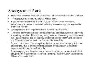
  • 1. Aneurysms of Aorta • Defined as abnormal localized dilatation of a blood vessel or wall of the heart. • True Aneurysms: Bound by arterial wall or heart. • False Aneurysms: Breach in wall of vessel, extravascular hematoma, connection with lumen is retained, pulsating hematoma e.g. post myocardial infarction rupture. • Aneurysms are most important clinically when involve aorta. • Two most important causes of aortic aneurysms are atherosclerosis and cystic medial degeneration. However any artery may be involved by this condition if wall gets weakened due to: trauma, congenital defects (Berry An), infections e.g. Mycotic, Syphilis, Systemic diseases like vasculitis. • Mycotic aneurysms: Due to septic embolization and weakening e.g. infective endocarditis, due to extension from adjacent process and by circulating organisms infecting the wall directly. • Macroscopic types: Saccular, are spherical involving a portion of wall, 5-20 cm, partially or completely filled with thrombus. Fusiform, may involve up to 20 cm of the aortic wall.
  • 2. Abdominal Aortic Aneurysms • Atherosclerosis in men above 50 years of age the most frequent cause which destroys the underlying tunica media thereby weakening the wall. • Abdominal aorta is most frequently affected, other parts including arch of aorta, common iliac arteries and thoracic aorta are also affected with less frequency. • Part of abdominal aorta most commonly involved is below the origin of renal arteries. • Aneurysm is usually fusiform measuring 15 x 25 cm. • Mural thrombosis is frequent which may be complete and may involve the lumen partially. Aortic aneurysms may also be accompanied by similar lesions in iliac arteries.
  • 3. • Occasionally the origins of renal and superior mesenteric artery may also be affected directly or by thrombosis. The atheromatous lesions in the aneurysms are ulcerated with mural thrombi. Atheroemboli can lodge in the vessels of kidneys or lower extremities. • Variants: two variants. 1- Inflammatory: characterized by dense periaortic fibrosis containing an abundant lymphoplasmacytic infiltration, many giant cells and macrophages. Cause is uncertain. • 2- Mycotic abdominal aneurysm: Aortic atherosclerotic aneurysms infected by deposition of circulating organisms in the wall, particularly salmonella gastroenteritis. This leads to suppuration and further destruction of media.
  • 4. Pathogenesis of Aneurysms. • Atherosclerosis along with other factors in men above 50 years of age. • Have been showing to be familial, in addition to the familial factors responsible for atherosclerosis and hypertension.. • Genetic defects e.g. Marfans syndrome in structural components of aorta can also cause aneurysms and dissections. • There is evidence that matrix metalloproteins and plasminogen activators, which degrade extracellular matrix contribute to aneurysm formation.
  • 5. Clinical Features • Depend on location and size. • Rupture into body cavities with massive hemorrhage. • Occlusion of a branch vessel. • Embolism from thrombosis or atheroma. • Compression of surrounding structures, e.g. ureter. • Presentation as abdominal mass.
  • 6. Syphilitic Aneurysms • Tertiary syphilis is marked by cardiovascular and nervous system complications. • Endarteritis obliterans involves small muscular arteries and arterioles; most devastating when it involves the vasa vasorum of aorta which can lead to aneurysm of thoracic aorta along with dilatation of aortic annulus.
  • 7. Morphologic Features. • Medial destruction is the key process in tertiary syphilis. • Inflammatory process starts in the aortic adventitia, esp. vasa vasorum, surrounded by lymphoplasmacytic infiltration. • There is ischemic injury to aortic media, patchy loss of elastic tissue and smooth muscle cells followed by inflammation and later on scarring. • Aorta loses its elastic support, becomes dilated. • Luetic involvement of aorta favors superimposed atherosclerosis inducing further florid atheromatosis. • Location in thoracic aorta an differentiate the luetic aneurysms from those purely due to atherosclerosis.
  • 8. • Luetic aortic aneurysms also cause, dilatation of aortic valve ring, producing aortic valve insufficiency, valves get damaged as well. • Left ventricle becomes hypertrophied with consequent marked cardiomegaly (cor bovinum).
  • 9. Clinical Features. • Both types of aneurysms produce signs and symptoms due to: • Encroachment on mediastinal structures. • Respiratory difficulties. • Swallowing difficulties. • Persistent cough- recurrent laryngeal nerve. • Pain following erosion of ribs and vertebral bodies. • Cardiac disease. • Rupture of aneurysm.
  • 10. Aortic Dissections • Aortic dissection is catastrophic disease, characterized by dissection of blood in between and along the laminar planes of media forming blood filled channel. Usually dilatation of aorta is minimal. • Two groups of patients are affected e.g. men between 40-60 years in whom chronic hypertension, second group consists of younger persons having systemic or localized abnormality of connective tissue of aorta e.g. Marfans syndrome. • Dissection may occur as complication of arterial catheterization. • Arterial dissections may occur in aorta or coronaries during pregnancy, reason is unknown. • Dissection is uncommon in advanced atherosclerosis due to scarring in the tunica media of aorta.
  • 11. Morphologic Features. • Starts mostly with intimal tear that occurs within 10 cm of aortic valve. • Tears are usually 1-5 cm in length. • Dissection may extend proximally or distally, may involve the aorta up to the iliacs and femoral artery. • Spreads along with laminar planes, between outer and middle one third of the wall of aorta. • The channel may again rupture in to the aorta through a secondary tear (double barrel aorta), this avoids fatal extra aortic rupture and fatal hemorrhage. • Most common cause of death is rupture into one of the body cavities.
  • 12. • Clinical manifestations may also be due to extension of the dissection to the origins of aortic branches, like coronary, renal, mesenteric or iliac arteries causing obstructions to vascular flow. • Histologically the most frequent and detectable lesion is cystic medial degeneration. The lesions consist of elastic tissue fragmentation and separation of elastic and fibromuscular elements of tunica media that creates cleft like or cystic spaces filled with amorphous extracellular matrix. Inflammation is absent. • Cystic medial degeneration is frequently found in patients with Marfans syndrome. • Cystic medial degeneration is frequently found at autopsy in persons who had experienced no symptoms of dissection.
  • 13. Pathogenesis • Hypertension is the major risk factor, but contribution to CMD is uncertain, no good correlation between CMD and dissection. • Some dissections are due to inherited connective tissue defects e.g. Marfans autosomal dominant disorder. • Complications of Marfans syndrome include; aortic dissection, aortic and mitral valve prolapse; other manifestations include; skeletal and occular. • The triggering factors for intimal tear and intramural hemorrhage is unknown. But once the process has started, hypertension plays an important role in the progression.
  • 14. Clinical Features. • Depend on level of aorta affected. • Sudden onset of excruciating pain, beginning in the front of chest, radiating to back, can be readily confused clinically with that of acute myocardial infarction. • The diagnosis may be based on various investigations, including angiography, two dimensional cardiac ultrasound, CT scan and MRI. • Prognosis has recently improved due to development of surgical techniques.
  • 15. Varicose Veins • Varicose veins have narrowed or abnormally dilated lumina along with incompetence of valves leading to venous stasis. These are tortuous, dilated, elongated and scarred. • Loss of support of vessel walls and increased intramural pressure. • Leg veins are affected due to posture, so occupations are important in its aetiology. • Prolonged erect posture also results in simple orthostatic oedema. • Condition is more common over 50 years, obese people and during pregnancy.
  • 16. Morphologic Features. • There is marked variation in the thickness of the wall. There may be thrombosis and valvular deformities. • Microscopically changes consist of variations in thickness of the wall caused by dilatations and compensatory hypertrophy of smooth muscle and subintimal fibrosis.
  • 17. Clinical Features • Venous stasis, congestion, oedema and thrombosis. • Embolism is rare in contrast to deep vein thrombosis. • Distension is painful. • Persistent varicose veins produce oedema of legs along with trophic changes leading to stasis dermatitis and ulcerations. • Varicose veins are also seen in lower esophagus, in patients of cirrhosis, due to portal hypertension.
  • 18.
  • 19. Aortic Dissection With Double Lumen
  • 20. Aortic Dissection aortic dissecting aneurysm from intimal tear, blood clot present in the adventitial layer may accumulate in the thorax, causing hemothorax.
  • 21. Intimal Tear in Aortic Dissection
  • 23. Aortic Dissection Aortic aneurysm dissecting into the muscular layer. The blood dissecting proximally may block the carotids and distally may shut the coronaries.