
Recomendados
Más contenido relacionado
Similar a Radiological Anatomy of Kidney, uteter and urinary bladder.pptx
Similar a Radiological Anatomy of Kidney, uteter and urinary bladder.pptx (20)
Último
Último (20)
Radiological Anatomy of Kidney, uteter and urinary bladder.pptx
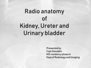
- 1. Radio anatomy of Kidney, Ureter and Urinary bladder Presented by Capt Alauddin MD residency phase A Dept of Radiologyand Imaging
- 2. •Radio anatomy means study of anatomy through the use of radiographic films. •The density differences in radiograph have to be described in terms of anatomical structures. Radio anatomy
- 3. Objective : To identify the Kidney, ureter and urinary bladder in different imaging modalities.
- 4. Kidney Retroperitonial organs that maintain electrolyte balance and function for waste excretion. location – T-12 to L-3 vertebra Rt kidney is slightly lower than the left kidney Long axis is directed downward and laterally
- 5. Kidneys have 2 poles: Upper and lower 2 borders : Medial and lateral 2 surfaces : Ant and post
- 6. Capsules of kidney: -Fibrous capsule : Covers the kidney -Perirenal fat: Layer of fat surrounding the fibrous capsule -Renal fascia of Gerota: Fibroareolar tissue surrounding the kidney and perirenal fat. -Pararenal fat: Fat that surrounds renal fascia
- 8. Internal structures Cortex: 2 parts: Cortical arch and renal collumn ( Collumn of Bartin). Medulla : About 10 conical masses called renal pyramids , the apices of which indent the minor calyces and bases are covered by cortical arches.
- 9. Contents of Cortex & Medulla:
- 10. Development of Kidney: Permanent kidney has 2 parts- A. Collecting system- It develops from ureteric bud which is an outgrowth of mesonephric duct. B. Excretory system- It develops from metanephric mesoderm.
- 11. Development of Kidney(cont) 9 th wk of dev
- 12. Ureters Connects the renal pelvis to the bladder and is 25–30 cm long. • Diameter : About 3 mm •3 Functionally narrow regions: -Junction of renal pelvis with ureter -As the ureter crosses bony pelvic brim -Intravesical ureter -about 2 cm and it runs obliquely through the muscular bladder wall where it has a valve like action
- 13. 3 ‘functionally’ narrow regions of ureter:
- 14. Urinary bladder Pyramidal muscular organ specially when empty. -The urine produced by kidney is temporarily stored in urinary bladder till it is cleared to the exterior through the urethra.
- 15. Imaging modalities for KUB: •Plain X ray •USG •IVU •CT scan •MRI •Renal angiography
- 16. In plain X ray: Only renal outline is seen. The renal edge may be visible, outlined by the surrounding perirenal fat. Renal size is 3and ½ vertebral bodies in height. Left kidney is usually larger(<2 cm). Bowel preparation is needed to reduce confusing gas shadows of stomach and
- 17. Plain X ray KUB (cont): The hilum of left kidney is closely approximate with the tip of left L1 transeverse process And hilum of right kidney is closely approximate with the tip of right L2 transeverse
- 18. Ureter in plain x ray: -not visible in normal cases. -its course is carefully observed over the tip of transeverse processes of L2-L5 vertebrae and anterior to SI joint specially when looking for radio opaque calculi .
- 19. Urinary bladder in plain X ray: May be identified specially when full as a round soft tissue density surrounded by a lucent line of perivesical fat. It should be smooth and symetrical.
- 20. USG: -Allows multiplanar evaluation of renal anatomy. Assessment of size, parenchyma, the pelvicalyceal system can be readily performed. -Cortex of the kidneys are less echogenic compared to liver parenchyma . Renal pyramids are relatively hypoechoic than cortex. -Outline: smooth -Cortical thickness: uniform - Renal sinus contains fat so those are echogenic.
- 21. Ureter : Proximal and distal ureter is seen when dilated . Urinary bladder: The bladder wall is best assessed with this modality - it should not exceed 3-5 mm in thickness. USG: (Cont)
- 22. IVU: Indications 1. Haematuria 2. Renal colic 3. Recurrent UTI 4. Loin pain 5. Suspected UT pathology. Contraindications : Patients with contrast medium allergies or with impaired renal function.
- 23. Contrast medium: Low osmolar contrast material (LOCM) 300–370 mg/ml Adult dose 50–100 ml. Paediatric dose 1 ml kg–1
- 24. Preliminary images: -Supine, full-length anterior posterior (AP) view of the abdomen, in inspiration , before the start of procedure of IVU.
- 25. After giving IV contrast , these images are taken: 1. Immediate film. shows the nephrogram phase where the renal parenchyma is opacified by contrast medium. 2. 5-min film : This film gives an initial assessment of pathology like obstruction before applying compression 3. 10-min film: Shows the pelvicalyceal system which is opacified due to urine containing contrast.
- 26. 4. Release film: To assess the ureters. 5. After micturition film: For assessment of urinary bladder emptying, tumour ,ureterovesical junction calculi and also urethral diverticulum(Female)
- 27. Dromedary hump/splenic hump is a prominence of lateral border of left kidney. It is a normal variant due to close proximity of spleen.
- 28. CT: Kidneys are isodense to liver before contrast. Hyperdense to liver post contrast. Collecting system visualized after 120 sec of applying contrast. Ureter : May be identified before contrast, well visualized 120 s after giving contrast .
- 29. MRI: Dependent on state of hydration. Cortex of higher signal intensity (SI) on T1W image (corticomeduallary junction may be seen) In T2w image , cortex is of lower signal intensity. Ureter : Hyperintense on T2W image T1 W T2 W
- 30. Renal vasculatures: Renal arteries branch from the bdominal aorta laterally between L1 and L2 -Rt renal artery passes posterior to the IVC Renal vein drain into the inferior venacava. Renal veins lie anterior to arteries.
- 33. •Renal angiography: •Allows assessment of vascular and other lesions of the kidneys, but is primarily used to facilitate interventional procedures such as renal artery angioplasty or stent placement .
- 34. Common anomalies of the kidneys: Anomaly 1.Renal agenesis 2.Pelvic kidney 3. Horseshoe kidney
- 35. 4. Pancake’ (discoid) kidney (Fused completely) 5. Crossed fused ectopia Common anomalies of the kidneys: (cont)
- 36. References: 1.Anatomy for Diagnostic Imaging, Stephanie Ryan FRCSI FFR(RCSI) Consultant Paediatric Radiologist, Children’s University Hospital, Temple Street, Dublin, Ireland 2.Weir & Abrahams’ Imaging Atlas of Human Anatomy 3.TEXTBOOK OF Radiology and imaging by DAVID SUTTON MD, FRcB FRcR, DMRD, FCan.AR (Hon) 4.CHAPMAN AND NAKIELNY’S GUIDE TO RADIOLOGICAL PROCEDURE 5. Radiopedia.org