SlideShare una empresa de Scribd logo
1 de 118
Describe the respiratory system

                 • Essential components
                   of respi system :
                   –   CNS
                   –   Respi center
                   –   Muscle
                   –   Lung tissue
Depict signs of respiratory distress in a real
                   patient
Respiratory distress
• It is an abnormal physiologic condition identified by
  increased work of breathing.

• The child in respiratory distress is effectively
  compensating to preserve adequate gas exchange.
Signs of Respiratory distress
• Tachypnea
• Tachycardia
• Retractions
  (supraclavicular, suprasternal, intercostal or
  subcostal)
• Use of accessory neck muscles
• Nasal flaring and grunting
• Pallor or cyanosis
• Altered mental status
  (restlessness, irritability, confusion, anxiety, inability
  to recognize parents)
Positioning
• With an upper airway obstruction, the patient
  characteristically assumes sniffing position (jaw thrust
  forward) and may drool due to inability to swallow
  secretions.

• A patient in severe respiratory distress may position
  himself in the tripod position (seated and leaning
  forward on outstretched arms), sitting upright and
  leaning forward to maximize use of accessory
  muscles.
Respiratory rate
• Abnormal respiratory rate is common finding in
  respiratory distress.
• Although normal respiratory rate varies with age, a
  rate greater than 60 breaths per minute is abnormal
  in any child.
• A slow respiratory rate might arise concerns as it
  may signifies respiratory fatigue and imminent
  respiratory arrest in an exhausted child.
Retractions
• Subcostal, intercostal and suprasternal retractions
  are manifestations of increased work of breathing as
  well as use of accessory muscles of respiration.

• Paradoxic breathing (abdomen retracts upon
  inspiration and expands upon expiration) may be
  noted.
Nasal flaring and grunting
• Flaring of the nostrils with inspiration is also a sign of
  respiratory distress.

• Grunting is a sound made by forceful expiration
  against a partially closed glottis.

• Head bobbing may be seen in infants.
Lung Sounds and Auscultation
• Decreased breath sounds and diminished air entry in
  the setting of increased respiratory effort suggest
  worsening respiratory disease.
Heart Rate
• Tachycardia is a common finding in children in
  respiratory distress and is not necessarily indicative
  of cardiovascular instability.

• Bradycardia, for instance, is indicative of severe
  hypoxemia and acidosis which require critical
  interventions.

• Bradycardia in a child with history of respiratory
  distress signifies imminent cardiopulmonary arrest.
Altered level of consciousness
• Some children might present with effortless
  tachypnea resulting from an increased respiratory
  drive caused by metabolic acidosis or central nervous
  system disease.

• Therefore, mental status is evaluated as a gauge of
  systemic oxygenation and critical management must
  be done if the patient appears lethargic or agitated.
Normal Respiratory Rate
•   Infant = 30 – 40 /min
•   Toddler = 24 – 40 /min
•   School age = 18 – 30 /min
•   Adolescent = 12 – 16 / min
Define respiratory failure
• Respiratory failure is a syndrome of inadequate gas
  exchange due to dysfunction of one or more essential
  components of the respiratory system:

• Type I : Hypoxaemic respiratory failure. PaO2 < 60 mm Hg
  with a normal or low PaCO2. This is caused by ventilation-
  perfusion mismatch with either/both:
   –  Under-ventilated alveoli (eg pulmonary oedema, pneumonia or
      acute asthma).
   – Venous blood bypasses ventilated alveoli (eg right to left cardiac
     shunts).

• Type II : Hypercapnic respiratory failure. PaCO2 > 50 mm Hg
  and indicates inadequate alveolar ventilation. Any
  ventilation-perfusion mismatch will affect PaO2 and
  therefore hypoxaemia is also common.
• Common causes of type • Common causes of type
  I respiratory failure   II respiratory failure
  – COPD.                        – Drug
  – Pneumonia.                     overdose, poisoning.
  – Pulmonary oedema.            – Myasthenia gravis.
  – Pulmonary fibrosis.          – Polyneuropathy.
  – Asthma.                      – Poliomyelitis.
  – Pneumothorax.                – Muscle disorders.
  – Pulmonary embolism.          – Head injuries and neck
  – Pulmonary hypertension.        injuries.
  – Cyanotic congenital heart    – Hypothyroidism.
    disease.
  – Bronchiectasis.
  – Acute respiratory distress
    syndrome.
Ix :
• Arterial blood gas analysis: confirmation of the diagnosis.
• CXR: often identifies the cause of respiratory failure.
• FBC: anaemia can contribute to tissue hypoxia; polycythaemia may
  indicate chronic hypoxaemic respiratory failure.
• Renal function tests and LFTs: may provide clues to the aetiology or
  identify complications associated with respiratory failure. Abnormalities in
  electrolytes such as potassium, magnesium, and phosphate may
  aggravate respiratory failure and other organ dysfunction.
• Serum creatine kinase and troponin I: to help exclude recent myocardial
  infarction. Elevated creatine kinase may also indicate myositis.
• TFTs (hypothyroidism may cause chronic hypercapnic respiratory failure).
• Spirometry: useful in the evaluation of chronic respiratory failure.
• Echocardiography: if a cardiac cause of acute respiratory failure is
  suspected.
• Pulmonary function tests are useful in the evaluation of chronic
  respiratory failure.
• ECG: to evaluate a cardiovascular cause; it may also detect dysrhythmias
  resulting from severe hypoxaemia or acidosis.
Management
• A patient with acute respiratory failure
  generally needs prompt hospital admission in
  an intensive care unit.
• Many patients with chronic respiratory failure
  can be treated at home, depending on the
  severity of respiratory failure, underlying
  cause, comorbidities and social circumstances.
• Immediate resuscitation may be required.
• Appropriate management of the underlying
  cause.
Describe obstructive and restrictive
      respiratory conditions
Obstructive disorders
• These are respiratory disease which make it more
  difficult to get air out of the lungs.

• This group of disorders is characterized by obstruction
  of normal airflow due to airway narrowing and
  includes: Asthma, COPD, bronchiectasis, cystic
  fibrosis and tumors.

• Mechanisms causing airway narrowing differ and may
  include obstruction by a mucus plug, airway
  compression and smooth muscle constriction.
Obstructive disorders
• In general, obstructive disorders lead to hyperinflation
  of the lungs as air is trapped behind closed airways.

• Residual volume is increased as in the ratio of RV:
  TLC. Functional residual capacity also increased.

• In patients with severe obstruction, air trapping is so
  extensive that vital capacity is decreased.
• In addition, dynamic compression of the airway
  prolongs the FEV1, as well as the forced expiratory
  flow rate measured over the middle half of expiration
  (FEF 25-75%)

• Both FEV1 and FVC are reduced, but the reduction in
  FEV1 is greater (airway narrowing reduces the speed
  of forced expiration such that FEV1 is reduced to an
  abnormally low fraction of FVC, perhaps to 40% or
  lower).

• Result: low FEV1 / FVC.
Obstructive disorders
• Extrathoracic- stridor

• Intrathoracic- wheezing, best heard during expiration

• Long deep breaths

• Hyperinflated and tympanitic to percussion
Bronchoconstriction causes obstructive
         airway problems
Restrictive disorders
• These are respiratory disease which makes it more
  difficult to get air into the lungs.

• They “restrict” inspiration.

• Examples include:
  Fibrosis, sarcoidosis, silicosis, asbestosis, muscular
  diseases and chest wall deformities.
Restrictive disorders
• In fibrosis or alveolar wall thickening, the increased
  connective tissue reduces lung compliance, making it
  difficult to expand the lung during inspiration.

• In condition such as alveolar edema, respiratory distress
  syndrome, or infiltrative interstitial lung diseases, the lungs
  are stiff decreasing its compliance.

• Extrapulmonary factors that can contribute to decrease
  vital capacity include limited thoracic expansion (Pleural
  fibrosis, Kyphoscoliosis) and nerve or muscular
  dysfunction.
• As a result, all lung volumes are reduced. In
  particular, there can be marked decreases in total lung
  capacity and functional vital capacity.

• FEV1 and FVC are reduced proportionately but the normal
  ratio is preserve.

• The patient might demonstrate rapid shallow breathing.
  He/she might also shows grunting.

• Percussion of the respiratory system demonstrate dull/
  hyper-resonance?
Differentiate clinical presentation of
              obstructive airway
Upper airways obstruction                       Lower airways obstruction
Stridor                                         Wheezing
• harsh sound                                   •Continuous expiratory sound
•Produced near larynx by the vibration of       •Due to turbulence airflow
upper airway structures                         •Musical
•Inspiratory ( expiratory – less common)        •Partial obstruction produces wheezing
•hoarseness – effect vocal chord                sound at later phase of expiration
•Supraglottic etiology – moves with child’s     •Harsh, monophonic and low pitch (usually
head                                            from large, central airways)
•Decreases while sleep – inspiration at lower   • high pitched and musical (from small and
rate during sleeping                            peripheral airways)
Suprasternal retraction                         Rhonchi
•Pressure gradient between trachea and          •Secretion in intrathoracic airways, resulting
atmosphere                                      in irregular sound
                                                Rales or crackles
                                                •Sound characterized by of crumpling
                                                celophane due to fluid or secretion in small
                                                airways
Describe conditions presented with
   wheezing in children and infants
• Acute wheezing :
  – Infection
  – Foreign body aspiration (FBA)


• Chronic/ recurrent wheezing
  – Anomalies of the tracheobronchial tree (ie Congenital
    tracheomalacia and bronchomalacia)
  – Cardiovascular disease (pulmonary artery dilation
    and/or left atrial enlargement, including large left to
    right shunts, can compress large airways and cause
    wheezing)
  – Mediastinal masses (ie tumors, cyst, LN enlargement)
Upper respiratory tract conditions -
to be presented in tabulated manner
            • common cold
           • acute pharyngitis
           • acute otitis media
            • acute sinusitis
common cold          acute pharyngitis        acute otitis media      acute sinusitis

Fever                  Inflamed pharynx and     fever,                Cough
Nasal discharge        soft palate              irritability,         Nasal symptoms
Loss of appetite       Enlarged and tender      headache,             Fever
Difficulty sleeping.   LN                       apathy,               Headache
                                                anorexia,             Facial pain and
                                                vomiting,             swelling
                                                diarrhea              Sore throat


Rhinovirus,            Adenovirus               RSV, rhinovirus,      Rhinovirus,
coronavirus, RSV       Enterovirus              Pneumococcus,         adenovirus, influenza,
                       Rhinovirus               H. influenzae         or parainfluenza
                       Gp A β-hemolytic
                       strepto


Treat fever and pain   Paracetamol              Paracetamol           Nebulized anti-
with paracetamol or    Penicillin (bact only)   Ibuprofen             microbials
ibuprofen
Pnuemococal     Staphylococcus        Mycoplasma           Viral pneumonia
pneumonia       pneumonia             pneumonia
S. Pneumoniae   Staphylococcus        Mycoplasma           Influenza virus
                aureus                pnuemoniae           Parainfluenza virus
                                      Community
                                      acquires
                Presentation          Presentation         Presentation
                •Skin infection       •Fever               •Fever
                •Soft tissue          •Malaise             •Chills
                infection –           •Headche             •Non productive
                pyomyositis, septic   •Persistent slowly   cough
                bursitis              worsening dry        •Rhinitis
                •Toxic shock          cough                •Myalgias
                syndrome              •Chills              •Headache
                •Endocarditis         •Sore throat         •fatigue
                •Osteromyelitis       •Pluertictic chest
                •Pneumonia            pain
                •Food poisoning
                •UTI
Tx              Tx                    Antibiotics          amantindine,
Antibiotics     Antimicrobial         Macrolides,          rimantidine,
                therapy               doxycyline           acylovir
Pertussis – whooping cough
Etiology
Caused by Bordetella pertussis
Infect among human through respiratory droplets (coughing)
Clinical presentation
Infants beyond neonatal period through school age
Catarrhal stage – injection, increased nasal secretion and low grade fever 1-2 weeks
Paroxysmal stage – coughing (expiration) to dislodge plugs of necrotic bronchial
epithelial tissues and thick mucus. Lasted 2-4 weeks
Followed by forceful inhalation against narrowed glottis – whoop characteristic
Convalescent stage – symptom gradually resolve 1-2 weeks, coughing less severe,
whooping cough dissapear.
CNS damage due to hypoxia, bacterial pnuemonia,
Adult – starts with broncholitis illness.
Tx
Erythromycin – early coarse, eradicates nasopharyngeal carriage within 3-4days,
ameliorate the effects of infection, ineffective at paroxysmal stage, , ass with pyloric
stenosis if given to neonates <4 weeks old
Azithromycin & clarithromycin – shorter duration, ass with gastrointestinal adverse
effects
Pertussis-specific immunoglobin – effectivelt reduced symptoms at paroxysmal stage
TMP-SMZ (trimethoprim/sulfamethozxazole)
Pulmonary tuberculosis
Etiology
Caused by Mycobacterium tuberculosis
Spread via respiratory droplets – become respiratory nuclei, <10um can reach alveoli
Clincal manifestation
•Asymptomatic in older and infants patient
•+ve tuberculin skin test
•Minimal abnormalities on CXR infiltration with hilar lymphadenopathy (Ghon complex)
•Malaise, low grade fever, erythema nodosum, lympahadenopathy symptoms.
Investigation
Tuberculin skin test (TST)
•T-cell-mediated hypersensitivity, 2-6 weeks
Mantoux test
•Intradermal injection of 5TU on volar surface of forearm, screen high-risk population and for
diagnosis in all ill patients or contacts.
•False –ve at early phase of illness or immunosuppression
Culture
Tissue, sputum, fluid
Treatment
Antibiotics – RIPES, 9-months of isoniazid&rifampin 98% case cured
Prevention - BCG
• Acute viral bronchiolitis
     • transient infant wheeze
• chronic lung diseases of prematurity
         • bronchiectasis.
Acute viral bronchiolitis
• lower respiratory tract infection (LRTI) that
  primarily affects the small airways
  (bronchioles)
  – RSV is the most common cause, followed by
    rhinovirus
  – Less common causes include parainfluenza
    virus, human metapneumovirus, influenza
    virus, adenovirus, coronavirus,
• S/s : fever (usually ≤38.3ºC), cough, mild
  respiratory distress, preceded by a one- to three-
  day history of upper respiratory tract
  symptoms, such as nasal congestion and/or
  discharge.
• PE :
   – tachypnea
   – intercostal and subcostal retractions, often with
     expiratory wheezing.
   – The chest may appear hyperexpanded with increased
     anteroposterior (AP) diameter and may be
     hyperresonant to percussion.
   – Findings on auscultation include any combination of
     expiratory wheeze, prolonged expiratory phase, and
     both coarse and fine crackles.
• Ix : Mild hypoxemia (oxygen saturation <95
percent) commonly is detected by pulse
oximetry, even without clinical signs of desaturation
• Mx :
  – SEVERITY ASSESSMENT — Severe bronchiolitis is
    indicated by persistently increased respiratory
    effort (tachypnea; nasal flaring;
    intercostal, subcostal, or suprasternal retractions;
    accessory muscle use;
    grunting), hypoxemia, apnea, or acute respiratory
    failure
  – In healthy infants and young
    children, bronchiolitis usually is a self-limited
    disease.
  – Maintain oxygenation and hydration.
  – Bronchodilator therapy may be beneficial in a
    subset of patients
Transient infant wheeze
• The successful transition from fetal to
  neonatal life at delivery requires a series of
  rapid physiologic changes of the
  cardiorespiratory system. These changes result
  in redirection of gas exchange from the
  placenta to the lung and comprise:
  – Replacement of alveolar fluid with air
  – Onset of regular breathing
  – Increase in pulmonary blood flow as a result of
    increased systemic vascular resistance and
    decreased pulmonary vascular resistance (PVR)
• difficulty may be a consequence of :
  – impaired lung function due to fluid retention,
  – airway obstruction associated with congenital
    anomalies,
  – persistent pulmonary hypertension,
  – apnea associated with lack of respiratory effort.
Respiratory distress syndrome
• Respiratory distress (ie, tachypnea and labored
  breathing) and cyanosis occur at or soon after birth.
• Typical signs include
   – grunting (which prevents end-expiratory alveolar collapse),
   – nasal flaring (which reduces nasal resistance and reflects
     increased use of accessory muscles of respiration),
   – and intercostal and subcostal retractions (due to decreased
     lung compliance and the highly compliant chest wall).
• Surfactant deficient
• inability to clear lung fluid from air spaces
Bronchiectasis
• Abnormal dilation and distortion of the
  bronchial tree, resulting in chronic obstructive
  lung disease.
• End result of a variety of pathophysiologic
  processes that render the bronchial walls
  weakened, easily collapsible, chronically
  inflamed, and plugged with mucus secretions.
• Associated findings include atelectasis,
  emphysema, fibrosis, and hypertrophy of the
  bronchial vasculature.
• S/s : productive cough, purulent sputum,
  fever, pleuritic chest pain, and dyspnea
• PE :
  – Crackles (inspiratory popping sounds) and rhonchi
    (coarse expiratory breath sounds),
  – Wheezing (less common)
  – Clubbing of the nailbed
  – Chest wall deformity, which can be seen in
    obstructive lung diseases (eg, cystic fibrosis)
• Ix :
   – CXR - linear atelectasis and dilated and thickened airways,
     HRCT
   – sweat chloride or DNA testing
   – blood count with differential, immunoglobulins, and IgG
     subclasses

• Mx : aimed at treating the underlying cause, improving
  mucocilliary clearance, treating and preventing
  infection, and controlling inflammation
   –   Antibiotic therapy
   –   Chest physiotherapy
   –   Mucolytic agents and airway hydration
   –   Bronchodilators
   –   Anti-inflammatory drugs
   –   Immunization
Laryngotracheobronchitis (Croup)
• It is a viral infection of the middle respiratory tract
  occurs from 6 months to 6 years of age with peak
  incidence in the 2nd year of life.

• There is mucosal inflammation and increased
  secretions affecting the airway, but it is the edema of
  the subglottic area that is potentially dangerous as it
  may result in critical narrowing of the trachea.
Laryngotracheobronchitis (Croup)
• A small decrease in diameter secondary to mucosal
  edema and inflammation exponentially increases
  airway resistance and significantly increases work of
  breathing.

• During inspiration, walls of the subglottic space are
  drawn together, aggravating the obstruction and
  producing stridor characteristic of croup.
Laryngotracheobronchitis (Croup)
• Symptoms are
    Low grade fever, cough and coryza for 12-72 hours followed
     by
    Stridor that may occur when excited, at rest or both
      (Harsh, high-pitched respiratory sound produced by
      turbulent airflow; usually inspiratory, but may be biphasic
      and is a sign of upper airway obstruction)
    hoarse voice, brassy or “barking” cough
    Respiratory distress of varying degree

• Onset of symptoms is usually over several days and often start
  and worse at night.

• Symptomatic reinfection is common but usually mild.
Laryngotracheobronchitis (Croup)
• Parainfluenza viruses (type 1,2,3) are the
  commonest cause but other viruses such as human
  metapneumovirus, RSV and influenza virus type A
  and B, adenovirus, enterovirus, measles, mumps
  and rhinoviruses can produce similar clinical picture.

• Chest X-ray reveals a characteristic narrowing of the
  subglottic region known as the “steeple sign”.
Assessment of severity
• a
Laryngotracheobronchitis (Croup)
• When the upper airway obstruction is mild, the
  stridor and chest recession disappear when the child
  is at rest.

• The child can usually be managed at home and
  need to be closely monitored for signs of increasing
  severity.

• Most cases of croup are mild and resolve within 1 to
  2 days.
Laryngotracheobronchitis (Croup)
• Indication for hospital admission
   Moderate and severe viral croup
   Poor oral intake
   Family lives a long distance from hospital, lacks
     reliable transport
   Toxic looking
   Age less than 6 months
   Unreliable caregivers at home
Management of Croup
Management of Croup
• Oxygen therapy indication include severe viral croup
  and percutaneous SaO2 <93%.

• Steroids are helpful in moderate to severe cases.

• Antibiotics may be indicated if the child fails to
  improve or if purulent secretions are present where
  coverage should be directed towards
  Staphylococcus and H.influenzae.
Acute epiglottitis (Supraglottitis)
• It is less common than croup, typically presents with
  a greater degree of airway compromise.

• H.influenzae type B is the major etiologic organism.

• The incidence of epiglottitis has decreased with the
  administration of H. influenzae vaccine.

• It is most common in children aged 1-6 years but
  affect all age groups.
Clinical manifestations of Acute epiglottitis

• Patients typically present with acute onset (over several hours)
  of
    High fever in an ill, toxic-looking child
    An intensely painful throat preventing child from speaking or
     swallowing with saliva drools down the chin
    Marked dysphagia
    Soft inspiratory stridor and rapidly increasing respiratory
     difficulty over hours
    Patients are often encountered leaning forward in a “tripod”
     position (Immobile, upright with an open mouth)

• Unlike croup, cough is frequently absent.
Acute epiglottitis (Supraglottitis)
• Attempts to lie the child down or examine the throat
  with a spatula or perform a lateral neck X-ray must
  not be undertaken as this may precipitate total
  obstruction and death.

• Instrumentation of the throat with tongue depressors
  is not advisable as this can precipitate a fatal
  laryngospasm.
Acute epiglottitis (Supraglottitis)
• If the diagnosis of epiglottitis is suspected, urgent hospital
  admission and treatment are required.
• The child should be intubated under controlled conditions
  with a general anesthetic.
• Rarely, this is impossible and urgent tracheostomy is life-
  saving.
• Only after securing the airway, blood is taken for culture
  and broad-spectrum IV antibiotics such as cefuroxime
  directed against H.influenzae and gram-positive
  organisms started.
• Tracheal tube can usually be removed after 24 hours and
  antibiotics given for 2-3 days.
• Prophylaxis with rifampicin is offered to close household
  contacts.
Clinical features of croup and epiglottitis
                     Croup             Epiglottitis
Onset                Over days         Over hours
Preceding coryza     Yes               No
Cough                Severe, barking   Absent or slight
Able to drink        Yes               No
Drooling saliva      No                Yes
Appearance           Unwell            Toxic, very ill
Fever                <38.5 Celsius     >38.5 Celsius
Stridor              Harsh, rasping    Soft, whispering
Voice, cry           Hoarse            Muffled, reluctant to
                                       speak
Laryngomalacia
• It is the most common congenital anomaly of the
  larynx, accounting for up to 60% of all anomalies.
• Inspiratory stridor is the hallmark of the condition.
• Symptoms are typically aggravated when the child is
  supine or crying.
• Examination reveals partial collapse of a flaccid
  supraglottic airway with inspiration.
• The condition is generally benign and self-limited, as
  most cases resolve by 18 months of age.
• Severe cases may require surgical intervention if the
  distress prevents adequate feeding.
Congenital subglottic stenosis
Etiology
•Can be iatrogenic
•Intubation and mechanical ventilation – residual damage to the larynx
•Down’s syndrome infants more susceptible due to smaller larynx
Clinical manifestation
•Subglottic obstruction produces stridor and frequently biphasic (expiration and
inspiration)
•Worsen with respiratory effort
•Viral infection exacerbate subglottic stenosis
Diagnostic studies
•Requires endoscopic evaluation
Treatment
•Tracheostomy and reconstructive surgery may be necessary
•Some improve with age as larynx grow and require no intervention
Retropharyngeal abscess & Peritonsilar abcess
 Retropharyngeal abscess                           Comparison          Peritonsilar abscess
<6 years                                Age                       >10 years
Posterior pharynx                       Location                  Oropharynx
S. aureus, anaerobes                    Etiology                  Group A streptococci, anaerobes

Insidious to sudden                     Prodrome onset            Biphasic with sudden worsening

None                                    Stridor                   No
Yes                                     Retraction                No
Muffled                                 Voice                     “Hot potato” muffled
Arching of neck or normal               Position and appearance   Normal
Drooling                                Swallowing (dysphagia)    Drooling, trismus
No                                      Barking cough             None
Severe                                  Toxicity                  Dyspnea
>101 F                                  Fever                     >101 F
Thickened retropharyngeal space         X Ray                     None needed
Leukocytosis with left shift            WBC count                 Leukocytosis with left shift
Antibiotics, surgical drain - abscess   Therapy                   Antibiotics, aspiration
None                                    Prevention                Treat Group A streptococci early
Diphtheria
Etiology
Caused by bacteria Corynebacterium diphtheriae
spread through respiratory droplets from infected person or carrier
Infects nose, throat causes gray to black, thick fiber like covering airway
Infects skin - Skin lesions
Produces toxins – spread via bloodstream to other organs
Signs and symptom
Mild fever, sore throat, chills – few days after infection
Breathing problems, croup-like (barking cough), drooling, hoarseness
Nasal discharge, fatigue
Dysphagia, paralysis, heart failure – serious
Tx
Antitoxin IV/IM
Antibiotics – penicillin and erythromycin
Diphtheria vaccine – lasted for 10 years (if in contact with infected person)
Chronic lung disease of prematurity
• Infants who still have an oxygen requirement at a
  post-menstrual age of 36 weeks are described as
  having bronchopulmonary dysplasia (BPD) or chronic
  lung disease.

• Criteria for diagnosis are oxygen requirement beyond
  36 weeks post conceptional age or beyond 28 days of
  life.
Pathophysiology of BPD
• The lung damage (alveolar cells, interstitium and
  blood vessels) comes from pressure and volume
  trauma from artificial ventilation, oxygen toxicity and
  infection.

• Oxygen concentration greater than 40% are toxic to
  the neonatal lung.

• Oxygen-mediated lung injury results from the
  generation of superoxides, hydrogen peroxide and
  oxygen free radicals which disrupt membrane lipids.
Pathophysiology of BPD

• Assisted ventilation with high peak pressures produces
  barotrauma, compounding the damaging effects of highly
  inspired oxygen levels.

• Inflammatory mediators are released and causing increase
  permeability of blood vessels leading to leakage of water
  and protein.

• In later stages, there is fibrosis and cellular hyperplasia.

• Severe lung damage can leads to respiratory failure.
Risk factors of BPD
• In most patients, BPD develops after ventilation for RDS
  that may have been complicated by PDA (Patent ductus
  arteriosus) or pulmonary interstitial emphysema.

• Inflammation from prolonged assisted ventilation and
  repeated systemic and pulmonary infections may play a
  major role.

• Failure of RDS to improve after 2 weeks, the need for
  prolonged mechanical ventilation and oxygen therapy
  required at 36 weeks’ postconceptional age are
  characteristics of patients with RDS in whom BPD
  develops.
Risk factors of BPD

• BPD also may develop in infants weighing less than
  1000g who require mechanical ventilation for poor
  respiratory drive in the absence of RDS.

• 50% of infants of 24 to 26 weeks’ gestational age
  require oxygen at 36 weeks’ corrected age.
Chest X-Ray of BPD

• The chest X-ray characteristically shows widespread
  areas of opacification, sometimes with cystic changes
  accompanied by areas of over-distention and
  atelectasis, giving the lung a sponge like appearance.

• Histopathology of BPD reveals interstitial
  edema, atelectasis, mucosal metaplasia, interstitial
  fibrosis, necrotizing obliterative bronchiolitis and
  overdistended alveoli.
Clinical manifestations of BPD
• The clinical manifestations of BPD are oxygen dependence,
  hypercapnia, compensatory metabolic alkalosis, pulmonary
  hypertension, poor growth and development of right-sided heart
  failure.

• Increased     airway    resistance with reactive airway
  bronchoconstriction also is noted and is treated with
  bronchodilating agents.

• Severe chest retractions produce negative interstitial pressure
  that draws fluid into the interstitial space.

• Together with cor pulmonale, these chest retractions cause fluid
  retention, necessitating fluid restriction and the administration of
  diuretics.
Management of BPD
• Some infants need prolonged artificial ventilation, but most are
  weaned onto continuous positive airways pressure (CPAP)
  followed by additional ambient oxygen, sometimes over several
  months.

• To reduce risk of subglottic stenosis, a tracheotomy may be
  indicated.

• To reduce oxygen toxicity and barotrauma, ventilator settings
  are reduced to maintain blood gases with slightly lower PaO2
  (50mmHg) and higher PaCO2 (50 to 75 mmHg) levels than for
  infants during the acute phase of RDS.
Management of BPD
• Dexamethasone therapy may reduce inflammation,
  improve pulmonary function and enhance weaning of
  patients from assisted ventilation.

• However, corticosteroid therapy may increase risk of
  abnormal neuro-development including cerebral palsy,
  thus limits use to those at highest risk and only short
  courses are given.
Management of BPD
• Some babies go home while still receiving additional
  oxygen.

• A few infants with severe disease may die of intercurrent
  infection or pulmonary hypertension.

• Some survivors of BPD have hyperinflation, reactive
  airways and developmental delay.

• Subsequent pertussis and RSV (Respiratory syncytial
  virus) infection may cause respiratory failure necessitating
  intensive care and thus prophylaxis against RSV should
  be given.
Inhaled foreign body
Epidemiology
•Common, 4 y/o
•Main right bronchus (direct continous with trachea
•May migrate on larynx (coughing) causes occlusing of airway

Clinical manifestation
•Cough, localized wheezing, unilateral absence of breath sounds, stridor, blood sputum (rare)
•fever, cough, sputum production, or chest pain – need medical attention
•Persistence wheezing unresponsive to bronchodilator therapy, persistent atelectasis, recurrent
or persistence pneumonia, persistent cough without other explanation – suspect foreign body
inhalation.
•Lodge in esophagus and compress trachea – produce respiratory symptoms


Diagnostic studies
•Radiographic studies – presence of radiopaque object or eveidence of air trapping
Prevention
•Education to parent and caregiver
•Small toys should kept away
Pneumothorax
• defined as a collection of air
that is located within the thoracic
cage between the visceral and
parietal pleura

  – Primary : no underlying lung disease
  – Secondary : complication of underlying lung disease
    (ie asthma, cystic fibrosis, necrotizing pneumonia, and
    interstitial lung disease)
• S/s : dyspnea and pleuritic chest pain that is
  described as sharp or stabbing. The pain
  typically is diffuse on the affected side with
  radiation to the ipsilateral shoulder. A dry or
  non-productive cough
• PE :
  – deviation of the trachea towards the contralateral
    side
  – diminished breath sounds
  – hyperresonant percussion
  – decreased vocal fremitus on the affected side.
  – Other signs : tachypnea, increased work of
    breathing, and cyanosis.
• Ix : ABG, CXR
• Mx : depends on the size of the
  pneumothorax, the extent of respiratory
  distress, and the presence or absence of
  underlying lung disease. The goals of
  treatment are to remove air from the pleural
  space and to prevent recurrence.
  – Supplemental oxygen
  – Needle aspiration
  – Thoracostomy tube
Empyema
• Early in the course of parapneumonic
effusion, the pleura becomes inflamed;
leakage of proteins, fluid, and leukocytes.
At the time of formation, the pleural effusion is usually
sterile with a low leukocyte count.
With time, bacteria invade the fluid, resulting in
empyema, which is defined as the presence of grossly
purulent fluid in the pleural cavity.
• S/s : persistent fever, cough, malaise, decreased
   appetite, chest pain, and dyspnea
• PE : tachypneic, but breaths may be shallow to minimize
   pain, mediastinal shift and tension
   hydrothorax, Hypoalbuminemia
• Ix : Pulse oximetry , Blood and sputum culture,
  CXR
• Management:
  – Main goals : sterilization of the pleural cavity,
    resolution of pleural fluid, and reexpansion of the
    lung
  – Antibiotics
  – Thoracocentesis
  – Chest tubes
  – Surgical (for symptoms for more than one week
    only)
Pneumatocele
• A pneumatocele is a cavity in the lung
  parenchyma filled with air (cyst) that may
  result from pulmonary trauma during
  mechanical ventilation
• Ddx : Lung ca, TB
• Mx : palliative care
Lung abscess
• Result from lung tissue necrosis from an
  infection (ie pneumonia)

• Conditions contributing to lung abscess
  – Aspiration of oropharyngeal or gastric secretion
  – Septic emboli
  – Vasculitis: Wegener's granulomatosis
  – Necrotizing tumors:
Congenital diaphragmatic hernia                   Congenital lobar emphysema (congenital
                                                  lobar over distention)
Etiology                                          Over inflation of one lobe during neonatal
Improper joining of structure during fetal dev.   period – left upper lobe (often)
Stomach, small intestine, spleen appear in
chest cavity,
Lung tissue not completely develop
Left side – common
Increase risk with +ve family hx

Symptom                                           •Produces respiratory distress because
Breathing difficulty after born due to            surrounding lung tissue has compressed
ineffective movement of diaphragm and             •Mediastinal shift
crowding of lung tissue, which causes collapse
Cyanosis, tachypnea, tachycardia
Irregular chest movement
Absence of breath sound on both side
Bowel sound heard in the chest
Palpation – abd feels less full
Xray – abd organs in chest cavity
Surgery - place abd organs back n repair          Lobectomy – if RDS is severe and progressive
diapgrmatic opening
Breathing support until recvers from surgery
H type- tracheoesophageal fistula -
• S/s : if the defect is large, with coughing and
  choking associated with feeding.
• Diagnosis — attempt to pass a catheter into
  the stomach. This finding can be confirmed
  with an anterior-posterior chest radiograph
  that demonstrates the catheter curled in the
  upper esophageal pouch
• Mx : surgical ligation of the fistula.
  – if the distance between esophageal segments is
    large, staged procedures are performed that
    include elongation of the esophagus.
Gastroesophageal reflux (GER)
 It is defined as the effortless retrograde movement of
  gastric contents into the esophagus.

 In infancy, GER is not always an abnormality.

 Distinction between physiologic GER and pathologic
  GER in infancy and childhood is determined by
  presence of reflux-related complications.
Physiologic GER
 Physiologic GER (Spitting up) is normal in infants
  younger than 8 to 12 months of age.
 Nearly half of all infants are reported to spit up at 2
  months of age.
 It is caused by inappropriate relaxation of the lower
  esophageal sphincter as result of functional
  immaturity.
 Infants who regurgitate stomach contents meet the
  criteria for physiologic GER, as long as they maintain
  adequate nutrition and have no signs of respiratory
  complications or esophagitis.
Clinical manifestations
 Presence of GER is easy to observe in an infant who spits up.

 In older children, the refluxate is usually kept down by
  reswallowing, but GER may be suspected by associated
  symptoms, such as:
  • Heartburn
  • Cough
  • Dysphagia
  • Wheezing
  • Aspiration pneumonia
  • Hoarse voice
  • Failure to thrive
  • Recurrent otitis media/ Sinusitis
Clinical manifestations
 GER can have both esophageal and extraesophageal
  manifestations.

 The esophageal manifestations are those caused
  directly by contact between refluxed gastric juice and
  the esophageal mucosa.

 The extra-esophageal manifestations are caused by
  contact between refluxed gastric juice and
  extraesophageal organs or by reflexes triggered by
  acid in the esophagus that affects other organs.
Extraesophageal complications
 Cardiac: Atypical chest pain, ischemia
 Vocal cords: Laryngitis, granuloma, polyps, ulcers,
  neoplasm
 Respiratory: asthma, recurrent bronchitis, aspiration
  or chronic interstitial pneumonia, sleep apnea,
  chronic or recurrent cough
 Pharyngeal: pharyngitis, hoarseness
 Oral: Erosions, burning mouth syndrome
 Other: sudden infant death syndrome, otitis media
Investigations
1. Barium upper gastrointestinal series

2. 24-hour esophageal pH probe monitoring

3. Esophageal impedance monitoring

4. Endoscopy
Relation to respiratory symptoms
 GER has been associated with significant respiratory
  symptoms in infants and children.

 The infant’s proximal airway and esophagus are lined
  with receptors that are activated by water, acid or
  distention.

 Activation of these receptors can increase airway
  resistance, leading to development of reactive airway
  disease.
Relation to respiratory symptoms
 One postulated mechanism for gastroesophageal reflux-
  mediated airway disease involves micro-aspiration of
  gastric contents that leads to inflammation and
  bronchospasm.

 However, experimental evidence also supports the
  involvement of esophageal acid-induced reflex
  bronchospasm in the absence of frank aspiration.

 Therapy using histamine 2 blockers or proton pump
  inhibitors has been shown to benefits patients with
  steroid-dependent asthma, nocturnal cough and reflux
  symptoms.
Asthma and GER
 In 1892, Sir William Osler first postulated a
  relationship between asthma and gastroesophageal
  reflux, manifested by a bi-directional cause-and-effect
  presentation.

 Prevalence of GER is more common in asthmatics
  compared to control populations ranging from 47 to
  75%.

 Severe studies suggest that severe nocturnal asthma
  is more likely to be associated with GER than daytime
  asthma.
Asthma and GER
 Not all asthmatics with GER will have esophageal
  symptoms.

 The mechanism of esophageal acid induced
  bronchoconstriction includes a vagally mediated
  esophageal    bronchial    reflex,  airway hyper
  responsiveness and microaspiration.
Asthma and GER
 It has been suggested that a vagally mediated reflex exists
  whereby acid reflux into the lower esophagus initiates
  bronchoconstriction via a shared autonomic innervation
  between esophagus and bronchi based on their common
  embryonic origin.

 Support is based on this two lines of evidence:
  • Acid infusion of the esophagus in asthmatic children results
     in increased airway resistance, which can be reversed with
     antacids.
  • Infusion of antacids into the distal esophagus of asthmatic
     children during sleep induces bronchoconstriction,
     suggesting presence of a protective mechanism.
Asthma and GER
 Esophageal acidification has also been associated with
  heightened non-specific hyperresponsivity of the airways to
  bronchoconstrictors     contributed      by    increased vagal
  tone, “priming” the airways to other triggers.
Asthma and GER
 Asthmatics have significant decreased lower esophageal sphincter
  pressures, greater esophageal acid exposure times, more frequent
  episodes of GER and longer esophageal acid clearance times.

 There may be increase in transient lower esophageal sphincter
  (LES) relaxations, LES hypotonia and esophageal due to autonomic
  dysfunction in asthmatics patient.

 Furthermore, LES pressure augmented by diaphragm normally
  prevents GER but it can be prevented by hyperinflation and hiatal
  hernia.

 LES pressure can also be overcome by increased negative
  intrathoracic pressures (bronchospasm, cough, upper airway
  resistance syndrome and obstructive apnea).
Asthma and GER
 Anti-reflux therapy results in improvement in asthma in
  approximately 70% of patients.

 Acid-suppression medication such as histamine 2 receptor
  antagonists (ranitidine) or proton pump inhibitors (omeprazole)
  are helpful in reducing volume of gastric contents.
  (Therapeutic trial)
Chronic cough and GER
 GER can potentially stimulate sensory limb of cough
  reflex in multiple ways.

 It can irritate upper respiratory tract without aspiration
  (such as larynx) and the lower respiratory tract by
  either macro-microaspiration.

 It can also cause cough by stimulating an esophagus
  bronchial cough reflex whereby refluxate into distal
  esophagus alone is a sufficient stimulus.
Chronic cough and GER
 GER related cough occurs predominantly during the
  day and in the upright position whereby some
  patients may have nocturnal symptoms.

 Cough may be the sole manifestation of GER and is
  often long standing.

 Diagnosis can be made with certainty only when
  cough are resolved with specific anti-reflux therapy.
Senario 3
  Samy, a 2 days old premature infant who was
   born at 30 weeks gestational age was noted
   to be cyanosed by a nurse. There was
   absence of chest movement.
  What is Samy’s clinical condition?
      Respiratory distress syndrome (RDS)
  What is your immediate action?
      surfactant replacement therapy
      IV glucocorticoid administration
  What is the mechanism of his respiratory
   failure?
      Immature lung  lack surfactant  atelectasis
Senario 5
 Aminah a 2 year old toddler has been having
  mild cough and rhinorrhea for the past 2
  days. It was associated with low grade fever.
  Suddenly her mother noticed that she breath
  noisily with fast breathing.
 What is Aminah’s clinical condition?
     Common cold
 What are the relevant physical finding that
  you would like to look for?
     Signs of respiratory distress
 Outline the principle management.
     Symptomatic therapy

Más contenido relacionado

La actualidad más candente

Dyspnea and Pulmonary Edema
Dyspnea and Pulmonary EdemaDyspnea and Pulmonary Edema
Dyspnea and Pulmonary Edemabajah423
 
Resp physiology & respiratory failure in children
Resp physiology & respiratory failure in childrenResp physiology & respiratory failure in children
Resp physiology & respiratory failure in childrenSameekshya Pradhan
 
Clinical vignette 3 (Causes of cough, dyspnea and tachypnea)
Clinical vignette 3 (Causes of cough, dyspnea and tachypnea)Clinical vignette 3 (Causes of cough, dyspnea and tachypnea)
Clinical vignette 3 (Causes of cough, dyspnea and tachypnea)Arwa H. Al-Onayzan
 
Shortness Of Breath
Shortness Of BreathShortness Of Breath
Shortness Of BreathAkmal Shah
 
Dyspnoea & Respiratory Failure
Dyspnoea & Respiratory FailureDyspnoea & Respiratory Failure
Dyspnoea & Respiratory FailureMuhammad Helmi
 
Acute respiratory distress syndrome (ards) pediatric oncall journal
Acute respiratory distress syndrome (ards)   pediatric oncall journalAcute respiratory distress syndrome (ards)   pediatric oncall journal
Acute respiratory distress syndrome (ards) pediatric oncall journalManjunath Vaddambal
 
Dyspnea redone
Dyspnea redoneDyspnea redone
Dyspnea redoneJohn Eod
 
Approach to dyspnea dr bikal
Approach to dyspnea dr bikalApproach to dyspnea dr bikal
Approach to dyspnea dr bikalBikal Lamichhane
 
Office Emergency Hyperventilation
Office Emergency  HyperventilationOffice Emergency  Hyperventilation
Office Emergency Hyperventilationmwhite17
 
A good respiratory case on post TB Fibrosis
A good respiratory case on post TB FibrosisA good respiratory case on post TB Fibrosis
A good respiratory case on post TB FibrosisKurian Joseph
 
aproch to patient with dyspnea
aproch to patient with dyspneaaproch to patient with dyspnea
aproch to patient with dyspneasamaramajid
 
Reexpansion pulmonary edema
Reexpansion pulmonary edemaReexpansion pulmonary edema
Reexpansion pulmonary edemaVijay Anand
 

La actualidad más candente (20)

dyspnea approach
dyspnea approachdyspnea approach
dyspnea approach
 
Dyspnea and Pulmonary Edema
Dyspnea and Pulmonary EdemaDyspnea and Pulmonary Edema
Dyspnea and Pulmonary Edema
 
Resp physiology & respiratory failure in children
Resp physiology & respiratory failure in childrenResp physiology & respiratory failure in children
Resp physiology & respiratory failure in children
 
Clinical vignette 3 (Causes of cough, dyspnea and tachypnea)
Clinical vignette 3 (Causes of cough, dyspnea and tachypnea)Clinical vignette 3 (Causes of cough, dyspnea and tachypnea)
Clinical vignette 3 (Causes of cough, dyspnea and tachypnea)
 
Shortness Of Breath
Shortness Of BreathShortness Of Breath
Shortness Of Breath
 
An approach to the patient of dyspnoea
An approach to the patient of dyspnoeaAn approach to the patient of dyspnoea
An approach to the patient of dyspnoea
 
evaluation of dyspnoea
evaluation of dyspnoeaevaluation of dyspnoea
evaluation of dyspnoea
 
Approach To Dyspnea
Approach To DyspneaApproach To Dyspnea
Approach To Dyspnea
 
Dyspnoea & Respiratory Failure
Dyspnoea & Respiratory FailureDyspnoea & Respiratory Failure
Dyspnoea & Respiratory Failure
 
Respiratory failure
Respiratory failureRespiratory failure
Respiratory failure
 
Acute respiratory distress syndrome (ards) pediatric oncall journal
Acute respiratory distress syndrome (ards)   pediatric oncall journalAcute respiratory distress syndrome (ards)   pediatric oncall journal
Acute respiratory distress syndrome (ards) pediatric oncall journal
 
Dyspnea redone
Dyspnea redoneDyspnea redone
Dyspnea redone
 
Approach to dyspnea dr bikal
Approach to dyspnea dr bikalApproach to dyspnea dr bikal
Approach to dyspnea dr bikal
 
Office Emergency Hyperventilation
Office Emergency  HyperventilationOffice Emergency  Hyperventilation
Office Emergency Hyperventilation
 
Dyspnea
DyspneaDyspnea
Dyspnea
 
A good respiratory case on post TB Fibrosis
A good respiratory case on post TB FibrosisA good respiratory case on post TB Fibrosis
A good respiratory case on post TB Fibrosis
 
aproch to patient with dyspnea
aproch to patient with dyspneaaproch to patient with dyspnea
aproch to patient with dyspnea
 
Reexpansion pulmonary edema
Reexpansion pulmonary edemaReexpansion pulmonary edema
Reexpansion pulmonary edema
 
Respiratory failure
Respiratory failureRespiratory failure
Respiratory failure
 
Dyspnea
DyspneaDyspnea
Dyspnea
 

Destacado

Histology epithelial tissue
Histology epithelial tissueHistology epithelial tissue
Histology epithelial tissueNancyDecker
 
Histology various type of epithelium
Histology   various type of epitheliumHistology   various type of epithelium
Histology various type of epitheliumMBBS IMS MSU
 
4. smooth muscle tissue
4.  smooth muscle tissue4.  smooth muscle tissue
4. smooth muscle tissueMichael Wrock
 

Destacado (6)

Tissue
TissueTissue
Tissue
 
Anatomy Tissues
Anatomy TissuesAnatomy Tissues
Anatomy Tissues
 
Epithelial tissue
Epithelial tissueEpithelial tissue
Epithelial tissue
 
Histology epithelial tissue
Histology epithelial tissueHistology epithelial tissue
Histology epithelial tissue
 
Histology various type of epithelium
Histology   various type of epitheliumHistology   various type of epithelium
Histology various type of epithelium
 
4. smooth muscle tissue
4.  smooth muscle tissue4.  smooth muscle tissue
4. smooth muscle tissue
 

Similar a Respiratory condition

Clinical History & General Examination.pptx
Clinical History & General Examination.pptxClinical History & General Examination.pptx
Clinical History & General Examination.pptxdrperumal
 
Acute Respiratory Failure PPT.pptx
Acute Respiratory Failure PPT.pptxAcute Respiratory Failure PPT.pptx
Acute Respiratory Failure PPT.pptxJayBhanushali32
 
Similarities and differences between asthma and asthma masqueraders
Similarities and differences between asthma and asthma masqueradersSimilarities and differences between asthma and asthma masqueraders
Similarities and differences between asthma and asthma masqueradersSaher Farghly
 
clinical features of tb.ppt
clinical features of tb.pptclinical features of tb.ppt
clinical features of tb.pptShakibSheikh5
 
Acute Respiratory malfunction presentation
Acute Respiratory malfunction presentationAcute Respiratory malfunction presentation
Acute Respiratory malfunction presentationcathelena1
 
Respiratory system by Dr. Shabnam rajput
Respiratory system   by Dr. Shabnam rajputRespiratory system   by Dr. Shabnam rajput
Respiratory system by Dr. Shabnam rajputshabnam rajput
 
Applied physiology of respiration
Applied  physiology of respirationApplied  physiology of respiration
Applied physiology of respirationpriyanka susruth
 
Breathlessness and tachycardia
Breathlessness and tachycardiaBreathlessness and tachycardia
Breathlessness and tachycardiaDr Rakesh Solanki
 
Hyperventilationsyndrome 160207131346
Hyperventilationsyndrome 160207131346Hyperventilationsyndrome 160207131346
Hyperventilationsyndrome 160207131346KelleyCollens
 
Hyperventilation syndrome
Hyperventilation syndromeHyperventilation syndrome
Hyperventilation syndromeSneha Arya
 
Pulmonology (resp emerg)
Pulmonology (resp emerg)Pulmonology (resp emerg)
Pulmonology (resp emerg)Ben Lesold
 
How to present a Thoracic Case | IACTS SCORE 2020
How to present a Thoracic Case | IACTS SCORE 2020How to present a Thoracic Case | IACTS SCORE 2020
How to present a Thoracic Case | IACTS SCORE 2020IACTSWeb
 
Respiratory diseases
Respiratory diseasesRespiratory diseases
Respiratory diseasesBhawna Rajput
 
6) respiratory failure.pptx
6) respiratory failure.pptx6) respiratory failure.pptx
6) respiratory failure.pptxssuser523204
 
Assessment of respiratory system 1
Assessment   of   respiratory   system  1Assessment   of   respiratory   system  1
Assessment of respiratory system 1SriRam071
 

Similar a Respiratory condition (20)

approach to dyspnea.pptx
approach to dyspnea.pptxapproach to dyspnea.pptx
approach to dyspnea.pptx
 
Clinical History & General Examination.pptx
Clinical History & General Examination.pptxClinical History & General Examination.pptx
Clinical History & General Examination.pptx
 
Acute Respiratory Failure PPT.pptx
Acute Respiratory Failure PPT.pptxAcute Respiratory Failure PPT.pptx
Acute Respiratory Failure PPT.pptx
 
Presentation1.pptx
Presentation1.pptxPresentation1.pptx
Presentation1.pptx
 
Similarities and differences between asthma and asthma masqueraders
Similarities and differences between asthma and asthma masqueradersSimilarities and differences between asthma and asthma masqueraders
Similarities and differences between asthma and asthma masqueraders
 
clinical features of tb.ppt
clinical features of tb.pptclinical features of tb.ppt
clinical features of tb.ppt
 
Acute Respiratory malfunction presentation
Acute Respiratory malfunction presentationAcute Respiratory malfunction presentation
Acute Respiratory malfunction presentation
 
COPD
COPDCOPD
COPD
 
4301187.ppt
4301187.ppt4301187.ppt
4301187.ppt
 
Respiratory system by Dr. Shabnam rajput
Respiratory system   by Dr. Shabnam rajputRespiratory system   by Dr. Shabnam rajput
Respiratory system by Dr. Shabnam rajput
 
Applied physiology of respiration
Applied  physiology of respirationApplied  physiology of respiration
Applied physiology of respiration
 
tb all in one.ppt
tb all in one.ppttb all in one.ppt
tb all in one.ppt
 
Breathlessness and tachycardia
Breathlessness and tachycardiaBreathlessness and tachycardia
Breathlessness and tachycardia
 
Hyperventilationsyndrome 160207131346
Hyperventilationsyndrome 160207131346Hyperventilationsyndrome 160207131346
Hyperventilationsyndrome 160207131346
 
Hyperventilation syndrome
Hyperventilation syndromeHyperventilation syndrome
Hyperventilation syndrome
 
Pulmonology (resp emerg)
Pulmonology (resp emerg)Pulmonology (resp emerg)
Pulmonology (resp emerg)
 
How to present a Thoracic Case | IACTS SCORE 2020
How to present a Thoracic Case | IACTS SCORE 2020How to present a Thoracic Case | IACTS SCORE 2020
How to present a Thoracic Case | IACTS SCORE 2020
 
Respiratory diseases
Respiratory diseasesRespiratory diseases
Respiratory diseases
 
6) respiratory failure.pptx
6) respiratory failure.pptx6) respiratory failure.pptx
6) respiratory failure.pptx
 
Assessment of respiratory system 1
Assessment   of   respiratory   system  1Assessment   of   respiratory   system  1
Assessment of respiratory system 1
 

Último

VIP Call Girls Mumbai Arpita 9910780858 Independent Escort Service Mumbai
VIP Call Girls Mumbai Arpita 9910780858 Independent Escort Service MumbaiVIP Call Girls Mumbai Arpita 9910780858 Independent Escort Service Mumbai
VIP Call Girls Mumbai Arpita 9910780858 Independent Escort Service Mumbaisonalikaur4
 
Call Girls Service Noida Maya 9711199012 Independent Escort Service Noida
Call Girls Service Noida Maya 9711199012 Independent Escort Service NoidaCall Girls Service Noida Maya 9711199012 Independent Escort Service Noida
Call Girls Service Noida Maya 9711199012 Independent Escort Service NoidaPooja Gupta
 
Glomerular Filtration and determinants of glomerular filtration .pptx
Glomerular Filtration and  determinants of glomerular filtration .pptxGlomerular Filtration and  determinants of glomerular filtration .pptx
Glomerular Filtration and determinants of glomerular filtration .pptxDr.Nusrat Tariq
 
Call Girls Service In Shyam Nagar Whatsapp 8445551418 Independent Escort Service
Call Girls Service In Shyam Nagar Whatsapp 8445551418 Independent Escort ServiceCall Girls Service In Shyam Nagar Whatsapp 8445551418 Independent Escort Service
Call Girls Service In Shyam Nagar Whatsapp 8445551418 Independent Escort Serviceparulsinha
 
Call Girls Hebbal Just Call 7001305949 Top Class Call Girl Service Available
Call Girls Hebbal Just Call 7001305949 Top Class Call Girl Service AvailableCall Girls Hebbal Just Call 7001305949 Top Class Call Girl Service Available
Call Girls Hebbal Just Call 7001305949 Top Class Call Girl Service Availablenarwatsonia7
 
Russian Call Girls in Pune Riya 9907093804 Short 1500 Night 6000 Best call gi...
Russian Call Girls in Pune Riya 9907093804 Short 1500 Night 6000 Best call gi...Russian Call Girls in Pune Riya 9907093804 Short 1500 Night 6000 Best call gi...
Russian Call Girls in Pune Riya 9907093804 Short 1500 Night 6000 Best call gi...Miss joya
 
Call Girls Hosur Just Call 7001305949 Top Class Call Girl Service Available
Call Girls Hosur Just Call 7001305949 Top Class Call Girl Service AvailableCall Girls Hosur Just Call 7001305949 Top Class Call Girl Service Available
Call Girls Hosur Just Call 7001305949 Top Class Call Girl Service Availablenarwatsonia7
 
Glomerular Filtration rate and its determinants.pptx
Glomerular Filtration rate and its determinants.pptxGlomerular Filtration rate and its determinants.pptx
Glomerular Filtration rate and its determinants.pptxDr.Nusrat Tariq
 
Housewife Call Girls Hsr Layout - Call 7001305949 Rs-3500 with A/C Room Cash ...
Housewife Call Girls Hsr Layout - Call 7001305949 Rs-3500 with A/C Room Cash ...Housewife Call Girls Hsr Layout - Call 7001305949 Rs-3500 with A/C Room Cash ...
Housewife Call Girls Hsr Layout - Call 7001305949 Rs-3500 with A/C Room Cash ...narwatsonia7
 
Russian Call Girl Brookfield - 7001305949 Escorts Service 50% Off with Cash O...
Russian Call Girl Brookfield - 7001305949 Escorts Service 50% Off with Cash O...Russian Call Girl Brookfield - 7001305949 Escorts Service 50% Off with Cash O...
Russian Call Girl Brookfield - 7001305949 Escorts Service 50% Off with Cash O...narwatsonia7
 
Call Girls Whitefield Just Call 7001305949 Top Class Call Girl Service Available
Call Girls Whitefield Just Call 7001305949 Top Class Call Girl Service AvailableCall Girls Whitefield Just Call 7001305949 Top Class Call Girl Service Available
Call Girls Whitefield Just Call 7001305949 Top Class Call Girl Service Availablenarwatsonia7
 
Call Girls Electronic City Just Call 7001305949 Top Class Call Girl Service A...
Call Girls Electronic City Just Call 7001305949 Top Class Call Girl Service A...Call Girls Electronic City Just Call 7001305949 Top Class Call Girl Service A...
Call Girls Electronic City Just Call 7001305949 Top Class Call Girl Service A...narwatsonia7
 
College Call Girls Pune Mira 9907093804 Short 1500 Night 6000 Best call girls...
College Call Girls Pune Mira 9907093804 Short 1500 Night 6000 Best call girls...College Call Girls Pune Mira 9907093804 Short 1500 Night 6000 Best call girls...
College Call Girls Pune Mira 9907093804 Short 1500 Night 6000 Best call girls...Miss joya
 
Book Call Girls in Kasavanahalli - 7001305949 with real photos and phone numbers
Book Call Girls in Kasavanahalli - 7001305949 with real photos and phone numbersBook Call Girls in Kasavanahalli - 7001305949 with real photos and phone numbers
Book Call Girls in Kasavanahalli - 7001305949 with real photos and phone numbersnarwatsonia7
 
Call Girls Kanakapura Road Just Call 7001305949 Top Class Call Girl Service A...
Call Girls Kanakapura Road Just Call 7001305949 Top Class Call Girl Service A...Call Girls Kanakapura Road Just Call 7001305949 Top Class Call Girl Service A...
Call Girls Kanakapura Road Just Call 7001305949 Top Class Call Girl Service A...narwatsonia7
 
call girls in green park DELHI 🔝 >༒9540349809 🔝 genuine Escort Service 🔝✔️✔️
call girls in green park  DELHI 🔝 >༒9540349809 🔝 genuine Escort Service 🔝✔️✔️call girls in green park  DELHI 🔝 >༒9540349809 🔝 genuine Escort Service 🔝✔️✔️
call girls in green park DELHI 🔝 >༒9540349809 🔝 genuine Escort Service 🔝✔️✔️saminamagar
 
Housewife Call Girls Bangalore - Call 7001305949 Rs-3500 with A/C Room Cash o...
Housewife Call Girls Bangalore - Call 7001305949 Rs-3500 with A/C Room Cash o...Housewife Call Girls Bangalore - Call 7001305949 Rs-3500 with A/C Room Cash o...
Housewife Call Girls Bangalore - Call 7001305949 Rs-3500 with A/C Room Cash o...narwatsonia7
 
Russian Call Girls Chickpet - 7001305949 Booking and charges genuine rate for...
Russian Call Girls Chickpet - 7001305949 Booking and charges genuine rate for...Russian Call Girls Chickpet - 7001305949 Booking and charges genuine rate for...
Russian Call Girls Chickpet - 7001305949 Booking and charges genuine rate for...narwatsonia7
 
VIP Call Girls Lucknow Nandini 7001305949 Independent Escort Service Lucknow
VIP Call Girls Lucknow Nandini 7001305949 Independent Escort Service LucknowVIP Call Girls Lucknow Nandini 7001305949 Independent Escort Service Lucknow
VIP Call Girls Lucknow Nandini 7001305949 Independent Escort Service Lucknownarwatsonia7
 
Call Girl Service Bidadi - For 7001305949 Cheap & Best with original Photos
Call Girl Service Bidadi - For 7001305949 Cheap & Best with original PhotosCall Girl Service Bidadi - For 7001305949 Cheap & Best with original Photos
Call Girl Service Bidadi - For 7001305949 Cheap & Best with original Photosnarwatsonia7
 

Último (20)

VIP Call Girls Mumbai Arpita 9910780858 Independent Escort Service Mumbai
VIP Call Girls Mumbai Arpita 9910780858 Independent Escort Service MumbaiVIP Call Girls Mumbai Arpita 9910780858 Independent Escort Service Mumbai
VIP Call Girls Mumbai Arpita 9910780858 Independent Escort Service Mumbai
 
Call Girls Service Noida Maya 9711199012 Independent Escort Service Noida
Call Girls Service Noida Maya 9711199012 Independent Escort Service NoidaCall Girls Service Noida Maya 9711199012 Independent Escort Service Noida
Call Girls Service Noida Maya 9711199012 Independent Escort Service Noida
 
Glomerular Filtration and determinants of glomerular filtration .pptx
Glomerular Filtration and  determinants of glomerular filtration .pptxGlomerular Filtration and  determinants of glomerular filtration .pptx
Glomerular Filtration and determinants of glomerular filtration .pptx
 
Call Girls Service In Shyam Nagar Whatsapp 8445551418 Independent Escort Service
Call Girls Service In Shyam Nagar Whatsapp 8445551418 Independent Escort ServiceCall Girls Service In Shyam Nagar Whatsapp 8445551418 Independent Escort Service
Call Girls Service In Shyam Nagar Whatsapp 8445551418 Independent Escort Service
 
Call Girls Hebbal Just Call 7001305949 Top Class Call Girl Service Available
Call Girls Hebbal Just Call 7001305949 Top Class Call Girl Service AvailableCall Girls Hebbal Just Call 7001305949 Top Class Call Girl Service Available
Call Girls Hebbal Just Call 7001305949 Top Class Call Girl Service Available
 
Russian Call Girls in Pune Riya 9907093804 Short 1500 Night 6000 Best call gi...
Russian Call Girls in Pune Riya 9907093804 Short 1500 Night 6000 Best call gi...Russian Call Girls in Pune Riya 9907093804 Short 1500 Night 6000 Best call gi...
Russian Call Girls in Pune Riya 9907093804 Short 1500 Night 6000 Best call gi...
 
Call Girls Hosur Just Call 7001305949 Top Class Call Girl Service Available
Call Girls Hosur Just Call 7001305949 Top Class Call Girl Service AvailableCall Girls Hosur Just Call 7001305949 Top Class Call Girl Service Available
Call Girls Hosur Just Call 7001305949 Top Class Call Girl Service Available
 
Glomerular Filtration rate and its determinants.pptx
Glomerular Filtration rate and its determinants.pptxGlomerular Filtration rate and its determinants.pptx
Glomerular Filtration rate and its determinants.pptx
 
Housewife Call Girls Hsr Layout - Call 7001305949 Rs-3500 with A/C Room Cash ...
Housewife Call Girls Hsr Layout - Call 7001305949 Rs-3500 with A/C Room Cash ...Housewife Call Girls Hsr Layout - Call 7001305949 Rs-3500 with A/C Room Cash ...
Housewife Call Girls Hsr Layout - Call 7001305949 Rs-3500 with A/C Room Cash ...
 
Russian Call Girl Brookfield - 7001305949 Escorts Service 50% Off with Cash O...
Russian Call Girl Brookfield - 7001305949 Escorts Service 50% Off with Cash O...Russian Call Girl Brookfield - 7001305949 Escorts Service 50% Off with Cash O...
Russian Call Girl Brookfield - 7001305949 Escorts Service 50% Off with Cash O...
 
Call Girls Whitefield Just Call 7001305949 Top Class Call Girl Service Available
Call Girls Whitefield Just Call 7001305949 Top Class Call Girl Service AvailableCall Girls Whitefield Just Call 7001305949 Top Class Call Girl Service Available
Call Girls Whitefield Just Call 7001305949 Top Class Call Girl Service Available
 
Call Girls Electronic City Just Call 7001305949 Top Class Call Girl Service A...
Call Girls Electronic City Just Call 7001305949 Top Class Call Girl Service A...Call Girls Electronic City Just Call 7001305949 Top Class Call Girl Service A...
Call Girls Electronic City Just Call 7001305949 Top Class Call Girl Service A...
 
College Call Girls Pune Mira 9907093804 Short 1500 Night 6000 Best call girls...
College Call Girls Pune Mira 9907093804 Short 1500 Night 6000 Best call girls...College Call Girls Pune Mira 9907093804 Short 1500 Night 6000 Best call girls...
College Call Girls Pune Mira 9907093804 Short 1500 Night 6000 Best call girls...
 
Book Call Girls in Kasavanahalli - 7001305949 with real photos and phone numbers
Book Call Girls in Kasavanahalli - 7001305949 with real photos and phone numbersBook Call Girls in Kasavanahalli - 7001305949 with real photos and phone numbers
Book Call Girls in Kasavanahalli - 7001305949 with real photos and phone numbers
 
Call Girls Kanakapura Road Just Call 7001305949 Top Class Call Girl Service A...
Call Girls Kanakapura Road Just Call 7001305949 Top Class Call Girl Service A...Call Girls Kanakapura Road Just Call 7001305949 Top Class Call Girl Service A...
Call Girls Kanakapura Road Just Call 7001305949 Top Class Call Girl Service A...
 
call girls in green park DELHI 🔝 >༒9540349809 🔝 genuine Escort Service 🔝✔️✔️
call girls in green park  DELHI 🔝 >༒9540349809 🔝 genuine Escort Service 🔝✔️✔️call girls in green park  DELHI 🔝 >༒9540349809 🔝 genuine Escort Service 🔝✔️✔️
call girls in green park DELHI 🔝 >༒9540349809 🔝 genuine Escort Service 🔝✔️✔️
 
Housewife Call Girls Bangalore - Call 7001305949 Rs-3500 with A/C Room Cash o...
Housewife Call Girls Bangalore - Call 7001305949 Rs-3500 with A/C Room Cash o...Housewife Call Girls Bangalore - Call 7001305949 Rs-3500 with A/C Room Cash o...
Housewife Call Girls Bangalore - Call 7001305949 Rs-3500 with A/C Room Cash o...
 
Russian Call Girls Chickpet - 7001305949 Booking and charges genuine rate for...
Russian Call Girls Chickpet - 7001305949 Booking and charges genuine rate for...Russian Call Girls Chickpet - 7001305949 Booking and charges genuine rate for...
Russian Call Girls Chickpet - 7001305949 Booking and charges genuine rate for...
 
VIP Call Girls Lucknow Nandini 7001305949 Independent Escort Service Lucknow
VIP Call Girls Lucknow Nandini 7001305949 Independent Escort Service LucknowVIP Call Girls Lucknow Nandini 7001305949 Independent Escort Service Lucknow
VIP Call Girls Lucknow Nandini 7001305949 Independent Escort Service Lucknow
 
Call Girl Service Bidadi - For 7001305949 Cheap & Best with original Photos
Call Girl Service Bidadi - For 7001305949 Cheap & Best with original PhotosCall Girl Service Bidadi - For 7001305949 Cheap & Best with original Photos
Call Girl Service Bidadi - For 7001305949 Cheap & Best with original Photos
 

Respiratory condition

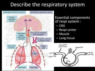
  • 1. Describe the respiratory system • Essential components of respi system : – CNS – Respi center – Muscle – Lung tissue
  • 2. Depict signs of respiratory distress in a real patient
  • 3. Respiratory distress • It is an abnormal physiologic condition identified by increased work of breathing. • The child in respiratory distress is effectively compensating to preserve adequate gas exchange.
  • 4.
  • 5. Signs of Respiratory distress • Tachypnea • Tachycardia • Retractions (supraclavicular, suprasternal, intercostal or subcostal) • Use of accessory neck muscles • Nasal flaring and grunting • Pallor or cyanosis • Altered mental status (restlessness, irritability, confusion, anxiety, inability to recognize parents)
  • 6. Positioning • With an upper airway obstruction, the patient characteristically assumes sniffing position (jaw thrust forward) and may drool due to inability to swallow secretions. • A patient in severe respiratory distress may position himself in the tripod position (seated and leaning forward on outstretched arms), sitting upright and leaning forward to maximize use of accessory muscles.
  • 7. Respiratory rate • Abnormal respiratory rate is common finding in respiratory distress. • Although normal respiratory rate varies with age, a rate greater than 60 breaths per minute is abnormal in any child. • A slow respiratory rate might arise concerns as it may signifies respiratory fatigue and imminent respiratory arrest in an exhausted child.
  • 8. Retractions • Subcostal, intercostal and suprasternal retractions are manifestations of increased work of breathing as well as use of accessory muscles of respiration. • Paradoxic breathing (abdomen retracts upon inspiration and expands upon expiration) may be noted.
  • 9. Nasal flaring and grunting • Flaring of the nostrils with inspiration is also a sign of respiratory distress. • Grunting is a sound made by forceful expiration against a partially closed glottis. • Head bobbing may be seen in infants.
  • 10. Lung Sounds and Auscultation • Decreased breath sounds and diminished air entry in the setting of increased respiratory effort suggest worsening respiratory disease.
  • 11. Heart Rate • Tachycardia is a common finding in children in respiratory distress and is not necessarily indicative of cardiovascular instability. • Bradycardia, for instance, is indicative of severe hypoxemia and acidosis which require critical interventions. • Bradycardia in a child with history of respiratory distress signifies imminent cardiopulmonary arrest.
  • 12. Altered level of consciousness • Some children might present with effortless tachypnea resulting from an increased respiratory drive caused by metabolic acidosis or central nervous system disease. • Therefore, mental status is evaluated as a gauge of systemic oxygenation and critical management must be done if the patient appears lethargic or agitated.
  • 13. Normal Respiratory Rate • Infant = 30 – 40 /min • Toddler = 24 – 40 /min • School age = 18 – 30 /min • Adolescent = 12 – 16 / min
  • 14. Define respiratory failure • Respiratory failure is a syndrome of inadequate gas exchange due to dysfunction of one or more essential components of the respiratory system: • Type I : Hypoxaemic respiratory failure. PaO2 < 60 mm Hg with a normal or low PaCO2. This is caused by ventilation- perfusion mismatch with either/both: – Under-ventilated alveoli (eg pulmonary oedema, pneumonia or acute asthma). – Venous blood bypasses ventilated alveoli (eg right to left cardiac shunts). • Type II : Hypercapnic respiratory failure. PaCO2 > 50 mm Hg and indicates inadequate alveolar ventilation. Any ventilation-perfusion mismatch will affect PaO2 and therefore hypoxaemia is also common.
  • 15. • Common causes of type • Common causes of type I respiratory failure II respiratory failure – COPD. – Drug – Pneumonia. overdose, poisoning. – Pulmonary oedema. – Myasthenia gravis. – Pulmonary fibrosis. – Polyneuropathy. – Asthma. – Poliomyelitis. – Pneumothorax. – Muscle disorders. – Pulmonary embolism. – Head injuries and neck – Pulmonary hypertension. injuries. – Cyanotic congenital heart – Hypothyroidism. disease. – Bronchiectasis. – Acute respiratory distress syndrome.
  • 16. Ix : • Arterial blood gas analysis: confirmation of the diagnosis. • CXR: often identifies the cause of respiratory failure. • FBC: anaemia can contribute to tissue hypoxia; polycythaemia may indicate chronic hypoxaemic respiratory failure. • Renal function tests and LFTs: may provide clues to the aetiology or identify complications associated with respiratory failure. Abnormalities in electrolytes such as potassium, magnesium, and phosphate may aggravate respiratory failure and other organ dysfunction. • Serum creatine kinase and troponin I: to help exclude recent myocardial infarction. Elevated creatine kinase may also indicate myositis. • TFTs (hypothyroidism may cause chronic hypercapnic respiratory failure). • Spirometry: useful in the evaluation of chronic respiratory failure. • Echocardiography: if a cardiac cause of acute respiratory failure is suspected. • Pulmonary function tests are useful in the evaluation of chronic respiratory failure. • ECG: to evaluate a cardiovascular cause; it may also detect dysrhythmias resulting from severe hypoxaemia or acidosis.
  • 17. Management • A patient with acute respiratory failure generally needs prompt hospital admission in an intensive care unit. • Many patients with chronic respiratory failure can be treated at home, depending on the severity of respiratory failure, underlying cause, comorbidities and social circumstances. • Immediate resuscitation may be required. • Appropriate management of the underlying cause.
  • 18. Describe obstructive and restrictive respiratory conditions
  • 19. Obstructive disorders • These are respiratory disease which make it more difficult to get air out of the lungs. • This group of disorders is characterized by obstruction of normal airflow due to airway narrowing and includes: Asthma, COPD, bronchiectasis, cystic fibrosis and tumors. • Mechanisms causing airway narrowing differ and may include obstruction by a mucus plug, airway compression and smooth muscle constriction.
  • 20. Obstructive disorders • In general, obstructive disorders lead to hyperinflation of the lungs as air is trapped behind closed airways. • Residual volume is increased as in the ratio of RV: TLC. Functional residual capacity also increased. • In patients with severe obstruction, air trapping is so extensive that vital capacity is decreased.
  • 21. • In addition, dynamic compression of the airway prolongs the FEV1, as well as the forced expiratory flow rate measured over the middle half of expiration (FEF 25-75%) • Both FEV1 and FVC are reduced, but the reduction in FEV1 is greater (airway narrowing reduces the speed of forced expiration such that FEV1 is reduced to an abnormally low fraction of FVC, perhaps to 40% or lower). • Result: low FEV1 / FVC.
  • 22. Obstructive disorders • Extrathoracic- stridor • Intrathoracic- wheezing, best heard during expiration • Long deep breaths • Hyperinflated and tympanitic to percussion
  • 24.
  • 25.
  • 26. Restrictive disorders • These are respiratory disease which makes it more difficult to get air into the lungs. • They “restrict” inspiration. • Examples include: Fibrosis, sarcoidosis, silicosis, asbestosis, muscular diseases and chest wall deformities.
  • 27. Restrictive disorders • In fibrosis or alveolar wall thickening, the increased connective tissue reduces lung compliance, making it difficult to expand the lung during inspiration. • In condition such as alveolar edema, respiratory distress syndrome, or infiltrative interstitial lung diseases, the lungs are stiff decreasing its compliance. • Extrapulmonary factors that can contribute to decrease vital capacity include limited thoracic expansion (Pleural fibrosis, Kyphoscoliosis) and nerve or muscular dysfunction.
  • 28. • As a result, all lung volumes are reduced. In particular, there can be marked decreases in total lung capacity and functional vital capacity. • FEV1 and FVC are reduced proportionately but the normal ratio is preserve. • The patient might demonstrate rapid shallow breathing. He/she might also shows grunting. • Percussion of the respiratory system demonstrate dull/ hyper-resonance?
  • 29.
  • 30.
  • 31.
  • 32. Differentiate clinical presentation of obstructive airway Upper airways obstruction Lower airways obstruction Stridor Wheezing • harsh sound •Continuous expiratory sound •Produced near larynx by the vibration of •Due to turbulence airflow upper airway structures •Musical •Inspiratory ( expiratory – less common) •Partial obstruction produces wheezing •hoarseness – effect vocal chord sound at later phase of expiration •Supraglottic etiology – moves with child’s •Harsh, monophonic and low pitch (usually head from large, central airways) •Decreases while sleep – inspiration at lower • high pitched and musical (from small and rate during sleeping peripheral airways) Suprasternal retraction Rhonchi •Pressure gradient between trachea and •Secretion in intrathoracic airways, resulting atmosphere in irregular sound Rales or crackles •Sound characterized by of crumpling celophane due to fluid or secretion in small airways
  • 33. Describe conditions presented with wheezing in children and infants • Acute wheezing : – Infection – Foreign body aspiration (FBA) • Chronic/ recurrent wheezing – Anomalies of the tracheobronchial tree (ie Congenital tracheomalacia and bronchomalacia) – Cardiovascular disease (pulmonary artery dilation and/or left atrial enlargement, including large left to right shunts, can compress large airways and cause wheezing) – Mediastinal masses (ie tumors, cyst, LN enlargement)
  • 34. Upper respiratory tract conditions - to be presented in tabulated manner • common cold • acute pharyngitis • acute otitis media • acute sinusitis
  • 35. common cold acute pharyngitis acute otitis media acute sinusitis Fever Inflamed pharynx and fever, Cough Nasal discharge soft palate irritability, Nasal symptoms Loss of appetite Enlarged and tender headache, Fever Difficulty sleeping. LN apathy, Headache anorexia, Facial pain and vomiting, swelling diarrhea Sore throat Rhinovirus, Adenovirus RSV, rhinovirus, Rhinovirus, coronavirus, RSV Enterovirus Pneumococcus, adenovirus, influenza, Rhinovirus H. influenzae or parainfluenza Gp A β-hemolytic strepto Treat fever and pain Paracetamol Paracetamol Nebulized anti- with paracetamol or Penicillin (bact only) Ibuprofen microbials ibuprofen
  • 36. Pnuemococal Staphylococcus Mycoplasma Viral pneumonia pneumonia pneumonia pneumonia S. Pneumoniae Staphylococcus Mycoplasma Influenza virus aureus pnuemoniae Parainfluenza virus Community acquires Presentation Presentation Presentation •Skin infection •Fever •Fever •Soft tissue •Malaise •Chills infection – •Headche •Non productive pyomyositis, septic •Persistent slowly cough bursitis worsening dry •Rhinitis •Toxic shock cough •Myalgias syndrome •Chills •Headache •Endocarditis •Sore throat •fatigue •Osteromyelitis •Pluertictic chest •Pneumonia pain •Food poisoning •UTI Tx Tx Antibiotics amantindine, Antibiotics Antimicrobial Macrolides, rimantidine, therapy doxycyline acylovir
  • 37. Pertussis – whooping cough Etiology Caused by Bordetella pertussis Infect among human through respiratory droplets (coughing) Clinical presentation Infants beyond neonatal period through school age Catarrhal stage – injection, increased nasal secretion and low grade fever 1-2 weeks Paroxysmal stage – coughing (expiration) to dislodge plugs of necrotic bronchial epithelial tissues and thick mucus. Lasted 2-4 weeks Followed by forceful inhalation against narrowed glottis – whoop characteristic Convalescent stage – symptom gradually resolve 1-2 weeks, coughing less severe, whooping cough dissapear. CNS damage due to hypoxia, bacterial pnuemonia, Adult – starts with broncholitis illness. Tx Erythromycin – early coarse, eradicates nasopharyngeal carriage within 3-4days, ameliorate the effects of infection, ineffective at paroxysmal stage, , ass with pyloric stenosis if given to neonates <4 weeks old Azithromycin & clarithromycin – shorter duration, ass with gastrointestinal adverse effects Pertussis-specific immunoglobin – effectivelt reduced symptoms at paroxysmal stage TMP-SMZ (trimethoprim/sulfamethozxazole)
  • 38. Pulmonary tuberculosis Etiology Caused by Mycobacterium tuberculosis Spread via respiratory droplets – become respiratory nuclei, <10um can reach alveoli Clincal manifestation •Asymptomatic in older and infants patient •+ve tuberculin skin test •Minimal abnormalities on CXR infiltration with hilar lymphadenopathy (Ghon complex) •Malaise, low grade fever, erythema nodosum, lympahadenopathy symptoms. Investigation Tuberculin skin test (TST) •T-cell-mediated hypersensitivity, 2-6 weeks Mantoux test •Intradermal injection of 5TU on volar surface of forearm, screen high-risk population and for diagnosis in all ill patients or contacts. •False –ve at early phase of illness or immunosuppression Culture Tissue, sputum, fluid Treatment Antibiotics – RIPES, 9-months of isoniazid&rifampin 98% case cured Prevention - BCG
  • 39. • Acute viral bronchiolitis • transient infant wheeze • chronic lung diseases of prematurity • bronchiectasis.
  • 40. Acute viral bronchiolitis • lower respiratory tract infection (LRTI) that primarily affects the small airways (bronchioles) – RSV is the most common cause, followed by rhinovirus – Less common causes include parainfluenza virus, human metapneumovirus, influenza virus, adenovirus, coronavirus,
  • 41. • S/s : fever (usually ≤38.3ºC), cough, mild respiratory distress, preceded by a one- to three- day history of upper respiratory tract symptoms, such as nasal congestion and/or discharge. • PE : – tachypnea – intercostal and subcostal retractions, often with expiratory wheezing. – The chest may appear hyperexpanded with increased anteroposterior (AP) diameter and may be hyperresonant to percussion. – Findings on auscultation include any combination of expiratory wheeze, prolonged expiratory phase, and both coarse and fine crackles. • Ix : Mild hypoxemia (oxygen saturation <95 percent) commonly is detected by pulse oximetry, even without clinical signs of desaturation
  • 42. • Mx : – SEVERITY ASSESSMENT — Severe bronchiolitis is indicated by persistently increased respiratory effort (tachypnea; nasal flaring; intercostal, subcostal, or suprasternal retractions; accessory muscle use; grunting), hypoxemia, apnea, or acute respiratory failure – In healthy infants and young children, bronchiolitis usually is a self-limited disease. – Maintain oxygenation and hydration. – Bronchodilator therapy may be beneficial in a subset of patients
  • 43. Transient infant wheeze • The successful transition from fetal to neonatal life at delivery requires a series of rapid physiologic changes of the cardiorespiratory system. These changes result in redirection of gas exchange from the placenta to the lung and comprise: – Replacement of alveolar fluid with air – Onset of regular breathing – Increase in pulmonary blood flow as a result of increased systemic vascular resistance and decreased pulmonary vascular resistance (PVR)
  • 44. • difficulty may be a consequence of : – impaired lung function due to fluid retention, – airway obstruction associated with congenital anomalies, – persistent pulmonary hypertension, – apnea associated with lack of respiratory effort.
  • 45. Respiratory distress syndrome • Respiratory distress (ie, tachypnea and labored breathing) and cyanosis occur at or soon after birth. • Typical signs include – grunting (which prevents end-expiratory alveolar collapse), – nasal flaring (which reduces nasal resistance and reflects increased use of accessory muscles of respiration), – and intercostal and subcostal retractions (due to decreased lung compliance and the highly compliant chest wall). • Surfactant deficient • inability to clear lung fluid from air spaces
  • 46. Bronchiectasis • Abnormal dilation and distortion of the bronchial tree, resulting in chronic obstructive lung disease. • End result of a variety of pathophysiologic processes that render the bronchial walls weakened, easily collapsible, chronically inflamed, and plugged with mucus secretions. • Associated findings include atelectasis, emphysema, fibrosis, and hypertrophy of the bronchial vasculature.
  • 47. • S/s : productive cough, purulent sputum, fever, pleuritic chest pain, and dyspnea • PE : – Crackles (inspiratory popping sounds) and rhonchi (coarse expiratory breath sounds), – Wheezing (less common) – Clubbing of the nailbed – Chest wall deformity, which can be seen in obstructive lung diseases (eg, cystic fibrosis)
  • 48. • Ix : – CXR - linear atelectasis and dilated and thickened airways, HRCT – sweat chloride or DNA testing – blood count with differential, immunoglobulins, and IgG subclasses • Mx : aimed at treating the underlying cause, improving mucocilliary clearance, treating and preventing infection, and controlling inflammation – Antibiotic therapy – Chest physiotherapy – Mucolytic agents and airway hydration – Bronchodilators – Anti-inflammatory drugs – Immunization
  • 49. Laryngotracheobronchitis (Croup) • It is a viral infection of the middle respiratory tract occurs from 6 months to 6 years of age with peak incidence in the 2nd year of life. • There is mucosal inflammation and increased secretions affecting the airway, but it is the edema of the subglottic area that is potentially dangerous as it may result in critical narrowing of the trachea.
  • 50. Laryngotracheobronchitis (Croup) • A small decrease in diameter secondary to mucosal edema and inflammation exponentially increases airway resistance and significantly increases work of breathing. • During inspiration, walls of the subglottic space are drawn together, aggravating the obstruction and producing stridor characteristic of croup.
  • 51. Laryngotracheobronchitis (Croup) • Symptoms are  Low grade fever, cough and coryza for 12-72 hours followed by  Stridor that may occur when excited, at rest or both (Harsh, high-pitched respiratory sound produced by turbulent airflow; usually inspiratory, but may be biphasic and is a sign of upper airway obstruction)  hoarse voice, brassy or “barking” cough  Respiratory distress of varying degree • Onset of symptoms is usually over several days and often start and worse at night. • Symptomatic reinfection is common but usually mild.
  • 52. Laryngotracheobronchitis (Croup) • Parainfluenza viruses (type 1,2,3) are the commonest cause but other viruses such as human metapneumovirus, RSV and influenza virus type A and B, adenovirus, enterovirus, measles, mumps and rhinoviruses can produce similar clinical picture. • Chest X-ray reveals a characteristic narrowing of the subglottic region known as the “steeple sign”.
  • 53.
  • 55. Laryngotracheobronchitis (Croup) • When the upper airway obstruction is mild, the stridor and chest recession disappear when the child is at rest. • The child can usually be managed at home and need to be closely monitored for signs of increasing severity. • Most cases of croup are mild and resolve within 1 to 2 days.
  • 56. Laryngotracheobronchitis (Croup) • Indication for hospital admission Moderate and severe viral croup Poor oral intake Family lives a long distance from hospital, lacks reliable transport Toxic looking Age less than 6 months Unreliable caregivers at home
  • 58. Management of Croup • Oxygen therapy indication include severe viral croup and percutaneous SaO2 <93%. • Steroids are helpful in moderate to severe cases. • Antibiotics may be indicated if the child fails to improve or if purulent secretions are present where coverage should be directed towards Staphylococcus and H.influenzae.
  • 59. Acute epiglottitis (Supraglottitis) • It is less common than croup, typically presents with a greater degree of airway compromise. • H.influenzae type B is the major etiologic organism. • The incidence of epiglottitis has decreased with the administration of H. influenzae vaccine. • It is most common in children aged 1-6 years but affect all age groups.
  • 60. Clinical manifestations of Acute epiglottitis • Patients typically present with acute onset (over several hours) of  High fever in an ill, toxic-looking child  An intensely painful throat preventing child from speaking or swallowing with saliva drools down the chin  Marked dysphagia  Soft inspiratory stridor and rapidly increasing respiratory difficulty over hours  Patients are often encountered leaning forward in a “tripod” position (Immobile, upright with an open mouth) • Unlike croup, cough is frequently absent.
  • 61. Acute epiglottitis (Supraglottitis) • Attempts to lie the child down or examine the throat with a spatula or perform a lateral neck X-ray must not be undertaken as this may precipitate total obstruction and death. • Instrumentation of the throat with tongue depressors is not advisable as this can precipitate a fatal laryngospasm.
  • 62. Acute epiglottitis (Supraglottitis) • If the diagnosis of epiglottitis is suspected, urgent hospital admission and treatment are required. • The child should be intubated under controlled conditions with a general anesthetic. • Rarely, this is impossible and urgent tracheostomy is life- saving. • Only after securing the airway, blood is taken for culture and broad-spectrum IV antibiotics such as cefuroxime directed against H.influenzae and gram-positive organisms started. • Tracheal tube can usually be removed after 24 hours and antibiotics given for 2-3 days. • Prophylaxis with rifampicin is offered to close household contacts.
  • 63. Clinical features of croup and epiglottitis Croup Epiglottitis Onset Over days Over hours Preceding coryza Yes No Cough Severe, barking Absent or slight Able to drink Yes No Drooling saliva No Yes Appearance Unwell Toxic, very ill Fever <38.5 Celsius >38.5 Celsius Stridor Harsh, rasping Soft, whispering Voice, cry Hoarse Muffled, reluctant to speak
  • 64. Laryngomalacia • It is the most common congenital anomaly of the larynx, accounting for up to 60% of all anomalies. • Inspiratory stridor is the hallmark of the condition. • Symptoms are typically aggravated when the child is supine or crying. • Examination reveals partial collapse of a flaccid supraglottic airway with inspiration. • The condition is generally benign and self-limited, as most cases resolve by 18 months of age. • Severe cases may require surgical intervention if the distress prevents adequate feeding.
  • 65.
  • 66.
  • 67.
  • 68. Congenital subglottic stenosis Etiology •Can be iatrogenic •Intubation and mechanical ventilation – residual damage to the larynx •Down’s syndrome infants more susceptible due to smaller larynx Clinical manifestation •Subglottic obstruction produces stridor and frequently biphasic (expiration and inspiration) •Worsen with respiratory effort •Viral infection exacerbate subglottic stenosis Diagnostic studies •Requires endoscopic evaluation Treatment •Tracheostomy and reconstructive surgery may be necessary •Some improve with age as larynx grow and require no intervention
  • 69. Retropharyngeal abscess & Peritonsilar abcess Retropharyngeal abscess Comparison Peritonsilar abscess <6 years Age >10 years Posterior pharynx Location Oropharynx S. aureus, anaerobes Etiology Group A streptococci, anaerobes Insidious to sudden Prodrome onset Biphasic with sudden worsening None Stridor No Yes Retraction No Muffled Voice “Hot potato” muffled Arching of neck or normal Position and appearance Normal Drooling Swallowing (dysphagia) Drooling, trismus No Barking cough None Severe Toxicity Dyspnea >101 F Fever >101 F Thickened retropharyngeal space X Ray None needed Leukocytosis with left shift WBC count Leukocytosis with left shift Antibiotics, surgical drain - abscess Therapy Antibiotics, aspiration None Prevention Treat Group A streptococci early
  • 70. Diphtheria Etiology Caused by bacteria Corynebacterium diphtheriae spread through respiratory droplets from infected person or carrier Infects nose, throat causes gray to black, thick fiber like covering airway Infects skin - Skin lesions Produces toxins – spread via bloodstream to other organs Signs and symptom Mild fever, sore throat, chills – few days after infection Breathing problems, croup-like (barking cough), drooling, hoarseness Nasal discharge, fatigue Dysphagia, paralysis, heart failure – serious Tx Antitoxin IV/IM Antibiotics – penicillin and erythromycin Diphtheria vaccine – lasted for 10 years (if in contact with infected person)
  • 71. Chronic lung disease of prematurity • Infants who still have an oxygen requirement at a post-menstrual age of 36 weeks are described as having bronchopulmonary dysplasia (BPD) or chronic lung disease. • Criteria for diagnosis are oxygen requirement beyond 36 weeks post conceptional age or beyond 28 days of life.
  • 72. Pathophysiology of BPD • The lung damage (alveolar cells, interstitium and blood vessels) comes from pressure and volume trauma from artificial ventilation, oxygen toxicity and infection. • Oxygen concentration greater than 40% are toxic to the neonatal lung. • Oxygen-mediated lung injury results from the generation of superoxides, hydrogen peroxide and oxygen free radicals which disrupt membrane lipids.
  • 73. Pathophysiology of BPD • Assisted ventilation with high peak pressures produces barotrauma, compounding the damaging effects of highly inspired oxygen levels. • Inflammatory mediators are released and causing increase permeability of blood vessels leading to leakage of water and protein. • In later stages, there is fibrosis and cellular hyperplasia. • Severe lung damage can leads to respiratory failure.
  • 74.
  • 75. Risk factors of BPD • In most patients, BPD develops after ventilation for RDS that may have been complicated by PDA (Patent ductus arteriosus) or pulmonary interstitial emphysema. • Inflammation from prolonged assisted ventilation and repeated systemic and pulmonary infections may play a major role. • Failure of RDS to improve after 2 weeks, the need for prolonged mechanical ventilation and oxygen therapy required at 36 weeks’ postconceptional age are characteristics of patients with RDS in whom BPD develops.
  • 76. Risk factors of BPD • BPD also may develop in infants weighing less than 1000g who require mechanical ventilation for poor respiratory drive in the absence of RDS. • 50% of infants of 24 to 26 weeks’ gestational age require oxygen at 36 weeks’ corrected age.
  • 77. Chest X-Ray of BPD • The chest X-ray characteristically shows widespread areas of opacification, sometimes with cystic changes accompanied by areas of over-distention and atelectasis, giving the lung a sponge like appearance. • Histopathology of BPD reveals interstitial edema, atelectasis, mucosal metaplasia, interstitial fibrosis, necrotizing obliterative bronchiolitis and overdistended alveoli.
  • 78.
  • 79.
  • 80. Clinical manifestations of BPD • The clinical manifestations of BPD are oxygen dependence, hypercapnia, compensatory metabolic alkalosis, pulmonary hypertension, poor growth and development of right-sided heart failure. • Increased airway resistance with reactive airway bronchoconstriction also is noted and is treated with bronchodilating agents. • Severe chest retractions produce negative interstitial pressure that draws fluid into the interstitial space. • Together with cor pulmonale, these chest retractions cause fluid retention, necessitating fluid restriction and the administration of diuretics.
  • 81. Management of BPD • Some infants need prolonged artificial ventilation, but most are weaned onto continuous positive airways pressure (CPAP) followed by additional ambient oxygen, sometimes over several months. • To reduce risk of subglottic stenosis, a tracheotomy may be indicated. • To reduce oxygen toxicity and barotrauma, ventilator settings are reduced to maintain blood gases with slightly lower PaO2 (50mmHg) and higher PaCO2 (50 to 75 mmHg) levels than for infants during the acute phase of RDS.
  • 82. Management of BPD • Dexamethasone therapy may reduce inflammation, improve pulmonary function and enhance weaning of patients from assisted ventilation. • However, corticosteroid therapy may increase risk of abnormal neuro-development including cerebral palsy, thus limits use to those at highest risk and only short courses are given.
  • 83. Management of BPD • Some babies go home while still receiving additional oxygen. • A few infants with severe disease may die of intercurrent infection or pulmonary hypertension. • Some survivors of BPD have hyperinflation, reactive airways and developmental delay. • Subsequent pertussis and RSV (Respiratory syncytial virus) infection may cause respiratory failure necessitating intensive care and thus prophylaxis against RSV should be given.
  • 84. Inhaled foreign body Epidemiology •Common, 4 y/o •Main right bronchus (direct continous with trachea •May migrate on larynx (coughing) causes occlusing of airway Clinical manifestation •Cough, localized wheezing, unilateral absence of breath sounds, stridor, blood sputum (rare) •fever, cough, sputum production, or chest pain – need medical attention •Persistence wheezing unresponsive to bronchodilator therapy, persistent atelectasis, recurrent or persistence pneumonia, persistent cough without other explanation – suspect foreign body inhalation. •Lodge in esophagus and compress trachea – produce respiratory symptoms Diagnostic studies •Radiographic studies – presence of radiopaque object or eveidence of air trapping Prevention •Education to parent and caregiver •Small toys should kept away
  • 85. Pneumothorax • defined as a collection of air that is located within the thoracic cage between the visceral and parietal pleura – Primary : no underlying lung disease – Secondary : complication of underlying lung disease (ie asthma, cystic fibrosis, necrotizing pneumonia, and interstitial lung disease)
  • 86. • S/s : dyspnea and pleuritic chest pain that is described as sharp or stabbing. The pain typically is diffuse on the affected side with radiation to the ipsilateral shoulder. A dry or non-productive cough • PE : – deviation of the trachea towards the contralateral side – diminished breath sounds – hyperresonant percussion – decreased vocal fremitus on the affected side. – Other signs : tachypnea, increased work of breathing, and cyanosis.
  • 87. • Ix : ABG, CXR • Mx : depends on the size of the pneumothorax, the extent of respiratory distress, and the presence or absence of underlying lung disease. The goals of treatment are to remove air from the pleural space and to prevent recurrence. – Supplemental oxygen – Needle aspiration – Thoracostomy tube
  • 88. Empyema • Early in the course of parapneumonic effusion, the pleura becomes inflamed; leakage of proteins, fluid, and leukocytes. At the time of formation, the pleural effusion is usually sterile with a low leukocyte count. With time, bacteria invade the fluid, resulting in empyema, which is defined as the presence of grossly purulent fluid in the pleural cavity. • S/s : persistent fever, cough, malaise, decreased appetite, chest pain, and dyspnea • PE : tachypneic, but breaths may be shallow to minimize pain, mediastinal shift and tension hydrothorax, Hypoalbuminemia
  • 89. • Ix : Pulse oximetry , Blood and sputum culture, CXR • Management: – Main goals : sterilization of the pleural cavity, resolution of pleural fluid, and reexpansion of the lung – Antibiotics – Thoracocentesis – Chest tubes – Surgical (for symptoms for more than one week only)
  • 90. Pneumatocele • A pneumatocele is a cavity in the lung parenchyma filled with air (cyst) that may result from pulmonary trauma during mechanical ventilation • Ddx : Lung ca, TB • Mx : palliative care
  • 91. Lung abscess • Result from lung tissue necrosis from an infection (ie pneumonia) • Conditions contributing to lung abscess – Aspiration of oropharyngeal or gastric secretion – Septic emboli – Vasculitis: Wegener's granulomatosis – Necrotizing tumors:
  • 92. Congenital diaphragmatic hernia Congenital lobar emphysema (congenital lobar over distention) Etiology Over inflation of one lobe during neonatal Improper joining of structure during fetal dev. period – left upper lobe (often) Stomach, small intestine, spleen appear in chest cavity, Lung tissue not completely develop Left side – common Increase risk with +ve family hx Symptom •Produces respiratory distress because Breathing difficulty after born due to surrounding lung tissue has compressed ineffective movement of diaphragm and •Mediastinal shift crowding of lung tissue, which causes collapse Cyanosis, tachypnea, tachycardia Irregular chest movement Absence of breath sound on both side Bowel sound heard in the chest Palpation – abd feels less full Xray – abd organs in chest cavity Surgery - place abd organs back n repair Lobectomy – if RDS is severe and progressive diapgrmatic opening Breathing support until recvers from surgery
  • 94. • S/s : if the defect is large, with coughing and choking associated with feeding. • Diagnosis — attempt to pass a catheter into the stomach. This finding can be confirmed with an anterior-posterior chest radiograph that demonstrates the catheter curled in the upper esophageal pouch • Mx : surgical ligation of the fistula. – if the distance between esophageal segments is large, staged procedures are performed that include elongation of the esophagus.
  • 95.
  • 96. Gastroesophageal reflux (GER)  It is defined as the effortless retrograde movement of gastric contents into the esophagus.  In infancy, GER is not always an abnormality.  Distinction between physiologic GER and pathologic GER in infancy and childhood is determined by presence of reflux-related complications.
  • 97. Physiologic GER  Physiologic GER (Spitting up) is normal in infants younger than 8 to 12 months of age.  Nearly half of all infants are reported to spit up at 2 months of age.  It is caused by inappropriate relaxation of the lower esophageal sphincter as result of functional immaturity.  Infants who regurgitate stomach contents meet the criteria for physiologic GER, as long as they maintain adequate nutrition and have no signs of respiratory complications or esophagitis.
  • 98. Clinical manifestations  Presence of GER is easy to observe in an infant who spits up.  In older children, the refluxate is usually kept down by reswallowing, but GER may be suspected by associated symptoms, such as: • Heartburn • Cough • Dysphagia • Wheezing • Aspiration pneumonia • Hoarse voice • Failure to thrive • Recurrent otitis media/ Sinusitis
  • 99. Clinical manifestations  GER can have both esophageal and extraesophageal manifestations.  The esophageal manifestations are those caused directly by contact between refluxed gastric juice and the esophageal mucosa.  The extra-esophageal manifestations are caused by contact between refluxed gastric juice and extraesophageal organs or by reflexes triggered by acid in the esophagus that affects other organs.
  • 100. Extraesophageal complications  Cardiac: Atypical chest pain, ischemia  Vocal cords: Laryngitis, granuloma, polyps, ulcers, neoplasm  Respiratory: asthma, recurrent bronchitis, aspiration or chronic interstitial pneumonia, sleep apnea, chronic or recurrent cough  Pharyngeal: pharyngitis, hoarseness  Oral: Erosions, burning mouth syndrome  Other: sudden infant death syndrome, otitis media
  • 101. Investigations 1. Barium upper gastrointestinal series 2. 24-hour esophageal pH probe monitoring 3. Esophageal impedance monitoring 4. Endoscopy
  • 102. Relation to respiratory symptoms  GER has been associated with significant respiratory symptoms in infants and children.  The infant’s proximal airway and esophagus are lined with receptors that are activated by water, acid or distention.  Activation of these receptors can increase airway resistance, leading to development of reactive airway disease.
  • 103. Relation to respiratory symptoms  One postulated mechanism for gastroesophageal reflux- mediated airway disease involves micro-aspiration of gastric contents that leads to inflammation and bronchospasm.  However, experimental evidence also supports the involvement of esophageal acid-induced reflex bronchospasm in the absence of frank aspiration.  Therapy using histamine 2 blockers or proton pump inhibitors has been shown to benefits patients with steroid-dependent asthma, nocturnal cough and reflux symptoms.
  • 104. Asthma and GER  In 1892, Sir William Osler first postulated a relationship between asthma and gastroesophageal reflux, manifested by a bi-directional cause-and-effect presentation.  Prevalence of GER is more common in asthmatics compared to control populations ranging from 47 to 75%.  Severe studies suggest that severe nocturnal asthma is more likely to be associated with GER than daytime asthma.
  • 105.
  • 106. Asthma and GER  Not all asthmatics with GER will have esophageal symptoms.  The mechanism of esophageal acid induced bronchoconstriction includes a vagally mediated esophageal bronchial reflex, airway hyper responsiveness and microaspiration.
  • 107.
  • 108.
  • 109. Asthma and GER  It has been suggested that a vagally mediated reflex exists whereby acid reflux into the lower esophagus initiates bronchoconstriction via a shared autonomic innervation between esophagus and bronchi based on their common embryonic origin.  Support is based on this two lines of evidence: • Acid infusion of the esophagus in asthmatic children results in increased airway resistance, which can be reversed with antacids. • Infusion of antacids into the distal esophagus of asthmatic children during sleep induces bronchoconstriction, suggesting presence of a protective mechanism.
  • 110. Asthma and GER  Esophageal acidification has also been associated with heightened non-specific hyperresponsivity of the airways to bronchoconstrictors contributed by increased vagal tone, “priming” the airways to other triggers.
  • 111. Asthma and GER  Asthmatics have significant decreased lower esophageal sphincter pressures, greater esophageal acid exposure times, more frequent episodes of GER and longer esophageal acid clearance times.  There may be increase in transient lower esophageal sphincter (LES) relaxations, LES hypotonia and esophageal due to autonomic dysfunction in asthmatics patient.  Furthermore, LES pressure augmented by diaphragm normally prevents GER but it can be prevented by hyperinflation and hiatal hernia.  LES pressure can also be overcome by increased negative intrathoracic pressures (bronchospasm, cough, upper airway resistance syndrome and obstructive apnea).
  • 112. Asthma and GER  Anti-reflux therapy results in improvement in asthma in approximately 70% of patients.  Acid-suppression medication such as histamine 2 receptor antagonists (ranitidine) or proton pump inhibitors (omeprazole) are helpful in reducing volume of gastric contents. (Therapeutic trial)
  • 113.
  • 114.
  • 115. Chronic cough and GER  GER can potentially stimulate sensory limb of cough reflex in multiple ways.  It can irritate upper respiratory tract without aspiration (such as larynx) and the lower respiratory tract by either macro-microaspiration.  It can also cause cough by stimulating an esophagus bronchial cough reflex whereby refluxate into distal esophagus alone is a sufficient stimulus.
  • 116. Chronic cough and GER  GER related cough occurs predominantly during the day and in the upright position whereby some patients may have nocturnal symptoms.  Cough may be the sole manifestation of GER and is often long standing.  Diagnosis can be made with certainty only when cough are resolved with specific anti-reflux therapy.
  • 117. Senario 3  Samy, a 2 days old premature infant who was born at 30 weeks gestational age was noted to be cyanosed by a nurse. There was absence of chest movement.  What is Samy’s clinical condition?  Respiratory distress syndrome (RDS)  What is your immediate action?  surfactant replacement therapy  IV glucocorticoid administration  What is the mechanism of his respiratory failure?  Immature lung  lack surfactant  atelectasis
  • 118. Senario 5  Aminah a 2 year old toddler has been having mild cough and rhinorrhea for the past 2 days. It was associated with low grade fever. Suddenly her mother noticed that she breath noisily with fast breathing.  What is Aminah’s clinical condition?  Common cold  What are the relevant physical finding that you would like to look for?  Signs of respiratory distress  Outline the principle management.  Symptomatic therapy