SlideShare una empresa de Scribd logo
1 de 86
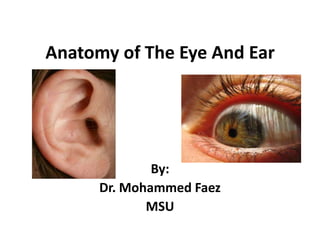
Anatomy of The Eye And Ear By: Dr. Mohammed Faez MSU
Orbit Cavity or socket of the skull which houses the eye  Protects and stabilizes the eye Serves as attachment site for extrinsic muscles
Orbit Orbital Margins – bases which open in the face        (4 borders)  Supraorbital margin – frontal bone  Infraorbitalmargin – zygomatic and maxilla bones Lateral margin – zygomatic and frontal bones Yellow – Frontal Bone Blue – Zygomatic Bone Purple – Maxilla Bone
Orbit Anterior aspect or roof Frontal Bone Posterior aspect Sphenoid Bone Medial aspect Lacrimal, ethmoid, maxillary, and sphenoid bones Lateral aspect Zygomatic and sphenoid bones Orbit is thickest Yellow – Frontal Bone Blue – Zygomatic Bone Purple – Maxilla Bone
Orbit Frontal Bone Ethmoid Bone Lacrimal Bone Sphenoid Bone Zygomatic Bone Maxilla Bone
Orbit Optic Canal Foramen which the optic nerve passes to reach the brain Optic Nerve Cranial nerve II Transmits visual information from the retina to the brain Superior Orbital Fissure Opening between lesser and greater wings of sphenoid bone Allows cranial nerves, arteries, and veins to communicate with eye
Surface Anatomy of The Eye  Major features of the eye include the sclera, cornea, iris, and pupil. The cornea is continuous with the sclera and is the clear circular region of the external covering of the eye through which the pupil and iris are visible.  The sclera is not transparent and is normally white.
Surface Anatomy of The Eye  The upper and lower eyelids of each eye enclose between them the palpebral fissure.  The eyelids come together at the medial and lateral palpebral commissures on either side of each eye.
Surface Anatomy of The Eye  At the medial side of the palpebral fissure and lateral to the medial palpebral commissure is a small triangular soft tissue structure (the lacrimal lake).  The elevated mound of tissue on the medial side of the lacrimal lake is the lacrimal caruncle, and the lateral margin overlying the sclera is the lacrimal fold.
Surface Anatomy of The Eye
The lacrimal apparatus The lacrimal apparatus consists of the lacrimal gland and the system of ducts and channels The lacrimal gland is associated with the upper eyelid and is in a small depression in the lateral roof of the orbit just posterior to the orbital margin.  The multiple small ducts of the gland open into the upper margin of the conjunctival sac, which is the thin gap between the deep surface of the eyelid and the cornea.
The lacrimal apparatus
The lacrimal apparatus Each punctum is on a small raised mound of tissue (a lacrimal papilla), and is the opening of a small canal (lacrimal canaliculus) that connects with the lacrimal sac. The lacrimal sac is in the lacrimal fossa on the medial side of the orbit. From the lacrimal sac, tears drain via the nasolacrimal duct into the nasal cavity.
Anatomy of the eye Eyeball  The globe-shaped eyeball occupies the anterior part of the orbit.  Its rounded shape is disrupted anteriorly, where it bulges outward.  This outward projection represents 1/6 of the total area of the eyeball
Anatomy of the eye
Walls of the eyeball The eye is made up of three tunics or layers of material. The outer fibrous layer The middle vascular layer The innersensory  layer
The Fibrous Layer (Tunic) The fibrous tunic is made up of the opaque white sclera and transparent cornea.  Sclera is the tough layer which covers most of the eye and is seen anteriorly as the white of the eye. The transparent cornea in the front.
Fibrous Tunic
The Vascular Layer (Tunic) The middle tunic or layer is the vascular tunic or uvea which is pigmented.  Structures which make up the vascular tunic include: the posterior choroid,  the ciliary body ,  the anterior iris.
The Vascular Layer (Tunic) Choroid Functions: Provides nutrients to all eye tunics. Absorbs light preventing reflecting & scattering of light within the eye. Ciliary Body Functions: Ciliary processes secrete aqueous humor. Suspensory ligaments hold lens in place. Ciliary muscles pull on the ligaments to change the thickness of the lens. ,[object Object]
Constricts or dilates to adjust the amount of light entering the eye.,[object Object]
The Lens It is a transparent body located behind the iris.  It is suspended by ligaments (called zonule fibers) attached to the anterior portion of the ciliary body.  The contraction or relaxation of these ligaments as a consequence of ciliary muscle actions, changes the shape of the lens, a process called accommodation that allows us to form a sharp image on the retina.
The sensory  layer (Tunic) The innermost tunic making up the eye is an out pocketing of the brain referred to as the sensory tunic. The specific name given to the sensory tunic  is the retina.
The sensory  layer (Tunic)
Chambers  of The Eye The eye has three chambers: Anterior chamber  Posterior chamber Vitreous Chamber
The Anterior Chamber The anterior chamber is between cornea and iris.
ThePosteriorchamber The Posterior chamber is between iris and zonule fibers and lens. Anterior and posterior chambers are filled with aqueous humor.
Vitreous Chamber The largest chamber of the eye,  between the lens and the retina filled with the amorphous and somewhat gelatinous material of the vitreous body.
External Ocular Muscles Medial Rectus Lateral Rectus Inferior Rectus Superior Rectus Inferior Oblique Superior Oblique
External Ocular Muscles Medial Rectus Strongest of the extra-ocular muscles Most mass of EOMs Most anterior insertion (extra leverage) Action – Adduction (eyes move towards the nose)
External Ocular Muscles Lateral Rectus Action - Abduction
External Ocular Muscles Superior Rectus Action – elevation, upward rotation Rotation – angles nasally toward site of origin Tendon of the Superior Oblique muscle passes underneath the SR
External Ocular Muscles Inferior Rectus Action – depression, downward rotation, adduction
External Ocular Muscles Superior Oblique Keeps the eyeballs level as the head tilts Longest of the EOMs Passes through a “pully” called the trochlea Redirects the action Action: Abduction of globe Depression of globe Rotation of globe
External Ocular Muscles Inferior Oblique Passes underneath the inferior rectus Action: Elevation of globe Adduction of globe Rotation of globe Keeps the eyeballs level as the head tilts
External Ocular Muscles
Blood Supply of The Eye The eye receives its arterial blood supply from the ophthalmic artery Most of the veins from the eye accompany the arteries and drain into the cavernous sinus by way of the ophthalmic veins.
General sensory innervation of the eye Sensory fibers from the cornea and uvea reach the nasociliary nerve of the ophthalmic nerve.
Anatomy of The Ear
Ear The ear is the organ of hearing and balance.  It has three parts: external ear middle ear internal ear
External Ear The external ear consists of two parts: auricle (pinna)  external acoustic meatus
Auricle The auricle is on the side of the head and assists in capturing sound.  It consists of cartilage covered with skin. Helix isthe large outside rim of the auricle. concha of auricle is the hollow center of the auricle.
Auricle Tragus isJust anterior to the opening of the external acoustic meatus Antitragusisopposite the tragus, and above the fleshy lobule Antihelixis a smaller curved rim, parallel and anterior to the helix .
Innervation of The Auricle   Outer more superficial surfaces of the auricle are supplied by the great auricular and lesser occipital nerves from the cervical plexus and the auriculotemporal branch of the mandibular nerve [V3].  Deeper parts of the auricle are supplied by branches from the facial nerve[vii] and the vagus nerve [x].
Blood Supply of The Auricle The external carotid artery supplies the posterior auricular artery  The superficial temporal artery supplies anterior auricular branches The occipital artery. Venous drainage is through vessels following the arteries.
External Acoustic Meatus  The external acoustic meatus extends from the deepest part of the concha to the tympanic membrane (eardrum), a distance of approximately 1 inch (2.5 cm). Its walls consist of cartilage and bone. The lateral one-third is formed from cartilaginous  The medial two-thirds is a bony tunnel in the temporal bone.
External Acoustic Meatus  It does not follow a straight course. From the external opening it passes upwards in an anterior direction, then turns slightly posteriorly still passing in an upwards direction, and finally, turns again in an anterior direction with a slight descent.
Sensory innervation of the external acoustic meatus From several of the cranial nerves The major sensory input travels through branches of the auriculotemporal nerve, a branch of the mandibular nerve [V3],  The auricular branch of the vagus nerve [X].  Minor sensory inputs may travel through branches of the facial nerve [vii].
Tympanic Membrane  It separates the external acoustic meatus from the middle ear. It is at an angle, sloping medially from top to bottom and posteriorly to anteriorly.  Its lateral surface faces inferiorly and anteriorly.
Innervation of The Tympanic Membrane sensory innervation of the skin on the outer surface of the tympanic membrane is primarily by the trigeminal nerve [V] with additional participation of the facial [VII] and vagus [X] nerves;  sensory innervation of the mucous membrane on the inner surface of the tympanic membrane is carried entirely by the glossopharyngeal [IX] nerve.
Middle ear The middle ear is an air-filled, mucous membrane-lined space in the temporal bone between the tympanic membrane laterally and the lateral wall of the internal ear medially.
Middle ear It is consisting of two parts: the tympanic cavity immediately adjacent to the tympanic membrane;  the epitympanic recess superiorly.
Boundaries of Middle ear The middle ear has a roof and a floor, and anterior, posterior, medial, and lateral walls. Tegmental wall  The tegmental wall (roof) of the middle ear consists of a thin layer of bone, which separates the middle ear from the middle cranial fossa. Jugular wall The jugular wall (floor) of the middle ear consists of a thin layer of bone that separates it from the internal jugular vein. Occasionally, the floor is thickened by the presence of mastoid air cells.
Boundaries of Middle ear Membranous wall  The membranous (lateral) wall of the middle ear consists almost entirely of the tympanic membrane and the bony lateral wall of the epitympanic recess. Mastoid wall The mastoid (posterior) wall of the middle ear is only partially complete.  The lower part of this wall consists of a bony partition between the tympanic cavity and mastoid air cells. Superiorly, the epitympanic recess is continuous with the aditus to the mastoid antrum.
Boundaries of Middle ear Anterior wall  partially complete.  The lower part consists of a thin layer of bone that separates the tympanic cavity from the internal carotid artery.  Superiorly, the wall is deficient due to the presence of:  a large opening for the entrance of the pharyngotympanic tube into the middle ear;  a smaller opening for the canal containing the tensor tympani muscle.
Boundaries of Middle ear Labyrinthine wall The labyrinthine (medial) wall of the middle ear is also the lateral wall of the internal ear. A prominent structure on this wall is a rounded bulge (the promontory) produced by the basal coil of the cochlea.
MiddleEar
Mastoid Antrum It is a cavity continuous with collections of air-filled spaces (the mastoid cells), throughout the mastoid part of the temporal bone, including the mastoid process.  The mastoid antrum is separated from the middle cranial fossa above by only the thin tegmen tympani.
Pharyngotympanic Tube It connects the middle ear with the nasopharynx and equalizes pressure on both sides of the tympanic membrane.  Its opening in the middle ear is on the anterior wall, and extends forward, medially, and downward to enter the nasopharynx just posterior to the inferior meatus of the nasal cavity.
Pharyngotympanic Tube It consists of two parts: a bony part (the one-third nearest the middle ear);  a cartilaginous part (the remaining two-thirds).
Auditory Ossicles The bones of the middle ear consist of the malleus, incus, and stapes.
Malleus The malleus is the largest of the auditory ossicles and is attached to the tympanic membrane.  It consistes of : head of malleus,  neck of malleus,  anterior and lateral processes,  handle of malleus Its posterior surface articulates with the incus.
Incus It is the second bone of auditory ossicles.  It consists of the body of incus, and long and short limbs
Stapes It is the most medial bone in the osseous chain.  It is attached to the oval window.  It consists of the head of stapes, anterior and posterior limbs, and the base of stapes
Read about ,[object Object]
Muscles associated with the auditory ossicles,[object Object]
Blood Supply of Middle Ear The two largest branches are the tympanic branch of the maxillary artery and the mastoid branch of the occipital or posterior auricular arteries.  Smaller branches come from the middle meningeal artery, the ascending pharyngeal artery, the artery of the pterygoid canal, and tympanic branches from the internal carotid artery. Venous drainage of the middle ear returns to the pterygoid plexus of veins and the superior petrosal sinus.
Internal Ear It consists of a series of bony cavities (the bony labyrinth) and membranous ducts and sacs (the membranous labyrinth) within these cavities. All these structures are in the petrous part of the temporal bone between the middle ear laterally and the internal acoustic meatus medially.
Internal Ear The cochlear duct is the organ of hearing.  The semicircular ducts, utricle, and saccule are the organs of balance. The nerve responsible for these functions is the vestibulocochlear nerve [VIII], which divides into vestibular (balance) and cochlear (hearing) parts after entering the internal acoustic meatus.
The Bony Labyrinth The bony labyrinth consists of the vestibule, three semicircular canals, and the cochlea. These bony cavities are lined with periosteum and contain a clear fluid (the perilymph).
The Membranous Labyrinth The membranous labyrinth consists of the semicircular ducts, the cochlear duct, and two sacs (the utricle and the saccule).  These membranous spaces are filled with endolymph.
The vestibule It is the central part of the bony labyrinth. It contains the oval window in its lateral wall. It communicates anteriorly with the cochlea and posterosuperiorly with the semicircular canals.
Semicircular canals They are projecting in a posterosuperior direction from the vestibule  They consist of anterior, posterior, and lateral semicircular canals. Each of these canals forms two-thirds of a circle connected at both ends to the vestibule and with one end dilated to form the ampulla. The canals are oriented so that each canal is at right angles to the other two.
Cochlea  It is projecting in an anterior direction from the vestibule is the cochlea.  It is a bony structure that twists on itself two and one-half to two and three-quarter times around a central column of bone (the modiolus). This arrangement produces a cone-shaped structure with a base of cochlea that faces posteromedially and an apex that faces anterolaterally.
Membranous Labyrinth The membranous labyrinth is a continuous system of ducts and sacs within the bony labyrinth.  It is filled with endolymph and separated from the periosteum that covers the walls of the bony labyrinth by perilymph.
Membranous Labyrinth It is consisting of two sacs (the utricle and the saccule) and four ducts (the three semicircular ducts and the cochlear duct). The membranous labyrinth has balance and hearing functions.
Membranous Labyrinth The general organization of the parts of the membranous labyrinth places:  the cochlear duct within the cochlea of the bony labyrinth, anteriorly;  the three semicircular ducts within the three semicircular canals of the bony labyrinth, posteriorly;  the saccule and utricle within the vestibule of the bony labyrinth, in the middle.
Innervation of Internal Ear  The vestibulocochlear nerve [VIII] carries special afferent fibers for hearing (the cochlear component) and balance (the vestibular component).  Inside the temporal bone, the vestibulocochlear nerve divides to form:  the cochlear nerve, the vestibular nerve.
Read about Blood supply to internal ear. Course of facial nerve [VII] in the temporal bone.
Thank You

Más contenido relacionado

La actualidad más candente (20)

Lacrimal apparatus
Lacrimal apparatusLacrimal apparatus
Lacrimal apparatus
 
3. pharynx
3. pharynx3. pharynx
3. pharynx
 
Pharynx
PharynxPharynx
Pharynx
 
Ears
EarsEars
Ears
 
Anatomy of the eye
Anatomy of the eyeAnatomy of the eye
Anatomy of the eye
 
Small intestine physiology
Small intestine physiologySmall intestine physiology
Small intestine physiology
 
Physiology of pharynx
Physiology of pharynxPhysiology of pharynx
Physiology of pharynx
 
Malerie's presentation#13
Malerie's presentation#13Malerie's presentation#13
Malerie's presentation#13
 
Physiology of hearing
Physiology of hearingPhysiology of hearing
Physiology of hearing
 
Tongue
TongueTongue
Tongue
 
anatomy of ear
 anatomy of ear anatomy of ear
anatomy of ear
 
Physiology of taste
Physiology of tastePhysiology of taste
Physiology of taste
 
Lps and orbicularis oculi
Lps and orbicularis oculiLps and orbicularis oculi
Lps and orbicularis oculi
 
Human ear and Physiology of Hearing
Human ear and Physiology of HearingHuman ear and Physiology of Hearing
Human ear and Physiology of Hearing
 
Physiology of deglutition
Physiology of deglutitionPhysiology of deglutition
Physiology of deglutition
 
The special senses - Taste
The special senses - TasteThe special senses - Taste
The special senses - Taste
 
Ear Anatomy
Ear AnatomyEar Anatomy
Ear Anatomy
 
2. paranasal sinuses
2. paranasal sinuses2. paranasal sinuses
2. paranasal sinuses
 
Physiology of ear
Physiology  of   earPhysiology  of   ear
Physiology of ear
 
Cranial Nerves for Medical Students by Dr Niazur Rahman
Cranial Nerves for Medical Students by Dr Niazur RahmanCranial Nerves for Medical Students by Dr Niazur Rahman
Cranial Nerves for Medical Students by Dr Niazur Rahman
 

Destacado (20)

Eye
EyeEye
Eye
 
Ear histology
Ear histologyEar histology
Ear histology
 
Histology of eye
Histology of eyeHistology of eye
Histology of eye
 
Vision in older adults fall 2013 abridged
Vision in  older adults fall 2013 abridgedVision in  older adults fall 2013 abridged
Vision in older adults fall 2013 abridged
 
04 tissues.doc
04 tissues.doc04 tissues.doc
04 tissues.doc
 
Lecture 23
Lecture 23Lecture 23
Lecture 23
 
special senses
special sensesspecial senses
special senses
 
53
5353
53
 
NB Ch 3 Tissues
NB Ch 3 TissuesNB Ch 3 Tissues
NB Ch 3 Tissues
 
Lec56
Lec56Lec56
Lec56
 
Eye
EyeEye
Eye
 
thyriod gd & parathyod
thyriod gd & parathyodthyriod gd & parathyod
thyriod gd & parathyod
 
Lec51()
Lec51()Lec51()
Lec51()
 
Lec52
Lec52Lec52
Lec52
 
Mediums of the Performing Arts
Mediums of the Performing ArtsMediums of the Performing Arts
Mediums of the Performing Arts
 
Attachment
AttachmentAttachment
Attachment
 
N 295 lecture 5 6 eye and ear student copy
N 295 lecture 5  6 eye and ear student copy N 295 lecture 5  6 eye and ear student copy
N 295 lecture 5 6 eye and ear student copy
 
Chapt14 lecture (4)
Chapt14 lecture (4)Chapt14 lecture (4)
Chapt14 lecture (4)
 
Constrictions of ureter
Constrictions of ureterConstrictions of ureter
Constrictions of ureter
 
Respiration8
Respiration8Respiration8
Respiration8
 

Similar a Anatomy of The Eye And Ear

Malerie's presentation#13
Malerie's presentation#13Malerie's presentation#13
Malerie's presentation#13mvega0722
 
Malerie's presentation#13
Malerie's presentation#13Malerie's presentation#13
Malerie's presentation#13mvega0722
 
Malerie's presentation#13
Malerie's presentation#13Malerie's presentation#13
Malerie's presentation#13mvega0722
 
Malerie's Presentation
Malerie's PresentationMalerie's Presentation
Malerie's Presentationmvega0722
 
Med Term Presentation #3
Med Term Presentation #3Med Term Presentation #3
Med Term Presentation #3nhalm
 
Unit 4; Special Senses, Educational Platform.pptx
Unit 4; Special Senses, Educational Platform.pptxUnit 4; Special Senses, Educational Platform.pptx
Unit 4; Special Senses, Educational Platform.pptxZiaUddin5613
 
ocular Anatomy lid +conj+lacr2 dr.mohammed
ocular Anatomy lid +conj+lacr2 dr.mohammedocular Anatomy lid +conj+lacr2 dr.mohammed
ocular Anatomy lid +conj+lacr2 dr.mohammedmohammed muneer
 
Surgical anatomy of cranial nerves pallav
Surgical anatomy of cranial nerves pallavSurgical anatomy of cranial nerves pallav
Surgical anatomy of cranial nerves pallavpallavkedia
 
anatomy - Scalp + face
anatomy - Scalp + faceanatomy - Scalp + face
anatomy - Scalp + faceMBBS IMS MSU
 
Med Term Presentation #3
Med Term Presentation #3Med Term Presentation #3
Med Term Presentation #3nhalm
 
Med Term Presentation #3
Med Term Presentation #3Med Term Presentation #3
Med Term Presentation #3nhalm
 
Bony orbit and its contents
Bony orbit and its contentsBony orbit and its contents
Bony orbit and its contentsmgmcri1234
 
Anatomy of orbit presentation.pptx
Anatomy of orbit presentation.pptxAnatomy of orbit presentation.pptx
Anatomy of orbit presentation.pptxMusaKhan67891
 

Similar a Anatomy of The Eye And Ear (20)

Malerie's presentation#13
Malerie's presentation#13Malerie's presentation#13
Malerie's presentation#13
 
Malerie's presentation#13
Malerie's presentation#13Malerie's presentation#13
Malerie's presentation#13
 
Malerie's presentation#13
Malerie's presentation#13Malerie's presentation#13
Malerie's presentation#13
 
Malerie's Presentation
Malerie's PresentationMalerie's Presentation
Malerie's Presentation
 
orofacial muscles.pptx
orofacial muscles.pptxorofacial muscles.pptx
orofacial muscles.pptx
 
Senses ppy
Senses ppySenses ppy
Senses ppy
 
Med Term Presentation #3
Med Term Presentation #3Med Term Presentation #3
Med Term Presentation #3
 
Unit 4; Special Senses, Educational Platform.pptx
Unit 4; Special Senses, Educational Platform.pptxUnit 4; Special Senses, Educational Platform.pptx
Unit 4; Special Senses, Educational Platform.pptx
 
Eye by meher
Eye by meherEye by meher
Eye by meher
 
ocular Anatomy lid +conj+lacr2 dr.mohammed
ocular Anatomy lid +conj+lacr2 dr.mohammedocular Anatomy lid +conj+lacr2 dr.mohammed
ocular Anatomy lid +conj+lacr2 dr.mohammed
 
Surgical anatomy of cranial nerves pallav
Surgical anatomy of cranial nerves pallavSurgical anatomy of cranial nerves pallav
Surgical anatomy of cranial nerves pallav
 
anatomy - Scalp + face
anatomy - Scalp + faceanatomy - Scalp + face
anatomy - Scalp + face
 
Scalp + face
Scalp + faceScalp + face
Scalp + face
 
SENSE ORGAN
SENSE ORGANSENSE ORGAN
SENSE ORGAN
 
Med Term Presentation #3
Med Term Presentation #3Med Term Presentation #3
Med Term Presentation #3
 
Med Term Presentation #3
Med Term Presentation #3Med Term Presentation #3
Med Term Presentation #3
 
Eye
EyeEye
Eye
 
Eye
EyeEye
Eye
 
Bony orbit and its contents
Bony orbit and its contentsBony orbit and its contents
Bony orbit and its contents
 
Anatomy of orbit presentation.pptx
Anatomy of orbit presentation.pptxAnatomy of orbit presentation.pptx
Anatomy of orbit presentation.pptx
 

Más de MBBS IMS MSU

Hema practical 05 hema staining
Hema practical 05 hema stainingHema practical 05 hema staining
Hema practical 05 hema stainingMBBS IMS MSU
 
Hema practical 03 coagulation
Hema practical 03 coagulationHema practical 03 coagulation
Hema practical 03 coagulationMBBS IMS MSU
 
Hema practical 02 hematology
Hema practical 02 hematologyHema practical 02 hematology
Hema practical 02 hematologyMBBS IMS MSU
 
Pharmacology anticoagulation
Pharmacology   anticoagulationPharmacology   anticoagulation
Pharmacology anticoagulationMBBS IMS MSU
 
Microbiology hiv-yf
Microbiology   hiv-yfMicrobiology   hiv-yf
Microbiology hiv-yfMBBS IMS MSU
 
Forensic medicine firearms and firearm injuries
Forensic medicine   firearms and firearm injuriesForensic medicine   firearms and firearm injuries
Forensic medicine firearms and firearm injuriesMBBS IMS MSU
 
Forensic medicine firearms and firearm injuries
Forensic medicine   firearms and firearm injuriesForensic medicine   firearms and firearm injuries
Forensic medicine firearms and firearm injuriesMBBS IMS MSU
 
Forensic medicine firearms and firearm injuries
Forensic medicine   firearms and firearm injuriesForensic medicine   firearms and firearm injuries
Forensic medicine firearms and firearm injuriesMBBS IMS MSU
 
Forensic medicine changes after death
Forensic medicine   changes after deathForensic medicine   changes after death
Forensic medicine changes after deathMBBS IMS MSU
 
Pharmacology cvs medicine
Pharmacology   cvs medicinePharmacology   cvs medicine
Pharmacology cvs medicineMBBS IMS MSU
 
Pharmacology antiarrhythmias
Pharmacology   antiarrhythmiasPharmacology   antiarrhythmias
Pharmacology antiarrhythmiasMBBS IMS MSU
 
Pharmacology angina
Pharmacology   anginaPharmacology   angina
Pharmacology anginaMBBS IMS MSU
 
Pathology hematology 3
Pathology   hematology 3Pathology   hematology 3
Pathology hematology 3MBBS IMS MSU
 
Forensic medicine the medico-legal autopsy
Forensic medicine   the medico-legal autopsyForensic medicine   the medico-legal autopsy
Forensic medicine the medico-legal autopsyMBBS IMS MSU
 
Forensic medicine post mortem artefact
Forensic medicine   post mortem artefactForensic medicine   post mortem artefact
Forensic medicine post mortem artefactMBBS IMS MSU
 
Pharmacology anemia and its treatment
Pharmacology   anemia and its treatmentPharmacology   anemia and its treatment
Pharmacology anemia and its treatmentMBBS IMS MSU
 
Pharmacology neuromuscular blockers & anemia
Pharmacology   neuromuscular blockers & anemiaPharmacology   neuromuscular blockers & anemia
Pharmacology neuromuscular blockers & anemiaMBBS IMS MSU
 
Pharmacology - Parkinsonism
Pharmacology - ParkinsonismPharmacology - Parkinsonism
Pharmacology - ParkinsonismMBBS IMS MSU
 
Forensic medicine medical negligence
Forensic medicine   medical negligenceForensic medicine   medical negligence
Forensic medicine medical negligenceMBBS IMS MSU
 
Forensic medicine medical negligence 2-bolam principle
Forensic medicine   medical negligence 2-bolam principleForensic medicine   medical negligence 2-bolam principle
Forensic medicine medical negligence 2-bolam principleMBBS IMS MSU
 

Más de MBBS IMS MSU (20)

Hema practical 05 hema staining
Hema practical 05 hema stainingHema practical 05 hema staining
Hema practical 05 hema staining
 
Hema practical 03 coagulation
Hema practical 03 coagulationHema practical 03 coagulation
Hema practical 03 coagulation
 
Hema practical 02 hematology
Hema practical 02 hematologyHema practical 02 hematology
Hema practical 02 hematology
 
Pharmacology anticoagulation
Pharmacology   anticoagulationPharmacology   anticoagulation
Pharmacology anticoagulation
 
Microbiology hiv-yf
Microbiology   hiv-yfMicrobiology   hiv-yf
Microbiology hiv-yf
 
Forensic medicine firearms and firearm injuries
Forensic medicine   firearms and firearm injuriesForensic medicine   firearms and firearm injuries
Forensic medicine firearms and firearm injuries
 
Forensic medicine firearms and firearm injuries
Forensic medicine   firearms and firearm injuriesForensic medicine   firearms and firearm injuries
Forensic medicine firearms and firearm injuries
 
Forensic medicine firearms and firearm injuries
Forensic medicine   firearms and firearm injuriesForensic medicine   firearms and firearm injuries
Forensic medicine firearms and firearm injuries
 
Forensic medicine changes after death
Forensic medicine   changes after deathForensic medicine   changes after death
Forensic medicine changes after death
 
Pharmacology cvs medicine
Pharmacology   cvs medicinePharmacology   cvs medicine
Pharmacology cvs medicine
 
Pharmacology antiarrhythmias
Pharmacology   antiarrhythmiasPharmacology   antiarrhythmias
Pharmacology antiarrhythmias
 
Pharmacology angina
Pharmacology   anginaPharmacology   angina
Pharmacology angina
 
Pathology hematology 3
Pathology   hematology 3Pathology   hematology 3
Pathology hematology 3
 
Forensic medicine the medico-legal autopsy
Forensic medicine   the medico-legal autopsyForensic medicine   the medico-legal autopsy
Forensic medicine the medico-legal autopsy
 
Forensic medicine post mortem artefact
Forensic medicine   post mortem artefactForensic medicine   post mortem artefact
Forensic medicine post mortem artefact
 
Pharmacology anemia and its treatment
Pharmacology   anemia and its treatmentPharmacology   anemia and its treatment
Pharmacology anemia and its treatment
 
Pharmacology neuromuscular blockers & anemia
Pharmacology   neuromuscular blockers & anemiaPharmacology   neuromuscular blockers & anemia
Pharmacology neuromuscular blockers & anemia
 
Pharmacology - Parkinsonism
Pharmacology - ParkinsonismPharmacology - Parkinsonism
Pharmacology - Parkinsonism
 
Forensic medicine medical negligence
Forensic medicine   medical negligenceForensic medicine   medical negligence
Forensic medicine medical negligence
 
Forensic medicine medical negligence 2-bolam principle
Forensic medicine   medical negligence 2-bolam principleForensic medicine   medical negligence 2-bolam principle
Forensic medicine medical negligence 2-bolam principle
 

Último

Call Girls in Dwarka Mor Delhi Contact Us 9654467111
Call Girls in Dwarka Mor Delhi Contact Us 9654467111Call Girls in Dwarka Mor Delhi Contact Us 9654467111
Call Girls in Dwarka Mor Delhi Contact Us 9654467111Sapana Sha
 
Holdier Curriculum Vitae (April 2024).pdf
Holdier Curriculum Vitae (April 2024).pdfHoldier Curriculum Vitae (April 2024).pdf
Holdier Curriculum Vitae (April 2024).pdfagholdier
 
Arihant handbook biology for class 11 .pdf
Arihant handbook biology for class 11 .pdfArihant handbook biology for class 11 .pdf
Arihant handbook biology for class 11 .pdfchloefrazer622
 
9548086042 for call girls in Indira Nagar with room service
9548086042  for call girls in Indira Nagar  with room service9548086042  for call girls in Indira Nagar  with room service
9548086042 for call girls in Indira Nagar with room servicediscovermytutordmt
 
1029 - Danh muc Sach Giao Khoa 10 . pdf
1029 -  Danh muc Sach Giao Khoa 10 . pdf1029 -  Danh muc Sach Giao Khoa 10 . pdf
1029 - Danh muc Sach Giao Khoa 10 . pdfQucHHunhnh
 
Nutritional Needs Presentation - HLTH 104
Nutritional Needs Presentation - HLTH 104Nutritional Needs Presentation - HLTH 104
Nutritional Needs Presentation - HLTH 104misteraugie
 
Sports & Fitness Value Added Course FY..
Sports & Fitness Value Added Course FY..Sports & Fitness Value Added Course FY..
Sports & Fitness Value Added Course FY..Disha Kariya
 
Sanyam Choudhary Chemistry practical.pdf
Sanyam Choudhary Chemistry practical.pdfSanyam Choudhary Chemistry practical.pdf
Sanyam Choudhary Chemistry practical.pdfsanyamsingh5019
 
General AI for Medical Educators April 2024
General AI for Medical Educators April 2024General AI for Medical Educators April 2024
General AI for Medical Educators April 2024Janet Corral
 
Paris 2024 Olympic Geographies - an activity
Paris 2024 Olympic Geographies - an activityParis 2024 Olympic Geographies - an activity
Paris 2024 Olympic Geographies - an activityGeoBlogs
 
BASLIQ CURRENT LOOKBOOK LOOKBOOK(1) (1).pdf
BASLIQ CURRENT LOOKBOOK  LOOKBOOK(1) (1).pdfBASLIQ CURRENT LOOKBOOK  LOOKBOOK(1) (1).pdf
BASLIQ CURRENT LOOKBOOK LOOKBOOK(1) (1).pdfSoniaTolstoy
 
Interactive Powerpoint_How to Master effective communication
Interactive Powerpoint_How to Master effective communicationInteractive Powerpoint_How to Master effective communication
Interactive Powerpoint_How to Master effective communicationnomboosow
 
Advanced Views - Calendar View in Odoo 17
Advanced Views - Calendar View in Odoo 17Advanced Views - Calendar View in Odoo 17
Advanced Views - Calendar View in Odoo 17Celine George
 
Q4-W6-Restating Informational Text Grade 3
Q4-W6-Restating Informational Text Grade 3Q4-W6-Restating Informational Text Grade 3
Q4-W6-Restating Informational Text Grade 3JemimahLaneBuaron
 
Disha NEET Physics Guide for classes 11 and 12.pdf
Disha NEET Physics Guide for classes 11 and 12.pdfDisha NEET Physics Guide for classes 11 and 12.pdf
Disha NEET Physics Guide for classes 11 and 12.pdfchloefrazer622
 
Russian Escort Service in Delhi 11k Hotel Foreigner Russian Call Girls in Delhi
Russian Escort Service in Delhi 11k Hotel Foreigner Russian Call Girls in DelhiRussian Escort Service in Delhi 11k Hotel Foreigner Russian Call Girls in Delhi
Russian Escort Service in Delhi 11k Hotel Foreigner Russian Call Girls in Delhikauryashika82
 
Beyond the EU: DORA and NIS 2 Directive's Global Impact
Beyond the EU: DORA and NIS 2 Directive's Global ImpactBeyond the EU: DORA and NIS 2 Directive's Global Impact
Beyond the EU: DORA and NIS 2 Directive's Global ImpactPECB
 
Unit-IV- Pharma. Marketing Channels.pptx
Unit-IV- Pharma. Marketing Channels.pptxUnit-IV- Pharma. Marketing Channels.pptx
Unit-IV- Pharma. Marketing Channels.pptxVishalSingh1417
 

Último (20)

Call Girls in Dwarka Mor Delhi Contact Us 9654467111
Call Girls in Dwarka Mor Delhi Contact Us 9654467111Call Girls in Dwarka Mor Delhi Contact Us 9654467111
Call Girls in Dwarka Mor Delhi Contact Us 9654467111
 
Holdier Curriculum Vitae (April 2024).pdf
Holdier Curriculum Vitae (April 2024).pdfHoldier Curriculum Vitae (April 2024).pdf
Holdier Curriculum Vitae (April 2024).pdf
 
Arihant handbook biology for class 11 .pdf
Arihant handbook biology for class 11 .pdfArihant handbook biology for class 11 .pdf
Arihant handbook biology for class 11 .pdf
 
9548086042 for call girls in Indira Nagar with room service
9548086042  for call girls in Indira Nagar  with room service9548086042  for call girls in Indira Nagar  with room service
9548086042 for call girls in Indira Nagar with room service
 
1029 - Danh muc Sach Giao Khoa 10 . pdf
1029 -  Danh muc Sach Giao Khoa 10 . pdf1029 -  Danh muc Sach Giao Khoa 10 . pdf
1029 - Danh muc Sach Giao Khoa 10 . pdf
 
Código Creativo y Arte de Software | Unidad 1
Código Creativo y Arte de Software | Unidad 1Código Creativo y Arte de Software | Unidad 1
Código Creativo y Arte de Software | Unidad 1
 
Nutritional Needs Presentation - HLTH 104
Nutritional Needs Presentation - HLTH 104Nutritional Needs Presentation - HLTH 104
Nutritional Needs Presentation - HLTH 104
 
Sports & Fitness Value Added Course FY..
Sports & Fitness Value Added Course FY..Sports & Fitness Value Added Course FY..
Sports & Fitness Value Added Course FY..
 
Advance Mobile Application Development class 07
Advance Mobile Application Development class 07Advance Mobile Application Development class 07
Advance Mobile Application Development class 07
 
Sanyam Choudhary Chemistry practical.pdf
Sanyam Choudhary Chemistry practical.pdfSanyam Choudhary Chemistry practical.pdf
Sanyam Choudhary Chemistry practical.pdf
 
General AI for Medical Educators April 2024
General AI for Medical Educators April 2024General AI for Medical Educators April 2024
General AI for Medical Educators April 2024
 
Paris 2024 Olympic Geographies - an activity
Paris 2024 Olympic Geographies - an activityParis 2024 Olympic Geographies - an activity
Paris 2024 Olympic Geographies - an activity
 
BASLIQ CURRENT LOOKBOOK LOOKBOOK(1) (1).pdf
BASLIQ CURRENT LOOKBOOK  LOOKBOOK(1) (1).pdfBASLIQ CURRENT LOOKBOOK  LOOKBOOK(1) (1).pdf
BASLIQ CURRENT LOOKBOOK LOOKBOOK(1) (1).pdf
 
Interactive Powerpoint_How to Master effective communication
Interactive Powerpoint_How to Master effective communicationInteractive Powerpoint_How to Master effective communication
Interactive Powerpoint_How to Master effective communication
 
Advanced Views - Calendar View in Odoo 17
Advanced Views - Calendar View in Odoo 17Advanced Views - Calendar View in Odoo 17
Advanced Views - Calendar View in Odoo 17
 
Q4-W6-Restating Informational Text Grade 3
Q4-W6-Restating Informational Text Grade 3Q4-W6-Restating Informational Text Grade 3
Q4-W6-Restating Informational Text Grade 3
 
Disha NEET Physics Guide for classes 11 and 12.pdf
Disha NEET Physics Guide for classes 11 and 12.pdfDisha NEET Physics Guide for classes 11 and 12.pdf
Disha NEET Physics Guide for classes 11 and 12.pdf
 
Russian Escort Service in Delhi 11k Hotel Foreigner Russian Call Girls in Delhi
Russian Escort Service in Delhi 11k Hotel Foreigner Russian Call Girls in DelhiRussian Escort Service in Delhi 11k Hotel Foreigner Russian Call Girls in Delhi
Russian Escort Service in Delhi 11k Hotel Foreigner Russian Call Girls in Delhi
 
Beyond the EU: DORA and NIS 2 Directive's Global Impact
Beyond the EU: DORA and NIS 2 Directive's Global ImpactBeyond the EU: DORA and NIS 2 Directive's Global Impact
Beyond the EU: DORA and NIS 2 Directive's Global Impact
 
Unit-IV- Pharma. Marketing Channels.pptx
Unit-IV- Pharma. Marketing Channels.pptxUnit-IV- Pharma. Marketing Channels.pptx
Unit-IV- Pharma. Marketing Channels.pptx
 

Anatomy of The Eye And Ear

  • 1. Anatomy of The Eye And Ear By: Dr. Mohammed Faez MSU
  • 2. Orbit Cavity or socket of the skull which houses the eye Protects and stabilizes the eye Serves as attachment site for extrinsic muscles
  • 3. Orbit Orbital Margins – bases which open in the face (4 borders) Supraorbital margin – frontal bone Infraorbitalmargin – zygomatic and maxilla bones Lateral margin – zygomatic and frontal bones Yellow – Frontal Bone Blue – Zygomatic Bone Purple – Maxilla Bone
  • 4. Orbit Anterior aspect or roof Frontal Bone Posterior aspect Sphenoid Bone Medial aspect Lacrimal, ethmoid, maxillary, and sphenoid bones Lateral aspect Zygomatic and sphenoid bones Orbit is thickest Yellow – Frontal Bone Blue – Zygomatic Bone Purple – Maxilla Bone
  • 5. Orbit Frontal Bone Ethmoid Bone Lacrimal Bone Sphenoid Bone Zygomatic Bone Maxilla Bone
  • 6. Orbit Optic Canal Foramen which the optic nerve passes to reach the brain Optic Nerve Cranial nerve II Transmits visual information from the retina to the brain Superior Orbital Fissure Opening between lesser and greater wings of sphenoid bone Allows cranial nerves, arteries, and veins to communicate with eye
  • 7.
  • 8. Surface Anatomy of The Eye Major features of the eye include the sclera, cornea, iris, and pupil. The cornea is continuous with the sclera and is the clear circular region of the external covering of the eye through which the pupil and iris are visible. The sclera is not transparent and is normally white.
  • 9. Surface Anatomy of The Eye The upper and lower eyelids of each eye enclose between them the palpebral fissure. The eyelids come together at the medial and lateral palpebral commissures on either side of each eye.
  • 10. Surface Anatomy of The Eye At the medial side of the palpebral fissure and lateral to the medial palpebral commissure is a small triangular soft tissue structure (the lacrimal lake). The elevated mound of tissue on the medial side of the lacrimal lake is the lacrimal caruncle, and the lateral margin overlying the sclera is the lacrimal fold.
  • 12. The lacrimal apparatus The lacrimal apparatus consists of the lacrimal gland and the system of ducts and channels The lacrimal gland is associated with the upper eyelid and is in a small depression in the lateral roof of the orbit just posterior to the orbital margin. The multiple small ducts of the gland open into the upper margin of the conjunctival sac, which is the thin gap between the deep surface of the eyelid and the cornea.
  • 14. The lacrimal apparatus Each punctum is on a small raised mound of tissue (a lacrimal papilla), and is the opening of a small canal (lacrimal canaliculus) that connects with the lacrimal sac. The lacrimal sac is in the lacrimal fossa on the medial side of the orbit. From the lacrimal sac, tears drain via the nasolacrimal duct into the nasal cavity.
  • 15. Anatomy of the eye Eyeball The globe-shaped eyeball occupies the anterior part of the orbit. Its rounded shape is disrupted anteriorly, where it bulges outward. This outward projection represents 1/6 of the total area of the eyeball
  • 17. Walls of the eyeball The eye is made up of three tunics or layers of material. The outer fibrous layer The middle vascular layer The innersensory layer
  • 18. The Fibrous Layer (Tunic) The fibrous tunic is made up of the opaque white sclera and transparent cornea. Sclera is the tough layer which covers most of the eye and is seen anteriorly as the white of the eye. The transparent cornea in the front.
  • 20. The Vascular Layer (Tunic) The middle tunic or layer is the vascular tunic or uvea which is pigmented. Structures which make up the vascular tunic include: the posterior choroid, the ciliary body , the anterior iris.
  • 21.
  • 22.
  • 23. The Lens It is a transparent body located behind the iris. It is suspended by ligaments (called zonule fibers) attached to the anterior portion of the ciliary body. The contraction or relaxation of these ligaments as a consequence of ciliary muscle actions, changes the shape of the lens, a process called accommodation that allows us to form a sharp image on the retina.
  • 24. The sensory layer (Tunic) The innermost tunic making up the eye is an out pocketing of the brain referred to as the sensory tunic. The specific name given to the sensory tunic is the retina.
  • 25. The sensory layer (Tunic)
  • 26. Chambers of The Eye The eye has three chambers: Anterior chamber Posterior chamber Vitreous Chamber
  • 27. The Anterior Chamber The anterior chamber is between cornea and iris.
  • 28. ThePosteriorchamber The Posterior chamber is between iris and zonule fibers and lens. Anterior and posterior chambers are filled with aqueous humor.
  • 29. Vitreous Chamber The largest chamber of the eye, between the lens and the retina filled with the amorphous and somewhat gelatinous material of the vitreous body.
  • 30. External Ocular Muscles Medial Rectus Lateral Rectus Inferior Rectus Superior Rectus Inferior Oblique Superior Oblique
  • 31. External Ocular Muscles Medial Rectus Strongest of the extra-ocular muscles Most mass of EOMs Most anterior insertion (extra leverage) Action – Adduction (eyes move towards the nose)
  • 32. External Ocular Muscles Lateral Rectus Action - Abduction
  • 33. External Ocular Muscles Superior Rectus Action – elevation, upward rotation Rotation – angles nasally toward site of origin Tendon of the Superior Oblique muscle passes underneath the SR
  • 34. External Ocular Muscles Inferior Rectus Action – depression, downward rotation, adduction
  • 35. External Ocular Muscles Superior Oblique Keeps the eyeballs level as the head tilts Longest of the EOMs Passes through a “pully” called the trochlea Redirects the action Action: Abduction of globe Depression of globe Rotation of globe
  • 36. External Ocular Muscles Inferior Oblique Passes underneath the inferior rectus Action: Elevation of globe Adduction of globe Rotation of globe Keeps the eyeballs level as the head tilts
  • 37.
  • 39. Blood Supply of The Eye The eye receives its arterial blood supply from the ophthalmic artery Most of the veins from the eye accompany the arteries and drain into the cavernous sinus by way of the ophthalmic veins.
  • 40. General sensory innervation of the eye Sensory fibers from the cornea and uvea reach the nasociliary nerve of the ophthalmic nerve.
  • 42. Ear The ear is the organ of hearing and balance. It has three parts: external ear middle ear internal ear
  • 43.
  • 44.
  • 45. External Ear The external ear consists of two parts: auricle (pinna) external acoustic meatus
  • 46.
  • 47. Auricle The auricle is on the side of the head and assists in capturing sound. It consists of cartilage covered with skin. Helix isthe large outside rim of the auricle. concha of auricle is the hollow center of the auricle.
  • 48. Auricle Tragus isJust anterior to the opening of the external acoustic meatus Antitragusisopposite the tragus, and above the fleshy lobule Antihelixis a smaller curved rim, parallel and anterior to the helix .
  • 49. Innervation of The Auricle Outer more superficial surfaces of the auricle are supplied by the great auricular and lesser occipital nerves from the cervical plexus and the auriculotemporal branch of the mandibular nerve [V3]. Deeper parts of the auricle are supplied by branches from the facial nerve[vii] and the vagus nerve [x].
  • 50. Blood Supply of The Auricle The external carotid artery supplies the posterior auricular artery The superficial temporal artery supplies anterior auricular branches The occipital artery. Venous drainage is through vessels following the arteries.
  • 51. External Acoustic Meatus The external acoustic meatus extends from the deepest part of the concha to the tympanic membrane (eardrum), a distance of approximately 1 inch (2.5 cm). Its walls consist of cartilage and bone. The lateral one-third is formed from cartilaginous The medial two-thirds is a bony tunnel in the temporal bone.
  • 52. External Acoustic Meatus It does not follow a straight course. From the external opening it passes upwards in an anterior direction, then turns slightly posteriorly still passing in an upwards direction, and finally, turns again in an anterior direction with a slight descent.
  • 53. Sensory innervation of the external acoustic meatus From several of the cranial nerves The major sensory input travels through branches of the auriculotemporal nerve, a branch of the mandibular nerve [V3], The auricular branch of the vagus nerve [X]. Minor sensory inputs may travel through branches of the facial nerve [vii].
  • 54. Tympanic Membrane It separates the external acoustic meatus from the middle ear. It is at an angle, sloping medially from top to bottom and posteriorly to anteriorly. Its lateral surface faces inferiorly and anteriorly.
  • 55. Innervation of The Tympanic Membrane sensory innervation of the skin on the outer surface of the tympanic membrane is primarily by the trigeminal nerve [V] with additional participation of the facial [VII] and vagus [X] nerves; sensory innervation of the mucous membrane on the inner surface of the tympanic membrane is carried entirely by the glossopharyngeal [IX] nerve.
  • 56. Middle ear The middle ear is an air-filled, mucous membrane-lined space in the temporal bone between the tympanic membrane laterally and the lateral wall of the internal ear medially.
  • 57. Middle ear It is consisting of two parts: the tympanic cavity immediately adjacent to the tympanic membrane; the epitympanic recess superiorly.
  • 58. Boundaries of Middle ear The middle ear has a roof and a floor, and anterior, posterior, medial, and lateral walls. Tegmental wall The tegmental wall (roof) of the middle ear consists of a thin layer of bone, which separates the middle ear from the middle cranial fossa. Jugular wall The jugular wall (floor) of the middle ear consists of a thin layer of bone that separates it from the internal jugular vein. Occasionally, the floor is thickened by the presence of mastoid air cells.
  • 59. Boundaries of Middle ear Membranous wall The membranous (lateral) wall of the middle ear consists almost entirely of the tympanic membrane and the bony lateral wall of the epitympanic recess. Mastoid wall The mastoid (posterior) wall of the middle ear is only partially complete. The lower part of this wall consists of a bony partition between the tympanic cavity and mastoid air cells. Superiorly, the epitympanic recess is continuous with the aditus to the mastoid antrum.
  • 60. Boundaries of Middle ear Anterior wall partially complete. The lower part consists of a thin layer of bone that separates the tympanic cavity from the internal carotid artery. Superiorly, the wall is deficient due to the presence of: a large opening for the entrance of the pharyngotympanic tube into the middle ear; a smaller opening for the canal containing the tensor tympani muscle.
  • 61. Boundaries of Middle ear Labyrinthine wall The labyrinthine (medial) wall of the middle ear is also the lateral wall of the internal ear. A prominent structure on this wall is a rounded bulge (the promontory) produced by the basal coil of the cochlea.
  • 63. Mastoid Antrum It is a cavity continuous with collections of air-filled spaces (the mastoid cells), throughout the mastoid part of the temporal bone, including the mastoid process. The mastoid antrum is separated from the middle cranial fossa above by only the thin tegmen tympani.
  • 64. Pharyngotympanic Tube It connects the middle ear with the nasopharynx and equalizes pressure on both sides of the tympanic membrane. Its opening in the middle ear is on the anterior wall, and extends forward, medially, and downward to enter the nasopharynx just posterior to the inferior meatus of the nasal cavity.
  • 65. Pharyngotympanic Tube It consists of two parts: a bony part (the one-third nearest the middle ear); a cartilaginous part (the remaining two-thirds).
  • 66. Auditory Ossicles The bones of the middle ear consist of the malleus, incus, and stapes.
  • 67. Malleus The malleus is the largest of the auditory ossicles and is attached to the tympanic membrane. It consistes of : head of malleus, neck of malleus, anterior and lateral processes, handle of malleus Its posterior surface articulates with the incus.
  • 68. Incus It is the second bone of auditory ossicles. It consists of the body of incus, and long and short limbs
  • 69. Stapes It is the most medial bone in the osseous chain. It is attached to the oval window. It consists of the head of stapes, anterior and posterior limbs, and the base of stapes
  • 70.
  • 71.
  • 72. Blood Supply of Middle Ear The two largest branches are the tympanic branch of the maxillary artery and the mastoid branch of the occipital or posterior auricular arteries. Smaller branches come from the middle meningeal artery, the ascending pharyngeal artery, the artery of the pterygoid canal, and tympanic branches from the internal carotid artery. Venous drainage of the middle ear returns to the pterygoid plexus of veins and the superior petrosal sinus.
  • 73. Internal Ear It consists of a series of bony cavities (the bony labyrinth) and membranous ducts and sacs (the membranous labyrinth) within these cavities. All these structures are in the petrous part of the temporal bone between the middle ear laterally and the internal acoustic meatus medially.
  • 74. Internal Ear The cochlear duct is the organ of hearing. The semicircular ducts, utricle, and saccule are the organs of balance. The nerve responsible for these functions is the vestibulocochlear nerve [VIII], which divides into vestibular (balance) and cochlear (hearing) parts after entering the internal acoustic meatus.
  • 75. The Bony Labyrinth The bony labyrinth consists of the vestibule, three semicircular canals, and the cochlea. These bony cavities are lined with periosteum and contain a clear fluid (the perilymph).
  • 76. The Membranous Labyrinth The membranous labyrinth consists of the semicircular ducts, the cochlear duct, and two sacs (the utricle and the saccule). These membranous spaces are filled with endolymph.
  • 77. The vestibule It is the central part of the bony labyrinth. It contains the oval window in its lateral wall. It communicates anteriorly with the cochlea and posterosuperiorly with the semicircular canals.
  • 78. Semicircular canals They are projecting in a posterosuperior direction from the vestibule They consist of anterior, posterior, and lateral semicircular canals. Each of these canals forms two-thirds of a circle connected at both ends to the vestibule and with one end dilated to form the ampulla. The canals are oriented so that each canal is at right angles to the other two.
  • 79. Cochlea It is projecting in an anterior direction from the vestibule is the cochlea. It is a bony structure that twists on itself two and one-half to two and three-quarter times around a central column of bone (the modiolus). This arrangement produces a cone-shaped structure with a base of cochlea that faces posteromedially and an apex that faces anterolaterally.
  • 80. Membranous Labyrinth The membranous labyrinth is a continuous system of ducts and sacs within the bony labyrinth. It is filled with endolymph and separated from the periosteum that covers the walls of the bony labyrinth by perilymph.
  • 81. Membranous Labyrinth It is consisting of two sacs (the utricle and the saccule) and four ducts (the three semicircular ducts and the cochlear duct). The membranous labyrinth has balance and hearing functions.
  • 82. Membranous Labyrinth The general organization of the parts of the membranous labyrinth places: the cochlear duct within the cochlea of the bony labyrinth, anteriorly; the three semicircular ducts within the three semicircular canals of the bony labyrinth, posteriorly; the saccule and utricle within the vestibule of the bony labyrinth, in the middle.
  • 83.
  • 84. Innervation of Internal Ear The vestibulocochlear nerve [VIII] carries special afferent fibers for hearing (the cochlear component) and balance (the vestibular component). Inside the temporal bone, the vestibulocochlear nerve divides to form: the cochlear nerve, the vestibular nerve.
  • 85. Read about Blood supply to internal ear. Course of facial nerve [VII] in the temporal bone.