1. I M M U N O H E M AT O L O G Y
Prokaryotic versus eukaryotic recombinant Lutheran blood
group protein for antibody identification
Axel Seltsam, Daniela Grüger, and Rainer Blasczyk
I
dentification of alloantibodies against high-
BACKGROUND: At present, identification of antibodies frequency red blood cell (RBC) antigens is still
against high-frequency antigens is limited to reference a major challenge for immunohematologic labora-
laboratories having panels of rare red blood cell (RBC) tories. Because high-frequency RBC antigens are
specimens in stock. Antibodies against Lub are among inherited traits occurring in 99 percent or more of the
the most frequent clinically relevant antibody specifici- general population,1 alloantibodies against them cannot
ties directed against high-frequency antigens. be easily specified by routine antibody identification tests
STUDY DESIGN AND METHODS: Soluble recombinant with standard test cell panels. The current procedure to
Lub fusion proteins consisting of the first three identify these types of antibodies is based on rare blood
N-terminal immunoglobulin superfamily domains and a specimens obtained from patients or donors lacking the
V5-His tag were generated. Eukaryotic recombinant Lub corresponding high-frequency antigens. Because such
proteins were isolated from cell culture supernatant of rare reference samples are usually reserved for reference
stably transfected HEK293 cells with anti-V5 laboratories, the occurrence of alloantibodies against
Sepharose. Prokaryotic Lub fusion proteins were high-frequency antigens generally leads to a delay in anti-
expressed in Escherichia coli, purified by Ni-NTA, and body identification and a corresponding delay in blood
refolded by chromatographic procedures. Ten anti-Lub transfusion. Our recent review of transfusion support for
serum samples, 6 anti-Lua serum samples, 30 serum hospitalized patients identified as having alloantibodies
samples directed against other blood group antigens, to high-frequency antigens showed that anti-Lub, anti-
10 serum samples from patients with RBC autoanti- Kpb, anti-Yta, and anti-Vel were identified in two-thirds of
bodies, and 100 serum samples from randomly these patients.2 Novel detection systems for rapid and easy
selected donors were used for antibody screening. identification of these antibody specificities could signifi-
RESULTS: Eukaryotic and prokaryotic recombinant Lub cantly accelerate the blood supply to patients immunized
proteins proved to be equally suited for identification of against high-frequency RBC antigens.
anti-Lub. Recombinant Lub protein–based enzyme-linked The principle that soluble forms of recombinant
immunosorbent assay correctly identified samples blood group proteins can be used in antibody detection
containing anti-Lub sera, and the titers were at least and identification procedures has already been demon-
two times higher than those measured by the gel strated with eukaryotically expressed soluble CR1 and
agglutination–based indirect antiglobulin test. In hemag- Lutheran proteins.3,4 The first attempt to use prokaryotic
glutination inhibition assays, recombinant Lub protein blood group proteins for antibody identification was
neutralized all anti-Lub, but none of the other alloanti-
bodies decreased in reactivity.
CONCLUSION: Antibody detection systems based on
soluble eukaryotic or prokaryotic recombinant blood
ABBREVIATION: IgSF = immunoglobulin superfamily.
group proteins have the potential to replace current
systems with rare RBCs for identification of alloantibod- From the Institute for Transfusion Medicine, Hannover Medical
ies against high- or low-frequency antigens. This inno- School, Hannover, Germany.
vation could bring routine laboratories one step closer Address reprint requests to: Prof Dr Med. Rainer Blasczyk,
to specialized antibody diagnostics. Institute for Transfusion Medicine, Hannover Medical School,
Carl-Neuberg-Strasse 1, D-30625 Hannover, Germany; e-mail:
blasczyk.rainer@mh-hannover.de.
Received for publication January 25, 2007; revision
received March 2, 2007, and accepted March 12, 2007.
doi: 10.1111/j.1537-2995.2007.01334.x
TRANSFUSION 2007;47:1630-1636.
1630 TRANSFUSION Volume 47, September 2007
2. RECOMBINANT Lub
described in a recently published abstract.5 It is well Expression of eukaryotic, soluble recombinant Lub
known that both eukaryotic and prokaryotic expression molecules
techniques have their advantages and disadvantages in Transfection. Human embryonic kidney 293
terms of protein integrity and expression efficiency. (HEK293) cells were transfected with the eukaryote
Therefore, it is essential to select the best-suited expres- pcDNA3.1/V5-His-Lub expression construct (12 mg) as
sion strategy with regard to quality and availability described previously.8 After 48 hours, part of the trans-
when developing a new recombinant protein–based anti- fected cells was harvested and analyzed for Lub protein
body identification assay. This study was therefore expression, and the other part was treated with G418 at a
designed to assess prokaryotically versus eukaryotically final concentration of 1000 mg per mL for selection of
expressed recombinant Lub proteins for their suitability stable transfectants. High-expression, G418-resistant
for use as antigens for detection of the clinically relevant clones were individually selected 10 to 16 days after the
anti-Lub. addition of selection medium to obtain stable cell lines.
The maximum yield from the cell culture supernatant, as
measured by sandwich enzyme-linked immunosorbent
MATERIALS AND METHODS assay (ELISA), was 250 mg per L. The ELISA procedure is
described below.
Generation of Lub expression constructs Purification of soluble proteins. Soluble recombi-
Lutheran blood group protein is a 85-kDa Type 1 single- nant Lub proteins were purified from a pooled cell culture
pass membrane protein composed of five disulfide- supernatant adjusted to pH 8.0 with anti-V5 Sepharose
bonded, extracellular immunoglobulin superfamily (IgSF) (Sigma-Aldrich, Munich, Germany). The recombinant
domains.6,7 The antithetical antigens Lua and Lub are asso- proteins were then eluted from the Sepharose with
ciated with His77Arg amino acid polymorphism in 0.1 mol per L glycine-HCl buffer (pH 2.7). Purity of the
N-terminal IgSF domain 1. The following cloning strategy recombinant proteins was assessed by sodium dodecyl
was used to generate expression constructs encoding for sulfate–polyacrylamide gel electrophoresis (SDS-PAGE)
C-terminally truncated Lutheran proteins carrying the Lub and immunoblot with horseradish peroxidase (HRP)-
antigen. conjugated anti-His (Invitrogen GmbH, Karlsruhe,
Total RNA was derived from peripheral blood cells Germany). The Lub proteins were further purified and
from a healthy Lu(a–b+) blood donor with a RNA blood concentrated by tangential-flow filtration with mem-
mini kit (QIAamp, Qiagen, Hilden, Germany); reverse branes with a molecular mass cutoff of 30 kDa (Millipore,
transcription in cDNA was subsequently performed (Pro- Schwalbach, Germany). Quantitative analysis was done
toskript, New England Biolabs, Frankfurt, Germany). To with a protein assay kit (BCA, Perbio Science, Bonn,
generate the eukaryotic expression construct encoding for Germany) and sandwich ELISA with anti-V5 (Serotec,
a soluble Lub fusion protein, LUB cDNA from exons 1 Cambridge, UK) and HRP–anti-His, respectively, as the
through 9 encoding the signal peptide, the first three capture and detection antibodies; predefined amounts of
N-terminal IgSF domains, and part of the fourth IgSF V5-His-tagged HLA Class I protein were used as the refer-
domain of the Lutheran protein were amplified with ence. The final concentration of the investigated eukary-
the primers LU03s (5′-AACATGGAGCCCCCGGACGCA- otic Lub protein fraction was adjusted to 1 mg per mL before
3′, nucleotides -3 to 18 of LUB cDNA) and LU26as testing.
(5′-GGAATCGAAGGTGATAGAACTG-3′, nucleotides 1233-
1254 of LUB cDNA) and then cloned into the mamma-
lian expression vector pcDNA3.1/V5-His (Invitrogen, Expression of prokaryotic, soluble recombinant
Karlsruhe, Germany). The eukaryotic expression con- Lub molecules
struct was used as a template to generate the prokaryotic Transformation. The prokaryotic expression plasmid
expression construct. LU37s (5′-ATggAggTgCgCTTg was used to transform BL21 (DE3) E. coli expression hosts
TCTgTACCC-3′, nucleotides 93-113 of LUB cDNA) and (Invitrogen). Ampicillin-resistant colonies were grown in
LU26as were used as the primer pair to amplify a 1164-bp liquid 2¥ YT medium, and expression of recombinant
fragment lacking the LUB cDNA signal sequence. The PCR protein was induced with 1 mmol per L isopropyl-b-d-
product was cloned into the prokaryote expression vector thiogalactopyranoside (Invitrogen). After 6 hours of agita-
pcRT7/CT (Invitrogen). tion at 37°C and 225 r.p.m. bacterial cultures were
The resulting eukaryotic and prokaryotic plasmids centrifuged at 3500 ¥ g for 10 minutes at 4°C.
encoded for a V5-His-tagged 46-kDa Lub protein com- Purification and refolding. Cells were solubilized
posed of the first three N-terminal IgSF domains and part with lysozyme (Sigma), and the pellet was washed with
of the fourth IgSF domain of the Lutheran glycoprotein. 1 mol per L urea buffer (Fluka, Sigma). Digestion of inclu-
All expression constructs were subcloned in Escherichia sion bodies was performed with 8 mol per L urea buffer.
coli and validated by nucleotide sequence analysis. The different bacterial compartments were checked for
Volume 47, September 2007 TRANSFUSION 1631
3. SELTSAM ET AL.
1 2 3 4 5 6 7 8 Lub proteins were analyzed for correct refolding by ELISA
(procedure described below) with the monoclonal anti-
body BRIC108 (mouse IgG1 anti-Lub) from the Interna-
tional Blood Group Reference Laboratory (IBGRL, Bristol,
UK). The yield of correctly folded prokaryotic Lub protein
was 5 mg per L. The final concentration of the investigated
prokaryotic Lub protein fractions was adjusted to 1 mg per
mL before testing.
100 kDa
70 kDa Recombinant Lub protein–based ELISA
55 kDa Polystyrene plates (Maxisorp, Nunc, Wiesbaden,
40 kDa Germany) were coated with 100 ng of protein per well,
incubated at 4°C overnight, and blocked with 5 percent
nonfat dry milk (blotting grade, Roth, Karlsruhe,
Germany) in phosphate-buffered saline (PBS) containing
0.05 percent Tween 20 (Fluka, Sigma) for 2 hours at room
temperature. After blocking, the plates were incubated
with patient and donor sera (diluted at least 1/200) for 2 h
at room temperature, and then washed and incubated
with HRP-conjugated anti-human-IgG (Serotec) for
1 hour at room temperature. Plates were washed and
developed with TMB plus substrate-chromogen (Dako,
Hamburg, Germany); extinction was read at 450 nm
(ELISA-reader ht3, Anthos Labtec Instruments, Salzburg,
Austria). Plates incubated with antisera without recombi-
P C I nant protein were used for background subtraction.
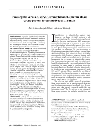
Fig. 1. Immunoblot analysis of soluble Lub proteins derived
from prokaryotic expression studies. Lanes 2 through 7 = bac- Hemagglutination inhibition tests on soluble
terial preparations from a prokaryotic expression study with recombinant Lub protein
an expression construct encoding for a 46-kDa Lub molecule. For inhibition studies, 0.5 to 3 mg of soluble recombinant
Lanes 2 and 3 = specimens from the periplasm (P); Lanes 4 Lub protein was added to 25 mL of patient sera containing
and 5 = specimens from the cytoplasm (C); and Lanes 6 and 7 anti-Lub or to 25 mL of reference sera and incubated for
= specimens from the intrabodies (I) of the bacteria. Lanes 1 30 minutes at room temperature. After incubation, 50 mL
and 8 = different molecular weight markers. The proteins were of a 0.8 percent antigen-positive donor RBC solution was
immunostained with monoclonal HRP-conjugated anti-His. added as an indicator, and the RBC-serum-recombinant
The size of the products (in kDa) is indicated on the right. protein mixture was incubated for an additional
30 minutes at 37°C. To distinguish inhibition of the anti-
the presence of the 46-kDa Lub protein by immunoblot Lub from a simple dilution effect, the anti-Lub sera were
analysis (Fig. 1). Protein renaturation and purification also tested by adding the same volume of PBS instead of
were performed on a Ni-NTA column with several buffer recombinant protein. A gel agglutination–based indirect
changes as previously described.9 Briefly, the proteins antiglobulin test (IAT) was used as the standardized sero-
were incubated in the presence of Ni-NTA agarose (Sigma) logic procedure for measurement of antibody reacti-
overnight. The proteins bound via their His-tags to the vity (Micro-Typing System, DiaMed, Cressier sur Morat,
Ni-NTA–agarose were then loaded in a PD10 column (GE Switzerland).
Healthcare, Uppsala, Sweden), and several washing steps
were performed with b-mercaptoethanol buffer. All rena-
turation steps were carried out with glutathione in Tris- Test samples
HCl buffer by gradually removing the urea used for Four commercially available polyclonal human anti-Lub
denaturation (from 8 to 0 mol/L). Refolded protein was serum samples (Optima, Rittersheim, Germany; Biolith
then eluted with a buffer supplemented with 250 mmol Diagnostica, Hann. Münden, Germany; Biotest, Dreieich,
per L imidazole. The eluted fractions containing soluble Germany; SD-nostik, Sinsheim, Germany) and 6 anti-Lub
refolded protein were again checked for the presence of serum samples from immunized in-house patients were
recombinant protein by SDS-PAGE and immunoblotting. tested. Thirty serum samples from patients containing
1632 TRANSFUSION Volume 47, September 2007
4. RECOMBINANT Lub
RBC alloantibodies of other common or rare specificities
(anti-K, anti-D, anti-c, anti-Jka, anti-Jkb, anti-Fya, anti-Fyb, TABLE 1. Eukaryotic versus prokaryotic
recombinant Lub protein–based ELISA*
anti-S, anti-s, anti-Doa, anti-Coa, anti-Yta, anti-Cha, anti-
Positive Negative
Rga, anti-Vel, anti-Lan, anti-Kpb, anti-Kna, anti-Jra, anti-
Antisera Eu Pro Eu Pro
Csa), 10 serum samples containing nonspecific warm
Anti-Lub 10 10 0 0
reactive RBC autoantibodies, and 100 serum samples Anti-Lua 0 0 6 6
obtained from randomly selected, healthy, nonimmu- Other specificities 0 0 30 30
Autoantibodies 0 0 10 10
nized donors were used as reference samples. Four com-
Donor sera 3 2 97 98
mercially available polyclonal human anti-Lua serum
* Eu = eukaryotic recombinant Lub protein; Pro = prokaryotic
samples (DiaMed, Biolith, SD-nostik, and Biotest) and 2 recombinant Lub protein.
anti-Lua serum samples obtained from in-house patients
were used to check the specificity of the recombinant Lub
protein–based antibody detection assays. Titration studies
were performed with double dilutions of the respective TABLE 2. Anti-Lub reactivity in the gel
antibody dissolved in PBS (pH 7.3) supplemented with agglutination–based IAT and ELISA
Anti-Lub titer
6 percent bovine serum albumin. The maximum dilution
Serum sample* IAT† ELISA‡
of serum samples used for antibody detection was 1/1024.
1 (Optima) 16 256
2 (Biotest) 64 >512
3 (Biolith) 32 >512
RESULTS 4 (SD-nostik) 32 >512
5 64 >512
ELISA 6 8 256
The ELISA technique was used to characterize the consti- 7 32 >512
tution of recombinant Lub proteins and their performance 8 2 256
9 2 128
as antigens in RBC antibody detection assays. The follow- 10 8 256
ing four recombinant Lub protein fractions were obtained * Samples 1 through 4 are commercial sera. Samples 5-10
from the expression studies and tested for correct folding were obtained from immunized patients and donors,
with monoclonal anti-Lub (BRIC108): 1) eukaryotic Lub, 2) respectively.
† IAT (gel agglutination technique) with Lu(a–b+) RBCs.
prokaryotic Lub stored under denaturating conditions, 3) ‡ ELISA with recombinant Lub proteins. Titration results were
prokaryotic Lub generated by a special refolding proce- identical for eukaryotic and prokaryotic Lub protein fractions.
dure, and 4) prokaryotic Lub dissolved in PBS but not sub-
jected to a special refolding procedure. The eukaryotic and
prokaryotic Lub fractions treated with refolding buffer protein–based ELISAs (Table 1). Of the 3 positive serum
tested positive by ELISA, whereas the prokaryotic Lub frac- samples, 2 tested positive in the eukaryotic as well as
tion stored under denaturating conditions tested nega- prokaryotic Lub protein–based ELISA, and 1 reacted posi-
tive. Surprisingly, a strongly positive reaction was also tive only to eukaryotic Lub protein. These 3 serum
obtained when prokaryotic Lub dissolved in PBS but not samples, which were obtained from healthy blood donors
subjected to refolding was used as the target antigen for with RBCs typed Lub-positive, had tested negative for RBC
monoclonal anti-Lub. These results indicate that the antibodies in IAT screening. DAT and IAT was negative for
correct Lub epitope was present in all recombinant Lub these donors at a 3-month follow-up, suggesting that the
fractions except the denaturated prokaryotic fraction. discrepancy in the results between IAT and ELISA in these
The correctly folded Lub protein fractions were cases was based on unspecific reactions rather than the
further used to repeatedly screen the patient and donor different sensitivity of the detection methods.
sera for the presence of anti-Lub. All 10 anti-Lub serum
samples reacted positive in the soluble Lub–based ELISA,
independently of whether eukaryotic Lub or one of the two Hemagglutination inhibition test
correctly folded prokaryotic Lub fractions was used as the As demonstrated by the gel agglutination–based IAT, all
antigen (Table 1). Titration studies with the anti-Lub sera 10 anti-Lub serum samples tested completely lost their
yielded equal results for the different Lub fractions. Anti- ability to agglutinate Lu(a–b+) test cells after incubation
Lub antibody titers determined by ELISA were consistently with 2.5 mg or more of soluble recombinant Lub protein,
at least four times higher than those measured in the gel irrespective of whether eukaryotic or prokaryotic Lub pro-
agglutination–based IAT test performed with Lu(a–b+) tein was used (Table 3). The inhibitory effect on anti-
RBCs (Table 2). Lub increased with increasing amounts of soluble Lub
All except 3 of the other serum samples (including the protein. Neither eukaryotic nor prokaryotic Lub protein
6 anti-Lua serum samples and 10 serum samples with RBC decreased the reactivity of anti-Lua, RBC alloantibodies
autoantibodies) tested negative in the recombinant Lub against other specificities, or warm-reactive RBC auto-
Volume 47, September 2007 TRANSFUSION 1633
5. SELTSAM ET AL.
high protein yield obtained by prokary-
TABLE 3. Inhibition study otic expression technology suggests that
Anti-Lub Amount of recombinant Lub protein used (mg)† the use of recombinant Lub protein
serum sample* 0 0.5 1 1.5 2 2.5 3 could be feasible for the majority of
1 (Optima) 3+ 2+ 1+ 0 0 0 0
immunohematologic laboratories.
2 (Biotest) 3.5+ 3.5+ 3+ 1+ 0.5+ 0 0
3 (Biolith) 3.5+ 3+ 2+ 0.5+ 0 0 0 In RBC serology, it is generally pre-
4 (SD-nostik) 4+ 3+ 2+ 0.5+ 0 0 0 sumed (though not systematically vali-
5 4+ 4+ 3+ 1+ 0.5+ 0 0
dated) that formation of a blood group
6 3+ 2+ 0 0 0 0 0
7 3+ 3+ 2+ 0.5+ 0 0 0 antigen mainly depends on the three-
8 1.5+ 1+ 0 0 0 0 0 dimensional structure of the blood
9 1.5+ 1+ 0 0 0 0 0
group proteins, which is determined by
10 2.5 2+ 0.5+ 0 0 0 0
the type of membrane protein (e.g.,
* Samples 1 through 4 are commercial sera. Samples 5 through 10 were obtained from
immunized patients and donors. single or multipass), the types of post-
† Results for eukaryotic and prokaryotic recombinant Lub proteins were identical. Sero- translational modifications (e.g., glyco-
logic results were obtained by IAT. Agglutination scores ranged from 0.5+ (very weak) sylation and disulfide bonding), and the
to 4+ (very strong). “0” indicates a negative IAT reflecting complete inhibition.
types of interactions between two or
more RBC membrane proteins.10
Although the serum of a person immu-
nized against a certain blood group
antigen may contain a mixture of anti-
bodies recognizing conformation- and
amino acid sequence–dependent linear
epitopes of the same antigen,11 the
usage of antigens in their native struc-
tural form may be crucial for the devel-
opment of antibody detection methods
that are reliable and sensitive enough to
b b a a b
aLu aLu aLu aLu a Kp b
aKp detect low RBC antibody titers. Our
findings strongly suggest that this is also
true for detection of anti-Lub. All of the
anti-Lub tested reacted exclusively with
b b b
+ rLu + rLu + rLu refolded recombinant Lub protein.
A major advantage of eukaryotic
protein expression systems is that
the proteins have the posttranslational
modifications required for proper pro-
tein folding and, thus, for the formation
Fig. 2. Representative hemagglutination inhibition tests with recombinant Lub of conformation-dependent, discon-
(rLub). Inhibition was analyzed with the gel agglutination–based IAT, with 3 mg of tinuous epitopes. Because posttrans-
the respective eukaryotic or prokaryotic recombinant Lub protein fractions for lational modifications in prokaryotic
inhibition. expression systems are not added by
bacteria, most of the expressed proteins
antibodies. Figure 2 shows representative inhibition test do not exhibit their native structure.12 Lutheran blood
results for anti-Lub, anti-Lua, and anti-Kpb sera. group protein is therefore an ideal candidate for prokary-
otic protein–based RBC antibody detection assays for two
reasons. First, its extracellular domain, which carries the
DISCUSSION
RBC antigens, is segmented into compact and structurally
Correct antigen presentation and protein availability are independent IgSF domains, suggesting that truncated
crucial parameters to consider when developing recombi- versions of this molecule may relatively easily adopt a
nant protein–based antibody detection systems. Both correct conformation under favorable conditions. Second,
parameters are mainly determined by the expression because its five potential N-linked glycosylation sites are
technique used and can vary for different specificities of located in the third and fourth IgSF domains, they may not
the target protein. This study demonstrated that eukary- affect the Lub epitope located in the unglycosylated
b
otic and prokaryotic recombinant Lu proteins are equally N-terminal IgSF domain 1.6,7 Indeed, our study results
suited for RBC antibody identification. In addition, the show that eukaryotic and prokaryotic recombinant Lub
1634 TRANSFUSION Volume 47, September 2007
6. RECOMBINANT Lub
proteins comprising the first three and a half IgSF glutination tests. This would lower the risk of delayed
domains are equally suited for detection and identifica- hemolytic transfusion reactions, which are one of the
tion of anti-Lub. The fact that high yields of prokaryotic Lub major causes of transfusion-related adverse events.16
protein were obtained within a relatively short period of Because most proteins carrying blood group anti-
time (around 7 working days) makes the prokaryotic gens can now be cloned, many can be produced in
expression procedure economically more attractive. soluble form. Of course, the ability to generate
Moreover, the fact that correct protein refolding occurred soluble forms greatly depends on the structure of the
spontaneously after removal of prokaryotic Lub molecules protein. Single-pass proteins such as Lutheran and
from the denaturing environment further simplifies the glycosylphosphatidylinositol-linked proteins with single
production of prokaryotic Lub protein. Both high yield and extracellular domains are generally good candidates for
easy manufacturing are important prerequisites for wide- expression as soluble recombinant molecules. In contrast,
spread use of recombinant proteins in immunohemato- it may be extremely difficult to impossible to achieve
logic diagnostics. soluble expression of antigens located on multipass
The hemagglutination inhibition test, a well- proteins with a complex conformation (e.g., RhD). Never-
established RBC serology method, is used to measure the theless, because the known blood group proteins greatly
amount of a specific antigen or to determine the identity differ from each other in terms of their topology, the
of antibodies in a blood serum sample.13 The latter appli- best expression strategy for each must be determined
cation is more relevant to this study. The hemagglutina- individually.
tion inhibition test works on the principle that the In a previous study, we demonstrated that alloanti-
reactivity of RBC antibodies with test cells in a hemagglu- bodies against Lub, Kpb, Yta, and Vel are the most frequent
tination assay can be neutralized by incubating the test specificities in patients with clinically relevant alloanti-
samples with various body fluids (plasma, urine, saliva, bodies against high-frequency antigens.2 Therefore, a
etc.) containing soluble antigenic substances. In the panel of recombinant Lub, Kpb, Yta, and Vel antigens could
Chido-Rodgers system, for example, antibodies are neu- help to facilitate the diagnosis of difficult-to-identify, clini-
tralized by human plasma containing C4, the complement cally relevant alloantibodies. An insect cell expression
system glycoprotein carrying the corresponding anti- system was recently used to express soluble Kell protein.17
gens.14,15 Similarly, soluble forms of recombinant RBC Yta may be predestined for soluble expression as it is
antigens can be applied to inhibition assays as part of the located on a protein with a large extracellular domain that
antibody detection and identification procedures.3 This is linked via a glycosylphosphatidylinositol anchor to the
would allow for easy and rapid identification of alloanti- cell surface. Although not all clinically relevant antigens
bodies in a one-well reaction system. In particular, it (e.g., Vel) have been cloned yet, the use of recombinant
would make advanced diagnostic tests for identification of blood group proteins in the near future to supplement the
RBC antibodies against high- or low-frequency antigens, use of rare RBCs for identification of alloantibodies against
which is now mainly reserved for reference laboratories, high-frequency antigens seems feasible.
available for routine laboratories. This is all the more sig-
nificant because the hemagglutination inhibition test is a
REFERENCES
standard method in blood group serology. Consequently,
soluble recombinant protein–based assays can easily be 1. Mollison PL, Engelfried CP, Contreras M. Blood transfusion
implemented in immunohematologic antibody identifi- in clinical medicine. Oxford: Blackwell; 1997.
cation procedures. In addition, our preliminary results on 2. Seltsam A, Wagner FF, Salama A, Flegel WA. Antibodies to
long-term storage of recombinant Lub protein suggest that high-frequency antigens may decrease the quality of trans-
the protein remains stable for months. The storage time of fusion support: an observational study. Transfusion 2003;
the protein may further be extended by cryopreservation. 43:1563-6.
Therefore, it might be practical even for smaller laborato- 3. Moulds JM, Rowe KE. Neutralization of Knops system anti-
ries to keep small stocks of the recombinant protein, bodies using soluble complement receptor 1. Transfusion
which can be used for detection of the rarely occurring 1996;36:517-20.
anti-Lub. 4. Ridgwell K, Dixey J, Parsons SF, Green CA, Scott ML.
It should also be possible to attach soluble blood Screening human sera for anti-Lu antibodies using soluble
group proteins to various surfaces to use them in other recombinant Lu antigens. Transfus Med 2003;Suppl:32.
common antibody identification systems, such as solid- 5. Sheffield WP, Bhakta V, Denomme GA. Use of recombinant
phase assays. Our ELISA results suggest that recombinant forms of Duffy blood group antigen to detect anti-Fy(a)
protein–based solid-phase assays have the potential to and anti-Fy(b). Transfusion 2003;43(Suppl):34A.
increase the sensitivity of RBC antibody detection and, 6. El Nemer W, Rahuel C, Colin Y, et al. Organization of the
thus, allow for identification of low-titer clinically relevant human LU gene and molecular basis of the Lu(a)/Lu(b)
antibodies which are under the detection limit of hemag- blood group polymorphism. Blood 1997;89:4608-16.
Volume 47, September 2007 TRANSFUSION 1635
7. SELTSAM ET AL.
7. Parsons SF, Mallinson G, Daniels GL, et al. Use of domain- frequently used expression systems for foreign genes.
deletion mutants to locate Lutheran blood group antigens J Biotechnol 2007;127:335-47.
to each of the five immunoglobulin superfamily domains 13. Brecher M. AABB technical manual. 15th ed. Bethesda:
of the Lutheran glycoprotein: elucidation of the molecular American Association of Blood Banks; 2005.
basis of the Lu(a)/Lu(b) and the Au(a)/Au(b) polymor- 14. Middleton J, Crookston MC. Chido-substance in plasma.
phisms. Blood 1997;89:4219-25. Vox Sang 1972;23:256-61.
8. Seltsam A, Das Gupta C, Wagner FF, Blasczyk R. Nondele- 15. Daniels G. Human blood groups. Oxford: Blackwell
tional ABO*O alleles express weak blood group A pheno- Scientific; 2002.
types. Transfusion 2005;45:359-65. 16. Serious Hazards of Transfusion Steering Committee.
9. Li M, Su ZG, Janson JC. In vitro protein refolding by chro- Serious hazards of transfusion: annual report 2005 [mono-
matographic procedures. Protein Expr Purif 2004;33:1-10. graph on the Internet]. Manchester (UK): SHOT Office;
10. Alzari PM, Lascombe MB, Poljak RJ. Three-dimensional 2006. Available from: http://www.shotuk.org/
structure of antibodies. Annu Rev Immunol 1988;6:555-80. SHOT%20report%202005.pdf
11. Issitt PD, Anstee DJ. Applied blood group serology. 4th ed. 17. Lee S, Lin M, Mele A, et al. Proteolytic processing of big
Durham (NC): Montgomery Scientific; 1998. endothelin-3 by the kell blood group protein. Blood
12. Yin J, Li G, Ren X, Herrler G. Select what you need: a com- 1999;94:1440-50.
parative evaluation of the advantages and limitations of
1636 TRANSFUSION Volume 47, September 2007