
Recomendados
Más contenido relacionado
La actualidad más candente
La actualidad más candente (20)
Destacado
Destacado (18)
Similar a Musculoskeletal systems
Similar a Musculoskeletal systems (20)
Último
Último (20)
Musculoskeletal systems
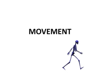
- 1. MOVEMENT
- 2. MAJOR FUNCTIONS 1. SUPPORT framework for body 2. MOVEMENT ~point of attachment for skeletal muscles ~joints
- 3. 3. PROTECTION ~ soft tissues (brain, spinal cord, heart, etc.)
- 4. 4. STORAGE ~ minerals: (Ca, P, Na, K ) for use when needed…
- 5. 5. FORMATION OF BLOOD CELLS ~Hematopoiesis: red marrow produces blood cells for circulation
- 6. TWO MAJOR TYPES OF BONE TISSUE • COMPACT BONE –Give bone hard, durable quality –Outer layers of bone –Functions served? –Composed of osteocytes packed into lacunae.
- 7. COMPACT BONE
- 8. • SPONGY BONE - Less dense than compact bone – Contains red marrow: stem cellsblood cell components – Yellow marrow: fat storage centers
- 11. Types of Joints • The type of joints that are particularly important for physical activity and sport are: – BALL AND SOCKET JOINT - allows a full range of movement. E.g. the hip and shoulder joints – HINGE JOINT - movement in one plane: flexion and extension.
- 12. – GLIDING JOINT - these occur in the many small bones of the hand and feet. They allow a slight sliding motion forwards and backwards and from side to side. – PIVOT JOINT - allows rotation. E.g. atlas and axis in the neck.
- 13. • TASK: • the picture shows: • 1: Shoulder joint -ball and socket • 2: Elbow joint - hinge joint • can you name another ball and socket and hinge joint?
- 15. FOUR MAJOR TYPES OF BONES 1. LONG BONES 2. SHORT BONES 3. FLAT BONES 4. IRREGULAR BONES
- 16. LONG BONES • Greater in length than width • Absorb stress from body weight • Upper/lower appendages
- 18. Long bone structure 1. Diaphysis- shaft with compact bone & medullary cavity 2. Epiphysis- ends, articular cartilage & compact bone covering cancellous bone 3. Epiphyseal line- between epiphysis & diaphysis- region of bone growth (epiphyseal plate) 4. Medullary cavity- central cavity within diaphysis
- 19. SHORT BONES • ~ equal length/width • Wrists, ankles
- 22. FLAT BONES • Thin, flat in structure • Skull, ribs, sternum
- 24. IRREGULAR BONES • Variety of shapes • Vertebral column / bones of face
- 28. Muscular System Functions • MOVEMENT • Maintain Posture • Stabilize Joints • Generate HEAT – 40% body mass – 80% body heat – Endothermy!
- 29. Muscle Tissue • Tissue Review: – Cardiac Cardiovascular System • Involuntary, Striated – Smooth Cardiovascular, Digestive, Reproductive, etc. • Involuntary, non-striated – Skeletal* MUSCULAR SYSTEM • Voluntary, striated
- 30. Muscles are… • Excitable (irritable) • Contractile • Extensible • Elastic • Myo-, Mys, Sarco- (muscle prefixes)
- 31. Skeletal Muscle Tissue • MUSCLE CELL STRUCTURE – Arrangement: large, long FIBERS • Fiber = muscle cell – Two major protein filaments present: • Actin • Myosin myofilaments
- 32. Skeletal Muscle Structure • Striated – Due to actin/myosin • Elongated – Varied lengths • Multinucleated
- 33. Muscle Anatomy
- 35. • http://www.bmb.psu.edu/courses/bisci004a/ muscle/b4muscle.htm
- 36. Skeletal Muscle Connections • Direct connection to Bone • Indirect connection via TENDON • ORIGIN: bone that does NOT move when muscle contracts • INSERTION: bone that MOVES when muscle contracts
- 37. Microscopic Structure of Muscle Fiber • Cell membrane = Sarcolemma • Cytoplasm = Sarcoplasm • Multiple Mitochondria = High E output • Fiber is filled with long myofibrils • Myofibrils filled with filaments arranged in contractile units called SARCOMERES. – Myosin (thick filament) – Actin (thin filament)
- 38. Sarcoplasmic Reticulum • Specialized ER connected to cell surface by T- tubules • Surrounds each myofibril • At rest, filled with Ca++ maintained by a calcium “pump”, uses ATP • When activated, pores open and release calcium, initiating contraction
- 40. Actin (thin) filament Composition • Long chains of actin globules in double spiral arrangement • Each actin contains binding site for myosin • Tropomyosin spiral around chain – blocks active site on actin • Troponin clustered along spiral – Binding site for calcium!
- 41. Actin Filament
- 42. Myosin (thick) filament Composition • Contains 2 tails each with globular heads • Heads have ATP binding sites and ATPase for splitting ATP • Heads attracted to active sites on actin molecules • Heads form cross-bridges with actin
- 43. Myosin Filament
- 45. Sarcomere structure • Alternating dark and light bands • Central H-zone contains MYOSIN only • Lateral A-bands contain both ACTIN and MYOSIN filaments • End in I-bands contain ACTIN only (with Z-line in center) Z LINE TO Z LINE = 1 SARCOMERE
- 47. Sarcomere
- 49. Nerve supply to Muscle Fiber • Each muscle fiber served by a motor neuron • Motor neuron ends in a pad filled with vacuoles packed with neurotransmitter • Pad sits above specialized piece of sarcolemma called motor end plate
- 51. Neuron pad + motor end plate = Neuromuscular junction (Space between called synaptic cleft )
- 53. Sliding Filament Theory of Muscle Contraction Sequence of Steps: 1. Neuron releases neurotransmitter, acetylcholine (ACh) into synaptic cleft 2. ACh diffuses to motor end plate 3. ACh binds to receptor on motor end plate 4. Gated channel protein opens, Na+ rushes into cell interior, upsets RMP!
- 55. Generation of Action Potential • RMP = -70 Mv • Sudden influx of Na+ generates Action Potential • RMP later restored to normal by sodium-potassium pump
- 56. 5. Action potential carried along the sarcolemma to transverse (“T”) tubules connected to Sarcoplasmic Reticulum 6. SR membrane becomes permeable to calcium 7. Sarcoplasm is flooded with calcium ions
- 58. 8. Ca++ binds to troponin 9. Ca/Troponin pulls tropomyosin out of the way, unmasks active site on actin molecules 10. Myosin heads attach to actin 11. Heads rotate, pull actin in to H-zone 12. Z lines get closer…
- 63. 13. Myosin splits ATP to recharge 14. Continues until Action Potential is restored and Ca++ is pumped back into SR 15.All sarcomeres shorten, shortening muscle cell
- 64. 16. Shortening cell pulls on tendons attached to bones, moving bone at articulation 17. Contraction of opposite muscle required to fully elongate shortened muscle
- 65. Reminders: • Refractory Period • All or None Effect • Breakdown of ACh by ACh-esterase
- 66. Disorders/Conditions of the Muscular System • Duchenne’s Muscular Dystrophy – Sex linked inheritance – Dystrophin protein deficiency – Tearing of sarcolemma – Accumulation of CT/fat – Muscular ATROPHY
- 67. Tetanus