Apidays New York 2024 - The value of a flexible API Management solution for O...
Melanoma Lipoblastic Cell Type
1. Images in Pathology
Primary Cutaneous Malignant Melanoma With
Lipoblast-like Cells
Joao Cruz, MD; Jorge S. Reis-Filho, MD; Jose Manuel Lopes, MD, PhD
˜ ´
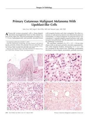
A 74-year-old woman presented with a dome-shaped, with irregular borders and color variegation. No other cu-
ulcerated, hyperpigmented lesion on the anterior as- taneous or subcutaneous lesions were detected on physical
pect of her right arm. Physical examination revealed a 1.5 examination. A clinical diagnosis of polypoid nevus was
1.2-cm, hyperpigmented, and partially ulcerated lesion considered. A spindle-shaped surgical excision with wide
margins was performed, and the specimen was submitted
for pathologic examination.
Accepted for publication October 1, 2002.
The specimen consisted of a 3.5 2.2 2.0-cm skin
From the Department of Pathology, Sao Joao Hospital, and Medical
˜ ˜
Faculty, University of Porto, Porto, Portugal (Drs Cruz and Lopes); and ellipse with an elevated, partially ulcerated, pigmented le-
IPATIMUP–Institute of Molecular Pathology and Immunology, Univer-
sion with irregular borders, measuring 1.5 1.2 0.8
sity of Porto, Porto, Portugal (Drs Reis-Filho and Lopes).
cm, localized in the central area. Histologic examination
Reprints: Jose Manuel Lopes, MD, PhD, IPATIMUP, R. Roberto Frias,
´
disclosed a dome-shaped, asymmetric, partially ulcerated
S/N, 4200 Porto, Portugal (e-mail: jreis@ipatimup.pt).
Primary Cutaneous Malignant Lymphoma—Cruz et al
370 Arch Pathol Lab Med—Vol 127, March 2003
2. melanocytic lesion extending from the epidermis to the place the nucleus to the periphery of the cell and also
confer a semilunar shape, and (2) a rarer variant, com-
deep dermis (Figure, A). The junctional component of the
posed of cells with intracytoplasmic vacuoles and scal-
neoplasm was asymmetric and composed of atypical ep-
loped eccentric nuclei.2,4 In such situations, the neoplastic
ithelioid melanocytes arranged in noncohesive nests in the
cells may resemble monovacuolated lipoblasts.2 Lipoblast-
dermal-epidermal junction; these nests were associated
like cells are very unusual features of metastatic MMs and
with a remarkable number of variably pigmented malig-
may be observed in metastatic deposits of signet ring and
nant cells arranged in a pagetoid fashion among epider-
in balloon cell melanomas.1–3 Owing to their rarity, the
mal cells (Figure, A, inset). The intradermal component of
prognostic significance of lipoblast-like cells in either pri-
the neoplasm was composed of medium-sized to large ep-
mary or metastatic malignant melanomas remains elu-
ithelioid cells with eosinophilic to vacuolated well-defined
sive.3 To the best of our knowledge, no report on primary
cytoplasm, large and irregular nuclei, clumped chromatin,
cutaneous MMs with both lipoblast-like and signet ring
and 1 to multiple prominent amphophilic to eosinophilic
cells has been published to date.
nucleoli. These cells were arranged in solid or noncohesive
The present case illustrates an epithelioid primary MM
sheets. Pleomorphic cells with a single, large, intracyto-
with signet ring cells and several foci composed of uni-
plasmic vacuole displacing the nuclei to the periphery,
vacuolated and multivacuolated cells with scalloped nu-
similar to signet ring cells, were observed (Figure, B). An
clei, which confer a remarkable resemblance to the epithe-
eye-catching feature was the presence of neoplastic cells
lioid variant of pleomorphic liposarcoma (PL).1–5 Pleomor-
with multiple cytoplasmic vacuoles of varying sizes; the
phic liposarcoma is the rarest variant of liposarcoma; it
vacuoles had an empty appearance, scalloping the atypical
usually affects deep soft tissues of the proximal extremi-
nuclei, remarkably resembling lipoblasts (Figure, C and
ties of elderly patients (median age, seventh decade); up
inset). Scattered cells with finely spider-webbed cyto-
to 20% of the cases may be superficial, and there are rare
plasm, balloon cells (Figure, B, arrow), as well as cells with
reports of PLs affecting children.5 Pleomorphic liposarco-
dusty Masson-Fontana–positive pigment and multinucle-
ma is characterized by the presence of adipocytic differ-
ated cells were detected. The tumor was rated Clark level
entiation, better exemplified by lipoblasts.5 The epithelioid
IV and was 5.75 mm thick. The mitotic index was 15.2 per variant of PL is composed of epithelioid cells with eosin-
10 high-power fields (10/mm2). Immunohistochemistry ophilic to vacuolated cytoplasm, round nuclei, and prom-
with antibodies for S100 protein, gp100 (HMB-45), vimen- inent nucleoli.5 In this variant, the presence of bona fide
tin, and cytokeratins (CAM5.2 and AE1/AE3) was per- lipoblasts may be very scanty, posing diagnostic difficul-
formed. Neoplastic cells were strongly and diffusely dec- ties to differentiate it from other epithelioid neoplasms
orated by S100 protein and vimentin, multifocally stained with lipoblast-like cells.5 The differentiation between an
by gp100 (HMB-45), and negative for cytokeratins. Lipo- epithelioid PL and an MM can be achieved by the pres-
blast-like cells and signet ring cells also showed immu- ence of a junctional component, as well as the expression
noreactivity for S100 protein (Figure, D) and focal im- of S100 protein and melanocytic markers, such as gp100,
munoreactivity for HMB-45. A diagnosis of malignant NKI-C3, MART1, and MiTF in the latter. A few cases of
melanoma with signet ring and lipoblast-like cells was es- S100 protein–negative signet ring cell melanomas have
tablished. The patient was discharged and remains free of been reported.2,4
disease 3 months after diagnosis. In our case, lipoblast-like cells and signet ring cells were
Malignant melanoma (MM) is well known for its pro- strongly decorated by S100 protein, but they were only
tean morphologic appearance.1–3 Besides the common cy- focally reactive for gp100. It should be noted that true
tomorphologic types (epithelioid and spindle cell), several lipoblasts are frequently immunoreactive for S100 protein
and, conversely, lipoblast-like and signet ring cells of MM
histopathologic variants have been reported, including
may lack gp100 reactivity.2,5 In conclusion, pathologists
desmoplastic, balloon cell, pleomorphic (fibrohistiocytic),
should be aware of the rare presence of lipoblast-like cells
hemangiopericytic, myxoid, small cell, and nevoid MM.1,3
in MM, to avoid misdiagnosing MM as an epithelioid var-
Interestingly, the most unusual morphologic appearances
iant of PL in small skin biopsies without adequate repre-
are more frequently observed in metastatic than in pri-
sentation of the epidermis and in metastatic deposits of
mary lesions,1–3 which may lead even experienced pathol-
unknown primary neoplasms.1,3
ogists to misdiagnoses of poorly differentiated carcinoma,
lymphoma, or sarcoma.1–3 Anecdotal cases of metastatic References
1. Lodding P, Kindblom LG, Angervall L. Metastases of malignant melanoma
MM mimicking several types of sarcomas, including fi-
simulating soft tissue sarcoma: a clinico-pathological, light- and electron micro-
brosarcoma, monophasic synovial sarcoma, malignant pe- scopic and immunohistochemical study of 21 cases. Virchows Arch A Pathol Anat
ripheral nerve sheath tumor, malignant fibrous histiocy- Histopathol. 1990;417:377–388.
2. Banerjee SS, Harris M. Morphological and immunophenotypic variations in
toma, leiomyosarcoma, and liposarcoma, have been re- malignant melanoma. Histopathology. 2000;36:387–402.
ported.1–3 3. Nakhleh RE, Wick MR, Rocamora A, Swanson PE, Dehner LP. Morphologic
Among the trickiest variants is signet ring melanoma, diversity in malignant melanomas. Am J Clin Pathol. 1990;93:731–740.
4. al-Talib RK, Theaker JM. Signet-ring cell melanoma: light microscopic, im-
which is frequently confounded with signet ring carcino- munohistochemical and ultrastructural features. Histopathology. 1991;18:572–
ma or signet ring lymphoma.1–3 Two distinctive types of 575.
5. Gebhard S, Coindre JM, Michels JJ, et al. Pleomorphic liposarcoma: clini-
signet ring cells may be observed in this variant: (1) the
copathologic, immunohistochemical, and follow-up analysis of 63 cases: a study
usual type, composed of polygonal cells with accumula- from the French Federation of Cancer Centers Sarcoma Group. Am J Surg Pathol.
tion of vimentin filaments in the cytoplasm, which dis- 2002;26:601–616.
Primary Cutaneous Malignant Lymphoma—Cruz et al 371
Arch Pathol Lab Med—Vol 127, March 2003