

Recomendados
Más contenido relacionado
La actualidad más candente
La actualidad más candente (20)
Destacado
Destacado (20)
Similar a Revision the nervous system
Similar a Revision the nervous system (20)
Revision the nervous system
- 3. The Nervous System What are its component parts? • Brain • Spinal cord • Nerves What does it do? • Enables you to control your body • Gives you feedback about the world
- 4. Look away if you’re squeamish… A dissected brain, spinal cord and sections of major nerves.
- 5. How the nervous system works For example; If you smell something burning… • Your nose (receptor) detects the stimulus receptor (smell) • Nerve fibres send the message to the brain • Your brain then sends a message to move your body away or to put out the fire! SIMPLES RIGHT!
- 6. Lets look at some bits more closely RECEPTORS • Receptors are sensors on the body that detect stimuli • They convert stimuli into electrical signals (messages) called impulses.
- 7. EFFECTORS An effector is any part of the body that produces the response. response Here are some examples of effectors: • a muscle contracting to move the arm • a muscle squeezing saliva from the salivary gland • a gland releasing a hormone into the blood
- 8. The Cells of the Nervous System - Neurones
- 9. NERVE FIBRES • Nerve fibres are bundles of nerve cells (neurones) that pass on electrical signals (impulses) to the brain. • From the brain, nerve fibres send impulses to effectors (muscles).
- 10. NEURONES There are three types of neurones 1. Sensory neurone – carries impulses from the receptors to the spinal cord. 2. Relay Neurone – carries impulses to and from the spinal cord and the brain 3. Motor Neurone – carries impulses from the brain to the effector
- 11. The route impulses take
- 12. The route impulses take Sensory Relay neurone neurones (in brain or spinal cord) Effectors – Motor neurone
- 13. Left: sensory neurone Right: motor neurone
- 14. Nerves – bundles of neurones
- 16. Sensory Receptors • Information from our environment is received via our senses. • The Eye receives light and transfers this information via our optic nerve to our brain. • Our Ear receives sound and via the auditory nerve sends messages to our brain • Our Skin receives messages about temperature, pain and pressure and via sensory nerves sends this information to our brain.
- 17. • The skin and body temperature – the skin helps to keep your body at a constant 37°C +/- 1˚C. • Sweating, shivering, body hair and fat beneath the skin all help this. Also, capillaries in the skin surface open or close to lose heat or keep heat in. • This vasodilation (opening) and vasoconstriction (closing) of blood vessels helps to lose or conserve body heat
- 18. cornea Inside the eye protects eye surface and focuses light rays suspensory ligaments retina hold lens in place senses light lens focuses light on retina iris regulates amount of light entering eye optic nerve ciliary muscles transmits impulses to the change shape of the lens brain
- 19. ACTION In bright In dim light The way the iris in our light eye adjusts the size •Radial muscles of •Radial muscles of of the pupil in the iris relax. the iris contract. response to bright •Circular muscles •Circular muscles or dim light is a of the iris contract. of the iris relax. reflex action. •Less light enters •More light enters the eye through the the eye through the contracted pupil. dilated pupil. http://www.bbc.co.uk/schools/gcsebitesize/science/aqa/human/thenervoussystem
- 21. Reflex Reactions – bypassing the brain for extra speed
- 22. Reflex Reactions Can you think of any? • Startle reflex – moving away, contraction of arm and leg muscles, blinking, breathing changes • Withdrawal reflex – moving away from potentially harmful influences (e.g. high temperature) • Iris reflex – pupil becomes smaller in bright light
- 23. Knee Jerk Reflex
- 24. Why the knee jerk reflex? • This reflex is quite useful for walking. Every time you put weight on your foot, your muscles contract to support you. Without this reflex, we would all look silly staggering around, having to consciously think about working our muscles for each step, but with the muscles reacting too hopelessly late to be useful. Chewing gum at the same time would be out of the question.
- 25. Endocrine System • As well as sending and receiving messages using our nervous system, we also use our endocrine system to do the same job. • Instead of using electrical impulses however, this system releases chemicals into our blood stem called hormones. • These hormones are not a fast acting as nervous impulses but are longer lasting.
- 26. Endocrine Glands
- 27. Hormones Pituitary gland – FSH, LH (stimulates the release and maturity of follicles during menstruation) Thyroid gland – Thyroxine (regulates the rate of metabolism) Adrenal gland - corticosteroids and catecholamines including cortisol and adrenaline and small amounts of testosterone (regulates stress levels) Pancreas – peptides (regulates the production of shorter active digestive enzymes) Ovary – oestrogen, progesterone and small amounts of testosterone (regulates the growth of eggs and stabilises the growing foetus during pregnancy) Testis – testosterone (plays a key role in the health and well-being of the man)
Notas del editor
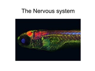
- The nervous system of this zebrafish is shown in green. I don’t know how they did this!
- (I don’t know why this image shows the pelvis – it isn’t part of the nervous system!) Ask students to discuss in groups what they think are the component parts of the nervous system. The brain and spinal cord make up the ‘central nervous system’ while the nerves comprise the ‘peripheral nervous system’. Students can then discuss what they think are the main functions of the nervous system before you reveal them.
- The nervous system – brain, spine and nerves - is made up of very specialised cells called nerve cells or neurones (also spelt ‘neurons’). They have all the main features of animal cells: cell membrane, cytoplasm, nucleus, etc. – but also some very specialised features which allow them to do their particular job. The large blob towards the lower left seems to be the cell body of a neurone, with an axon branching off to the right.
- Overview of the route signals take through the nervous system. The brain or spinal cord is sometimes referred to as the ‘co-ordinator’ in this process.
- Students must be clear about the types of neurones, in particular sensory neurones (from receptors to spine/brain, giving feedback on the outside world) and motor neurones (from brain/spine to effectors, which are usually muscles, enabling the brain/spine to control the body). These neurones are one-way only. For non-reflex reactions, there would be many relay neurones involved on the right, as the impulse from the sensory neurone enters the brain and the brain then makes decisions on how to respond, and sends impulses to the appropriate muscles.
- Not clear enough for students to copy, but gives them an idea of the two neurone types. Students could be asked to pick out similarities and differences between the two.
- Again, notice that the colours do not necessarily reflect reality. Individual neurones are bundled together in nerves. The neurones do not interfere with each other, but all act independently, and each neurone is one-way only. A single nerve contains both motor neurones and sensory neurones. A nerve is a bit like a bundle of optical fibres used in telecommunications. Like all cells, nerve cells need energy to function. So the nerves also contain tiny blood vessels (capillaries) to carry oxygen and glucose to the cells.
- Reflex reactions do not involve the brain; the brain is simply informed of them afterwards. The receptor causes an impulse along a sensory neurone, which connects to a relay neurone in the spine. Rather than taking the impulse up to the brain, it passes it directly to a motor neurone which causes a reaction in an appropriate muscle. Reflex reactions are usually there to protect the body from sources of danger, e.g. heat or injury. The body is full of ‘reflex arcs’ like this, ready for action should the need arise.
- Students can test out the iris reflex.
- Students could (carefully!) try this on themselves or each other. A hammer is not needed – the side of the hand works just as well. Gently tap just below the knee, and if you hit the right spot, the leg will jerk forwards. This movement is involuntary.