SlideShare una empresa de Scribd logo
1 de 8
Descargar para leer sin conexión
J. Agric. Food Chem. 2008, 56, 7773–7780       7773


                    Toxicity and Antioxidant Activity in Vitro and in Vivo
                             of Two Fucus vesiculosus Extracts
                          M. C. ZARAGOZA,† D. LOPEZ,† M. P. SAIZ,† M. POQUET,‡ J. PEREZ,‡
                                          ´        ´           ´                   ´
                     P. PUIG-PARELLADA,§ F. MARMOL,§ P. SIMONETTI,| C. GARDANA,| Y. LERAT,⊥
                                                 `
                     P. BURTIN,⊥ C. INISAN,# I. ROUSSEAU,# M. BESNARD,# AND M. T. MITJAVILA*,†
                     Departments of Physiology and Cellular Biology, Faculty of Biology, University of Barcelona, Av.
                     Diagonal 645, 08028-Barcelona, Spain, Unit of Pharmacology, Faculty of Medicine, University of
                    Barcelona, Casanova, 143, 08036-Barcelona, Spain, Department of Food Science and Microbiology,
                                                                                                                  ´
                    Division of Human Nutrition, University of Milan, Via Celoria 2, 20133-Milan, Italy, Centre d’Etude
                         et de Valorisation des Algues (CEVA), BP 3, 22610-Pleubian, France, and Diana Naturals,
                                            Phytonutriance, La gare, BP15, 35560-Antrain, France


             The consumption of seaweeds has increased in recent years. However, their adverse and beneficial
             effects have scarcely been studied. Two extracts from the brown seaweed Fucus vesiculosus
             containing 28.8% polyphenols or 18% polyphenols plus 0.0012% fucoxanthin have been obtained
             and studied to determine their toxicity in mice and rats and also their antioxidant activity. Both extracts
             were shown to lack any relevant toxic effects in an acute toxicity test following a 4 week daily treatment
             in rats. The extracts exhibited antioxidant activity in noncellular systems and in activated RAW 264.7
             macrophages, as well as in ex vivo assays in plasma and erythrocytes, after the 4 week treatment
             in rats. Our ex vivo results indicated that compounds from extract 2 may be more easily absorbed
             and that the antioxidants in their parent or metabolized form are more active. These findings support
             the view that the daily consumption of F. vesiculosus extract 2 (Healsea) would have potential benefits
             to humans.

             KEYWORDS: Fucus vesiculosus; polyphenols; phloroglucinol; fucoxanthin; antioxidant activity; super-
             oxide anion; nitric oxide; oxidative stress

INTRODUCTION                                                            macrophages, and mast cells) to the inflamed area. These
                                                                        inflammatory cells are triggered by mediators of inflammation
   The Western diet is rich in polyphenols and carotenoids.
                                                                        and generate superoxide anion (O2•-) and nitric oxide (NO)
Seaweeds, which are widely consumed in Asia, are an important
source of such compounds. A variety of in vitro studies have            radicals. Thus, antioxidant and anti-inflammatory activities are
shown that polyphenols and carotenoids exhibit antioxidant              very close to each other.
activity (1, 2). Flavonoids act either by blocking the generation          Fucus Vesiculosus is a brown seaweed species rich in
of hypervalent metal forms (3), by scavenging free radicals (4),        phlorotannins (polyphenols present in brown seaweeds that
or by breaking lipid peroxidation chain reactions (5). Caro-            consist of oligomers or polymers of phloroglucinol or 1,3,5-
tenoids are also capable of reacting with radical species (2).          trihydroxybenzene) (6), which also contains fucoxanthin, a major
Such antioxidant activities of flavonoids and carotenoids may            marine carotenoid. F. Vesiculosus is considered a source of
protect cell constituents against oxidative damage, thereby             minerals, iodine, proteins, and fiber. However, no toxicological
limiting the risk of diseases associated with oxidative stress and,     studies on this seaweed or derived extracts have been performed
thus, the inflammatory processes when involved.                          to date, and very few studies have been conducted on the
   An inflammatory response is characterized by the attraction           antioxidant activity of phlorotannins (7-9) and fucoxanthin (10, 11)
of large amounts of leukocytes (neutrophyles, monocytes-                from seaweeds. Thus, we hypothesized that on the basis of
                                                                        polyphenol and carotenoid contents, F. Vesiculosus extracts
  * To whom correspondence should be addressed. Tel: +34-93-            might have an antioxidant effect. To test this hypothesis, from
4021530. Fax: +34-93-4110358. E-mail: mmitjavila@ub.edu.                an initial screening, we selected two F. Vesiculosus extracts,
  †
    Department of Physiology, University of Barcelona.                  one rich in phlorotannins and the other rich in phlorotannins
  ‡
    Department of Cellular Biology, University of Barcelona.            and fucoxanthin, to compare their effects. We then evaluated
  §
    Unit of Pharmacology, University of Barcelona.
  |
    University of Milan.                                                the acute and 4 week toxicities and the antioxidant properties
  ⊥           ´                                                         via three approaches, namely, reducing power, free radical
    Centre d’Etude et de Valorisation des Algues (CEVA).
  #
    Diana Naturals.                                                     trapping, and enzymatic activities, by several noncellular
                                    10.1021/jf8007053 CCC: $40.75  2008 American Chemical Society
                                                       Published on Web 08/07/2008
7774    J. Agric. Food Chem., Vol. 56, No. 17, 2008                                                                                Zaragoza et al.
                                                                                                                                          ´

(chemically generated oxidants), cellular (macrophages), and                colorimetric Folin-Ciocalteau method (12), and the molecular weights
ex vivo (in plasma and erythrocytes after treating rats with two            distribution was performed using Centricon cartridges with cutoffs of
F. Vesiculosus extracts or with phloroglucinol) tests. The ex vivo          3, 10, 30, and 50 kDa and an ultracentrifugation with ceramic
tests allowed us to assess differences resulting from the presence          membranes with a cutoff of 300 kDa.
of metabolized forms of the parent antioxidants in blood.                      EValuation of Phloroglucinol and Its DeriVatiVes. Sixty milligrams
                                                                            of each F. Vesiculosus extract was sonicated in 10 mL of water, and
                                                                            after 10 min, the mixture was filtered, and the resulting solution was
MATERIALS AND METHODS                                                       adjusted to 20 mL. The mixture was centrifuged at 1000g for 2 min,
    Apparatus, Reagents, and Biological Material. Centricon car-            and the supernatant (20 µL) was injected into the HPLC-DAD-MS
tridges were acquired from Millipore (Bedford, MA). Ceramic mem-            system. The chromatographic system consisted of an Alliance 2695
branes were from Sepra (Cesano Maderno, Italy). The high-performance        equipped with a DAD 2996 and a Quattromicro triple quadrupole mass
liquid chromatograph (HPLC) Alliance and the diode array detection          spectrometer. A 3.5 µm C18 Symmetry column 150 mm × 2.0 mm
(DAD) system were from Waters (Milford, MA), and the Quattromicro           was used for the separation at 0.25 mL/min. The column was maintained
triple quadrupole mass spectrometer and the Masslinx 4.0 software were      at 30 °C, and the separation was performed by means of a linear gradient
from Micromass (Beverly, MA). The Symmetry columns were acquired            elution (eluent A, 0.1% HCOOH in water; eluent B, 0.1% HCOOH in
from Waters, and the Synergy Hydro columns were from Phenomenex             acetonitrile). The gradient was as follows: 0% B at time zero, 0-20%
(Torrence, CA). Cell counter CelltacR was acquired from Nihon               B in 20 min, 20-80% B in 5 min, and 80% B for 20 min.
(Kohden, Japan). An Easy Lyte Na/K/Cl analyzer was from Medica              Chromatographic data were acquired in the 200-450 nm range and
(Bedford, MA). The MicroOsmometer was acquired from Advanced                were integrated at 275 nm. The mass spectrometer was operated in
Instruments (Norwood, MA). The Iso-NOP sensor was from World                negative full-scan mode in the range 100-2000 Da. The capillary
Precision Instruments (Saratosa, FL). Fucoxanthin, violaxanthin, ast-       voltage was set to 3.0 kV, and the cone voltage was 30 V. The source
haxanthin, and echinenone were purchased from DHI (Hoersholm,               temperature and the desolvating temperature were 130 and 300 °C,
Denmark). The protein assay was from BioRad (Hercules, CA).                 respectively. Data were acquired by using Masslinx 4.0.
Biochemical kits were acquired from Biosystems (Barcelona, Spain),             QualitatiVe and QuantitatiVe EValuation of Carotenoids. One gram
Olympus (Hamburg, Germany), and Randox (Crumlin, United King-               of each F. Vesiculosus extract was sonicated in 70 mL of methanol,
dom). Folin-Ciocalteau reagent, quercetin, phloroglucinol, xanthine         and after 30 min, the mixture was filtered, and the resulting solution
(X), xanthine oxidase (XO), nitroblue tetrazolium salts, 2,2-diphenyl-      was adjusted to 100 mL with methanol. The mixture was centrifuged
1-picrylhydrazyl (DPPH), ascorbic acid, 2,2′-azo-bis-2-amidinopropane       at 1600g for 10 min, and the supernatant (10 µL) was injected into the
(ABAP), luminol, 2,2′-azinobis(3-ethylbenzothiazoline-6-sulfonic acid)      HPLC-DAD-MS system. The chromatographic system was the same
(ABTS), Cu-Zn superoxide dismutase (Cu-Zn SOD), cytochrome c                as for phloroglucinol evaluation. A 4 µm C18 Synergy Hydro column
(horse heart type VI), phorbol 12-myristate 13-acetate (PMA), li-           150 mm × 2.0 mm was used for the separation at 0.3 mL/min. The
popolysaccharide (LPS), paraoxon, R-phycoerythrin, and pyrogallol           column was maintained at 30 °C, and the separation was performed
were purchased from Sigma-Aldrich (St. Louis, MO). Trolox and chloral       by means of a linear gradient elution (eluent A, 2 mM CH3COONH4,
hydrate were acquired from Fluka Chemie (Buchs, Switzerland). N5-           pH 5.2; eluent B, methanol). The gradient was as follows: 80% B at
(1-iminoethyl)-L-ornithine dihydrochloride (L-NIO) was provided by          time zero, 80-100% B in 8 min, and 100% B for 7 min. Chromato-
A.G. Scientific, (San Diego, CA). F. Vesiculosus was obtained from           graphic data were acquired in the 200-700 nm range and were
                                                ´
Agrimer (Plougerneau, France) and Centre d’Etude et de Valorisation         integrated at 445 nm. The mass spectrometer was operated in positive
des Algues (CEVA, Pleubian, France). Swiss mice and Sprague-Dawley
                                                                            full-scan mode in the range 200-800 Da. The capillary voltage was
rats were provided by Harlan Interfauna Iberica (Barcelona, Spain).
                                              ´
                                                                            set to 3.0 kV, and the cone voltage was 20-60 V. The source
RAW 264.7 macrophages were from American Type Culture Collection
                                                                            temperature and the desolvating temperature were 130 and 350 °C,
(Rockville, MD).
                                                                            respectively. Data were acquired by using Masslinx 4.0 with the Quan-
    Plant Material. F. Vesiculosus raw material was obtained at two
                                                                            Optimize option for cone voltage and collision energy optimization.
different sites from the French North Coast. F. Vesiculosus was
harvested in late winter/spring and in late summer, as the highest and         Antioxidant Activity of the Extracts in Noncellular Systems.
similar contents in polyphenolic compounds were found in both areas.        Although many techniques can be used to test for antioxidants, there
Once harvested, the seaweeds were quickly washed with soft water,           are no approved standard methods. The significance and relevance of
dried at 100 °C for 1 h, and then finely ground (120-250 µm).                natural antioxidants, which are often multifunctional, depend strongly
    Preparation of the F. Wesiculosus Extracts. Extract 1. A hydro-         on the test applied. Thus, it is commonly accepted that several tests
ethanolic extraction (30-35% ethanol) of F. Vesiculosus (10% w/w)           should be used to correctly assess the capacity of compounds to respond
was carried out at room temperature under mechanical stirring for 4 h.      to oxidative stress. We performed some tests simultaneously with the
After filtration on a filter press, the liquid phase underwent an initial     two extracts, with phloroglucinol and when possible with a positive
purification step to remove the alginates by precipitating them in the       control and a negative control (13).
presence of excess CaCl2. Following filtration, the liquid phase                Reducing Power. To evaluate the electron donor capacity (14), the
underwent a second purification step involving diafiltration to remove        seaweed extracts, phloroglucinol and quercetin (positive control), were
the iodine and the low molecular weight compounds and was freeze-           mixed with 0.2 M phosphate buffer, pH 6.6, and 1% potassium
dried to obtain a powder extract.                                           ferricyanide, incubated at 50 °C for 20 min, and centrifuged at 2250g
    Extract 2. A hydroethanolic extraction with high-percent ethanol        for 10 min after adding 10% trichloroacetic acid. The upper layer was
(50-70%) of F. Vesiculosus (Healsea) (10% w/w) was conducted to             mixed with 0.1% FeCl3, and the optical density was measured at 690
solubilize a higher amount of carotenoids at room temperature under         nm and reported to quercetin activity.
mechanical stirring for 2 h. After filtration on a filter press, the liquid      O2•- ScaVenging ActiVity. This test was based on the reduction of
phase underwent an initial purification step to remove the alginates by      the nitroblue tetrazolium salt to water-soluble formazan by the O2•-
precipitating them in the presence of excess CaCl2. After solid-liquid      generated by the xanthine/xanthine oxidase (X/XO) system (15). The
separation, a second extraction was performed under the same condi-         reaction was initiated by adding XO, and we measured the difference
tions. The two liquid phases were then blended, submitted to a              in absorbance at 560 nm between 0 and 10 min at 25 °C. The percentage
diafiltration to remove the iodine and the low molecular weight              of O2•- inhibition was then evaluated. The concentration of the sample
compounds, and freeze-dried to obtain a powder extract.                     that inhibited the nitroblue tetrazolium salt reduction by 50% (IC50)
    Characterization of the F. Wesiculosus Extracts. Chemical               was calculated, and lower IC50 values pointed to stronger activity.
Composition. The chemical composition of the F. Vesiculosus extracts           DPPH Radical ScaVenging ActiVity. DPPH radical is one of the
was determined by standard methods. Iodine evaluation in seaweed            major tests commonly used to evaluate the antioxidant activity of
extracts was performed by the method described in the Food Chemical         phenolic compounds. The method is based on the reaction of antioxi-
Codex V. The total polyphenols content was evaluated using the              dants with this stable radical in an alcoholic solution (16).
Toxicity and Antioxidant Activity of Fucus vesiculosus                                       J. Agric. Food Chem., Vol. 56, No. 17, 2008         7775

   Dilutions of F. Vesiculosus extracts were mixed with 100 µM DPPH          to collect urine and feces, fasted for 24 h, and anesthetized with chloral
solution in ethanol for 30 min at 37 °C. The sample backgrounds were         hydrate. The rats were then exsanguinated from the heart, and the most
evaluated by adding ascorbic acid (250 µM final concentration). The           important organs were removed, weighed, and histologically examined.
scavenging potential was compared with a solvent control (0% radical         The hematological and clinical chemistry parameters evaluated were
scavenging) and ascorbic acid (100% radical scavenging). The IC50            as follows:
was then calculated.                                                         In blood: WBC, RBC, platelet count, hemoglobin, hematocrit, MCH,
   Total Radical Antioxidant Parameter (TRAP). This test used peroxyl        MCHC, and MCV (counter CelltacR); prothrombine time, prothrombine
radicals generated by thermal homolysis of ABAP to oxidize antioxi-          activity, and cephaline time
dants (17) and luminol as an amplifying signal. Trolox, a water-soluble      In plasma: proteins, uric acid, urea, glucose and R-amylase (Biosys-
analogue of vitamin E, was used as a positive control. When a stable         tems); transaminases (Olympus); alkaline phosphatase, bilirubin, total
chemiluminescence signal was obtained, varying concentrations of             cholesterol, and triglycerides (Randox); electrolytes such as Cl-, Na+,
the antioxidant solution (F. Vesiculosus extracts or phloroglucinol) were    and K+ (Easy Lyte); and osmolarity (MicroOsmometer model 3300)
added in a volume of 20 µL. The time required to reach 50% of the            In urine: diuresis; pH; excretion of Cl-, Na+, and K+ (Easy Lyte);
initial lecture was registered and reported to trolox activity.              osmolarity (MicroOsmometer model 3300); and creatinine (Biosystems)
   ABTS Radical Cation ScaVenging ActiVity. The ABTS radical cation          In feces: occult blood (21)
decolorization test reflected the ability of hydrogen- or electron-donating   In tissues: Rats were perfused through the left ventricle with 4%
antioxidants to scavenge this radical (18). ABTS radical cation was          formaldehyde in 0.1 M PBS. Organs were embedded in paraffin and
produced by reacting 7 mM ABTS with 2.45 mM potassium persulfate             cut. Obtained sections (7 µm thick) were stained with hematoxylin-eosin.
(final concentration) and allowing the mixture to stand in the dark at        Histopathological changes were evaluated, taking into account the cell
room temperature for 12-16 h before use. The ABTS radical cation             types affected and the degree of alteration involved.
solution was diluted with ethanol to an absorbance of 0.70 ( 0.02 at
734 nm and equilibrated at 30 °C. Dilutions (10 µL) of F. Vesiculosus           Phloroglucinol and Fucoxanthin EValuation in Rat Plasma. Analyses
extracts, ethanol (negative control), or standard solutions of Trolox        were carried out using plasma from 24 h-fasted male rats daily treated
(positive control) were mixed for 30 s with 1 mL of diluted ABTS             with the two F. Vesiculosus extracts (200 mg/kg body weight) for 4
radical cation solution, and the absorbance at 734 nm was taken at 30        weeks. Phloroglucinol was evaluated in plasma samples (100 µL) to
°C exactly 50 s after initial mixing. The percentage inhibition was          which 100 µL of 1% HCOOH in methanol was added, and the resulting
calculated and reported to trolox activity.                                  mixture was vortexed for 1 min. The mixture was centrifuged at 10000g
   Antioxidant Activity of the Extracts in a Cellular System. RAW            for 1 min, and the upper layer was dried under N2. The residue was
264.7 cells (1 × 10⁶ cells per tube), a mouse macrophage-like cell line      dissolved in 50 µL of methanol:0.1% HCOOH in water (50:50, v/v),
transformed with the Abelson leukemia virus, were incubated for 1 h          and 20 µL was injected into the HPLC-MS/MS system. The cone
in phosphate-buffered saline (PBS), pH 7.4, and 100 ng/mL PMA or             voltage was 30 V, the collision energy was 30 V, and argon was used
100 ng/mL bacterial LPS in the presence of different concentrations          at 2 × 10⁻³ mbar to improve fragmentation in the collision cell. The
of the extracts, and the IC50 was calculated.                                fragmentation transition for the multiple reaction monitoring experiment
   O2•- Generation. This was assayed by measuring the Cu-Zn SOD-             was (m/z)- 125f57, with a dwell time of 0.8 s. Phloroglucinol was
inhibitable reduction of 0.15 mM cytochrome c (horse heart type VI)          used as the external standard, and its primary stock solution (1 mg/
in the presence of 0.6 mM L-NIO (19).                                        mL) was diluted to yield working solutions in methanol:0.1% HCOOH
   NO Generation. Nitrites were measured as indicators of NO                 in water (50:50, v/v) in the range 5-50 ng/mL. The stock solution and
generation using Griess reagent (19), and assays were performed in           the working solutions were stored at -80 and 4 °C, respectively.
the presence of 0.6 mM L-arginine and 60 U/mL Cu-Zn SOD. In                     Fucoxanthin was evaluated in plasma samples (100 µL) to which
addition, 0.6 mM L-NIO was used in some tubes to evaluate the degree         200 µL of ethanol-containing echinenone as internal standard was
of L-NIO inhibitable NO production.                                          added, and the resulting solution was extracted by 300 µL of hexane.
   Animals Studies with the Extracts. Swiss mice and Sprague-Dawley          The upper layer was dried under N2, the residue was dissolved in 50
rats were kept at 22 ( 2 °C with 55 ( 10% relative humidity and              µL of methanol, and 20 µL was injected into the HPLC-MS/MS system.
controlled lighting (12 h light/dark cycle) throughout the study. Animals    The cone voltage was 30 V, the collision energy was 35 V, and argon
were fed with standard feedstuff. The diet and water were both               was used at 3.2 × 10⁻³ mbar to improve fragmentation in the collision
consumed ad libitum. The procedures and animal care were performed           cell. The fragmentation transitions for the multiple reaction monitoring
in compliance with the appropriate laws and European Union guidelines.       experiment were as follows: (m/z)+ 659f109, (m/z)+ 641f109, and
The Ethic Committee of the University of Barcelona approved the              (m/z)+ 581f109, with a dwell time of 0.2 s per transition. Fucoxanthin
experiments.                                                                 was used as an external standard, and its primary stock solution (1.13
   Toxicity Studies. For the acute toxicity assay, we followed the up-       µg/mL) was diluted to yield working solutions in methanol in the range
and-down method according to OCDE 425 guidelines. The F. Vesicu-             6.5-260 ng/mL. The stock solution and the working solutions were
losus extracts were resuspended in 1% carboximethylcelulose and orally       stored at -80 and 4 °C, respectively.
administered by intragastric canula or by an intraperitoneal route to           Nonenzymatic Protection in Rat Plasma. Tests were carried out using
rats and mice of both sexes. The intraperitoneal route was used to           plasma from 24 h-fasted male rats daily treated by oral route with F.
increase plasma and tissues levels of the compounds absorbed. An Irwin       Vesiculosus extracts (200 mg/kg body weight) at the end of the 4 week
test (20) was performed at 1 and 5 h after administration of the extracts    toxicity and also with plasma from male rats treated with 43 mg/kg
to detect motor, respiratory, temperature, circulatory, behavior, or other   body weight of phloroglucinol in similar conditions to F. Vesiculosus-
alterations. Animals were observed over a period of 7 days, and the          treated rats. The dose of phloroglucinol was chosen to administer a
approximate lethal dose 50 (LD50, dose by which half of the animals          similar dose of polyphenols in the three groups of rats. These assays
died) was established.                                                       were performed only in males, as it was assumed that both sexes would
   A 4 week toxicity study was carried out in rats with an initial weight    respond similarly (13).
of 150 g. Five groups of seven males and seven females were daily               The reducing power was measured as previously described (14)
administered either 1% carboximethylcelulose (control group) or extract      although using 30% diluted plasma. The concentration of the total
1 or 2 at two doses by an intragastric canula. Taking into account the       sulfydryl (-SH) groups was measured in 50 µL of plasma using
approximate LD50 by oral route for rats, we chose 200 and 750 mg/kg          glutathione as a positive control (22).
as the low dose and the high dose, respectively, to be administered to          Oxidative stress induces lesions to cells and tissues and also to
the rats. This latter dose was used to observe the potential toxicity.       molecules and complex macromolecules such as low-density lipopro-
Because of the characteristics of the extracts, it was not advisable to      teins. Besides antioxidants, paraoxonase-1 (PON-1) present in high-
daily administer a higher dose. The food and water intake, as well as        density lipoproteins protects low-density lipoproteins against oxidation.
the body weight, were checked twice a week. At the end of the                   The PON-1 activity was determined using paraoxon as a substrate
experimental period, rats were housed in individual metabolic cages          (23). Plasma was incubated at 25 °C in 100 mM Tris buffer, pH 8.0,
7776     J. Agric. Food Chem., Vol. 56, No. 17, 2008                                                                                      Zaragoza et al.
                                                                                                                                                 ´

Table 1. Chemical Composition (g/100 g) of F. vesiculosus Extracts 1
and 2a

                                    extract 1                 extract 2
       water                        5.3 ( 0.0                9.1 ( 0.0***
       ashes                       13.7 ( 0.1               13.0 ( 0.1**
       proteins                     3.6 ( 0.0                5.4 ( 0.0***
       total lipids                 1.2 ( 0.0                7.5 ( 0.6***
       carbohydrates               17.8 ( 0.5               11.1 ( 0.4***
       uronic acids                 9.9 ( 0.4               10.1 ( 0.6
       total fiber                  20.1 ( 1.5                1.3 ( 0.1***
       iodine (mg/kg)               113 ( 1                   77 ( 2***

   a
      Results are expressed as means ( SEM. Differences between extracts by
the Student’s t test are given by the following: **p < 0.01; ***p < 0.001; n ) 3
replicates.

in the presence of 2 mM CaCl2 and 2 mM paraoxon. Paraoxon
degradation to p-nitrophenol was monitored by the absorbance increase
at 412 nm (extinction coefficient 18290 L/mol/cm) for 5 min. One unit
of PON-1 hydrolyzed 1 nmol of paraoxon/min.
   To evaluate the O2•- scavenging activity, we used the same
procedure (15) as described in the noncellular system, albeit with serial
dilutions of plasma. An oxygen radical absorbance capacity (ORAC)
assay was used to elucidate the antioxidant behavior of plasma against
the peroxyl radicals with R-phycoerythrin serving as a redox-sensitive
fluorescent indicator and ABAP as a chemical peroxyl radical generator
(24). Trolox was used as a positive control. R-Phycoerythrin fluores-
cence decline was measured as the difference between 5 and 30 min at
37 °C (excitation wavelength, 530 nm; emission wavelength, 585 nm).
   The thiobarbituric acid-reactive substances (TBARS) in plasma as
indicative of malondialdehyde presence were determined by the method
of Yagi (25) with one minor modification: Butylated hydroxytoluene                  Figure 1. (A) LC-DAD-MS chromatogram of a F. vesiculosus extract.
and ethylenediaminetetraacetic acid were added to the reaction mixture             (B) Ions found under the peak I (RT ) 26 min) using cone voltage values
as antioxidants at a final concentration of 0.01% and 1.3 µM,                       higher than 30 V. The ions with (m/z)- 125 correspond to phloroglucinol,
respectively.                                                                      while ions with (m/z)- 83, 69, and 57 were its main ions product.
   The NO released in plasma was oxidized to nitrites. NO was                      Phloroglucinol (RT ) 5.6 min) was not found. (C) LC-MS/MS analysis in
measured in deproteinized samples (100 µL) by 20% trichloroacetic
                                                                                   parent scan mode. The ions found correspond to the parents of ion
acid (1:1 v/v) at 4 °C and centrifuged at 18600g for 15 min.
Supernatants (50 µL) were incubated in 5 mL of a solution (10 mM                   products with (m/z)- 125.
KI and 10 mM H2SO4) converting nitrites to NO, which was evaluated
by a 2.0 mm Iso-NOP sensor. The maximal signal was recorded and                    Expert Committee on Food Additives is 1000 µg/day. A new
compared to a NaNO2 standard.                                                      European Union regulation (2006/352) sets to 150 µg/day the
   Enzymatic Protection in Rat Erythrocytes. Tests were carried out in             maximum iodine allowed in food supplements. Thus, the iodine
erythrocytes from 24 h-fasted male rats daily treated by oral route with           content in the extracts should be taken into account in order
F. Vesiculosus extracts (200 mg/kg body weight) at the end of the 4                not to exceed the limits allowed when administered to humans.
week toxicity study and with phloroglucinol (43 mg/kg body weight)                    Total polyphenols (g/100 g extract) expressed as gallic acid
in the same conditions as F. Vesiculosus-treated rats.                             and phloroglucinol equivalents were higher in extract 1 (28.8
   To evaluate the Cu-Zn SOD activity, 750 µL of hemolysed
                                                                                   ( 1.7 and 27.7 ( 1.5, respectively) than in extract 2 (18.0 (
erythrocytes was used according to a technique based on the inhibition
of pyrogallol auto-oxidation (26). The catalase activity was measured
                                                                                   1.6 and 16.3 ( 0.3, respectively) (Table 2). The molecular
in 20 µL of hemolysed erythrocytes at room temperature, and the                    masses of the polyphenolic fractions of the two extracts
decomposition of H2O2 at 240 nm was evaluated (27). The rate constant              demonstrate that they were similar with about 40% > 300 kDa,
(k ) 107 L/mol s) for the first 30 s was calculated.                                43% between 50 and 300 kDa, 7% between 30 and 50 kDa,
   Statistical Analysis. The statistical analysis was conducted by using           10% between 10 and 30 kDa, and traces with molecular masses
the SPSS version 12 statistical analysis packages. Results were                    <10 and <3 kDa. The LC-DAD-MS(MS) chromatograms of
expressed as the means ( standard errors of the mean (SEM). Data                   the F. Vesiculosus extract 2 are reported in Figure 1. The UV
were evaluated using either the unpaired Student’s t test or one-factor            spectrum of peak I (Figure 1A) suggests a structure of a
analysis of variance. The DMS test for multiple comparisons was used               phloroglucinol derivative, while the mass spectrum at high cone
to detect differences among groups (p < 0.05).
                                                                                   voltage values yields ions with (m/z)- 125, 83, 69, and 57
                                                                                   (Figure 1B). The ions with (m/z)- 83, 69, and 57 represent
RESULTS AND DISCUSSION
                                                                                   ions product of phloroglucinol, suggesting that peak I may
   Characterization of the F. Wesiculosus Extracts. Although                       contain a phloroglucinol moieties. Figure 1C reports the LC-
extract 1 contained less proteins and lipids than extract 2, it                    MS/MS chromatogram obtained when acquiring the parents of
was richer in carbohydrates and total fiber. The presence of total                  ions product with (m/z)- 125. It should be emphasized that peak
fiber in the extracts is inversely related to the percentage of                     I was not present in the fraction lower than 10 kDa and that
ethanol used in the extraction procedure (Table 1). The iodine                     phloroglucinol was not found in the F. Vesiculosus extracts.
content (Table 1) was greater in extract 1 than in extract 2.                      Thus, the results seem to indicate that the polyphenolic fractions
The European Scientific Committee on Food suggests an upper                         are high molecular weight forms of polymeric phloroglucinol.
intake of 600 µg of iodine/day whether the limit set by the Joint                  Although there were differences in the condensed forms,
Toxicity and Antioxidant Activity of Fucus vesiculosus                                           J. Agric. Food Chem., Vol. 56, No. 17, 2008     7777

Table 2. Polyphenols and Fucoxanthin in F. vesiculosus Extracts 1 and 2a           serve as ingredients for the formation of highly pro-oxidant
                                                                                   radical species such as hydroxyl radical and peroxynitrite (a
                                                    extract 1      extract 2
                                                                                   potent oxidant generated by the mol:mol reaction of O2•- and
total polyphenols g gallic ac. equiv/100 g     28.8 ( 1.7 18.0 ( 1.6**             NO) (29) that lead to severe damage to host cells and tissues.
                  g phloroglucinol equiv/100 g 27.7 ( 1.5 16.3 ( 0.3**
                                                                                   The extracts had a similar effect on reducing O2•- production
fucoxanthin       mg/100 g                     0.26 ( 0.02 1.24 ( 0.06***
                                                                                   when RAW 264.7 macrophages were stimulated with PMA
   a
      Results are expressed as means ( SEM. Differences between extracts by        (Table 4). Indeed, extract 1 proved to be more effective than
the Student’s t test are given by the following: **p < 0.01; ***p < 0.001; n ) 3   extract 2 when LPS was used (Table 4). With regard to
replicates.                                                                        decreasing NO production, extract 1 was more effective than
                                                                                   extract 2 (Table 4). A reduction of NO production by RAW
structures, and molecular weights, the chemical properties were                    264.7 macrophages by phloroglucinol derivatives was also
similar. Thus, it was difficult, and also not particularly useful,                  observed by Ishii et al. (30). Furthermore, our group has
to evaluate single phlorotannins in the two extracts. Moreover,                    previously demonstrated that NO inhibits the O2•- production
monomeric forms of polyphenols were not present.                                   by inflammatory exudate-derived polymorphonuclear leukocytes
   Although fucoxanthin levels were low in both extracts, these                    (31) by inactivating the NAD(P)H oxidase. Thus, it is likely
levels were higher in extract 2 (1.24 ( 0.06 mg/100 g) (Table                      that the final O2•- measurement may be the result of NAD(P)H
2). The extracts also contained astaxanthin and violaxanthin in                    inactivation by NO and the O2•- scavenging activity, which is
low concentrations (<0.05 mg/100 g).                                               supported by the reduction of IC50 in the X/XO assay. Reduced
   Antioxidant Activity of the Extracts in Noncellular Sys-                        O2•- and NO production can indicate that the generation of
tems. The two extracts from F. Vesiculosus showed a good                           either hydroxyl radicals or peroxynitrite will be reduced. The
activity in a broad spectrum of the antioxidant chemical tests.                    capacity of the two extracts to protect against oxidative damage
Both extracts had important reducing power (Table 3); thus,                        caused by free radicals may be of great interest in the prevention
they are likely to contain molecules that act as electron donors,                  of the acute and chronic inflammatory processes that occur in
converting them into more stable products, thereby terminating                     many diseases and are concomitant with the activation of
the radical chain reaction. Although this approach is being used                   polymorphonuclear leukocytes and macrophages. However,
in the evaluation of antioxidant activity of dietary polyphenols,                  these cells also have an antimicrobial function because of their
caution is necessary to extrapolate the results to the physiological               phagocytic nature and capacity to release reactive species into
environment, as this assay is performed in acidic pH value.                        the phagosome. The measurement of O2•- and NO production
   Extract 1 proved to be a more potent antioxidant in terms of                    by RAW 264.7 macrophages indicated that extract 1 exhibited
a variety of radical-generating systems such as O2•- (381%),                       higher antioxidant activity than extract 2, which may be related
DPPH radical (218%), peroxyl radicals (71%), and also in the                       to the 60% higher polyphenols or polyphenols with higher
ABTS radical cation (80%) than did extract 2 (Table 3). The                        antioxidant activity.
O2•- scavenging activity is relevant because although this radical
                                                                                      As observed with extracts from seaweed species other than
does not directly initiate lipid oxidation, but it does generate
                                                                                   F. Vesiculosus obtained by 50% ethanol, the greater total
hydroxyl radicals by the Fenton reaction. No assays on the
                                                                                   polyphenol content correlates with greater O2•- scavenging
antioxidant activity of F. Vesiculosus have been performed to
                                                                                   activity in vitro (9). However, evidence of antioxidant activity
date. However, assays using other brown seaweeds rich in
                                                                                   in vitro cannot be easily extrapolated into an in vivo setting
phloroglucinol or in phloroglucinol derivatives showed great
                                                                                   due to the fact that once ingested polyphenols and carotenoids
reducing power and DPPH radical scavenging activity (7) and
                                                                                   undergo extensive modification during first-pass metabolism and
O2•- (8, 9) and DPPH radical scavenging activities (8),
                                                                                   also because of the concentrations tested (32).
respectively. Azo compounds such as ABAP generate peroxyl
                                                                                      Toxicity Studies. The LD50 by the oral route in rats for
radicals at a reproducible and constant rate. Phenols are the most
                                                                                   extracts 1 and 2 (Table 5) ranged between 1000 and 2000 and
common antioxidant compounds to readily scavenge peroxyl
                                                                                   >2000 mg/kg, respectively, while with female mice, they ranged
radicals by donating hydrogen atoms as well as by stabilizing
                                                                                   between 1000 and 2000 and >750 mg/kg, respectively. As
the resulting antioxidant radical by electron delocalization and/
                                                                                   expected, administration of the two extracts by an intraperitoneal
or intramolecular hydrogen bonding or by further oxidation (28).
                                                                                   route resulted in higher toxicity than by the oral route. Moreover,
The higher activity of extract 1 is likely to be related to the
                                                                                   the extracts were more toxic in mice than in rats (Table 5).
greater content of total polyphenols. In addition, polyphenols
                                                                                   The Irwin test did not show important alterations even by the
and lipophilic antioxidants such as carotenoids can be measured
                                                                                   intraperitoneal route.
with the ABTS radical cation test. Fucoxanthin has also been
described to be a scavenger of DPPH (10, 11) and ABTS                                 Treated female rats ingested approximately less food (13%
radicals (11). The higher levels of fucoxanthin present in extract                 reduction along the 4 week toxicity study) than their respective
2 should be partly responsible for the antioxidant activity (10).                  control; thus, their weight gain was also reduced. The hema-
   The capacity of the two extracts from F. Vesiculosus to                         tological values did not reveal any pathological modification
scavenge O2•-, peroxyl radicals, or artificial radicals provides                    and were found to range within the normal limits for rats. We
support for antioxidant efficacy in vitro. However, the cell-based                  only detected a decrease in the white cell count in those groups
in vitro results, as well as those obtained ex vivo after                          treated with extract 2 (about 50%) and in the group treated with
administering the extracts, are considered more predictive than                    the high dose of extract 1 (about 30%). However, the differential
the present results based on chemically generated oxidants.                        white cell percentage was maintained. No differences were
   Antioxidant Activity of the Extracts in a Cellular System.                      detected in any of the coagulation parameters assayed.
Activated inflammatory cells, such as macrophages, contain                             We have also observed an increase of R-amylase in rats
NAD(P)H-oxidase. The NAD(P)H-oxidase and the inducible                             treated either with the high dose of extract 1 (44%) or with
NO synthase are strongly induced upon exposure to bacterial                        both doses of extract 2 (about 36%), although these values were
endotoxin and inflammatory cytokines. These enzymes mediate                         within the normal limits for rats. In addition, we observed an
the massive production of O2•- and NO, respectively, which                         increase in Na+ urine excretion when extracts 1 and 2 (68 and
7778      J. Agric. Food Chem., Vol. 56, No. 17, 2008                                                                                                Zaragoza et al.
                                                                                                                                                            ´

Table 3. Reducing Power and Antioxidant Activities of F. vesiculosus Extracts 1 and 2 by Noncellular Systemsa

                                                                                    extract 1                      extract 2                     phloroglucinol
          reducing power (mmols quercetin equiv/g extract)                          1.5 ( 0.1 b                   1.0 ( 0.0 a                     8.5 ( 0.1 c
          O2•- scavenging activity (IC50, µg/mL)                                   182 ( 7 b                      694 ( 35 c                      86 ( 4 a
          DPPH (IC50, µg/mL)                                                      11.9 ( 0.1 a                     26 ( 1 c                      18.0 ( 0.9 b
          TRAP (mmols trolox equiv/g extract)                                       2.4 ( 0.1 a                    1.4 ( 0.1 a                   28.7 ( 1.8 b
          ABTS scavenging activity (mmols trolox equiv/g extract)                   1.8 ( 0.1 b                    1.0 ( 0.1 a                   17.0 ( 0.2 c

   a
     Significant differences were evaluated by the DMS test. Mean values within a row with unlike superscript letters were significantly different, p < 0.05. Results are
expressed as means ( SEM; n ) 3 replicates.

Table 4. O2•- and NO Production by F. vesiculosus Extracts 1 and 2 in                  plasma following the daily 4 week treatment at 200 mg/kg,
RAW 264.7 Macrophagesa                                                                 probably due to their low systemic uptake (32), and, thus, small
                                                                                       reached concentrations.
                            inducible agent      extract 1          extract 2
                                                                                          Nonenzymatic Antioxidant Protection in Rat Plasma.
  O2•- (IC50, µg/mL)        PMA                  38 ( 4           31 ( 4
                            LPS                  39 ( 5           68 ( 6**
                                                                                       Knowledge-based antioxidant efficacy must drive from in vivo/
  NO (IC50, µg/mL)          PMA                  37 ( 5          not inhibition        ex vivo data, and its activity can be through a direct scavenging
                            LPS                  95 ( 10        >100                   of free radicals. The increased reducing power (29%), PON-1
                                                                                       activity (33%), and O2•- scavenging activity (25%) in the plasma
   a
     Results are expressed as means ( SEM. Differences between extracts by             of rats treated with the extract 2 (Table 7) indicate that some
Student’s t test are given by the following: **p < 0.01; n ) 4 replicates.             compounds were absorbed and their presence in the parent or
Table 5. Approximate LD50 Values of F. vesiculosus Extracts 1 and 2
                                                                                       metabolized forms will exhibit antioxidant activity in the
                                                                                       organism although the -SH groups were maintained at levels
                                                 LD50 (mg/kg body weight)              similar to those of the control. An increase in the reducing power
   animal         route           sex           extract 1           extract 2          has also been observed in rats a short time after the administra-
                                                                                       tion of a very high single dose (2000 mg/kg containing 600
   rat            oral         female         1000-2000             >2000
                               male           1000-2000             >2000              mg/kg polyphenols) of brown seaweeds extract (VNP) (7). The
                  i.p.         female         250                   >500               fact that plasma nitrite concentration remained constant while
                               male           1000-2000             500                plasma exhibited a higher O2•- scavenging activity may indicate
   mice           oral         female         1000-2000             <750               that less peroxynitrite is being produced. This observation is
                               male           1000                  500
                                                                                       corroborated by the significantly reduced levels of TBARS
                  i.p.         female         150-200               250-500
                               male           150-200               250-500            (17%) (Table 7), which most likely reflects not only plasma’s
                                                                                       increased ability to scavenge free radicals but also PON-1’s
                                                                                       greater hydrolytic activity. PON-1 protects low-density lipo-
90%, respectively) were administered at the highest dose.                              proteins from oxidative modification by reactive oxygen species
However, we have not observed a decrease in plasma Na+                                 and thus significantly contributes to the atheroprotective effect
concentration. This may be explained by an effect of extracts                          of high-density lipoproteins. It hydrolyzes phospholipid hydro-
on Na+ release from the metabolism of organic molecules such                           peroxides and cholesterol ester hydroperoxides (esterase activity)
as uronic acids, as NaCl was not present in the extracts. Fecal                        and reduces lipid hydroperoxides to the respective hydroper-
blood yielded negative results throughout the 4 week of daily                          oxides as well as degrades hydrogen peroxide (peroxidase
oral doses of F. Vesiculosus extracts, indicating an absence of                        activity) (23). Thus, antioxidants in extract 2 maintain ex vivo
lesions in the gastrointestinal tract.                                                 their reducing capacity and O2•- scavenging activity, in addition
   In terms of organ weight (Table 6), we found an increase in                         to other activities. It is likely that the increased activity of extract
the relative liver weight in male rats treated with the high dose                      2 ex vivo is caused by a distinct phlorotannin content and to
of extract 2 (25%) and in the kidneys of male rats treated with                        the presence of fucoxanthin, as carotenoids react with peroxyl
the high dose of extract 1 (21%). Increased liver weight stemmed                       radicals by forming an adduct.
not from fat accumulation, since this was not observed in the                             Enzymatic Antioxidant Protection in Rat Erythrocytes.
anatomopathological study, nor was it due to alcohol, as the                           The Cu-Zn SOD activity was increased (about 32%) in rats
latter was absent from extracts. Moreover, the absence of any                          treated with the extract 2 and phloroglucinol (Figure 2A). The
changes in liver and kidney functional parameters (transami-                           catalase activity (Figure 2B), however, remained in both groups
nases, alkaline phosphatase, and bilirubin and urea and creati-                        of extract-treated rats at levels similar to those of the control
nine, respectively) supports a lack of toxicity in these organs.                       group. An increased Cu-Zn SOD activity can be seen as an
In general, the anatomopathological changes observed were                              additional protective mechanism against oxidative stress if
slight or moderate and neither matched the severity of acute                           catalase activity is sufficiently high enough to counteract the
alterations nor were dose-dependent, and they were slightly                            H2O2 generated by the SOD. Depending upon the phenolic
greater in rats treated with extract 2. Small alterations in both                      compound involved (its structure, specificity to the tissue, and
extract treated rats groups were observed in lungs that may be                         the degree of systemic bioavailability), its effect on rat anti-
related to a hypersensitivity reaction stemming from factors other                     oxidant enzymes is likely to be different (33).
than extract administration. The overall results obtained from                            Our results indicate that the oxidative stress observed in
the 4 week toxicity study indicate that even at the daily dose of                      plasma and erythrocytes is reduced when rats are treated with
750 mg/kg body weight for 4 weeks, no relevant signs of toxicity                       the extract 2 for a period of 4 weeks, although extract 1
occurred with the two F. Vesiculosus extracts studied.                                 contained 60% more total polyphenols than extract 2, which is
   Phloroglucinol and Fucoxanthin Evaluation in Rat Plasma.                            probably a reflection of absorption and the rate of metabolism
Phloroglucinol and fucoxanthin studied by HPLC-MS/MS, in                               and also of the levels of fucoxanthin, although compounds were
their either parent or metabolized forms, were not found in rat                        not detected. One possibility is that the antioxidant effects ex
Toxicity and Antioxidant Activity of Fucus vesiculosus                                                         J. Agric. Food Chem., Vol. 56, No. 17, 2008                 7779

Table 6. Body and Relative Organ Weights of Female Rats after a Daily Dose of F. vesiculosus Extracts 1 and 2 during a 4 Week Perioda

                              body weight       heart            stomach              liver                 spleen         lungs             kidneys           ovaries/testes
 females
 control                      211 ( 6 a     0.44 ( 0.02 a     1.07 ( 0.08 a     3.41 ( 0.25 a         0.30 ( 0.01 a    0.68 ( 0.06 a      0.54 ( 0.03 a        0.15 ( 0.03 a
 extract 1,     200   mg/kg   201 ( 9 a     0.43 ( 0.03 a     0.94 ( 0.07 a     3.79 ( 0.07 a         0.34 ( 0.02 a    0.88 ( 0.23 a      0.51 ( 0.02 a        0.13 ( 0.01 a
 extract 1,     750   mg/kg   190 ( 5 a     0.44 ( 0.03 a     1.01 ( 0.03 a     3.79 ( 0.38 a         0.34 ( 0.02 a    0.70 ( 0.10 a      0.49 ( 0.03 a        0.13 ( 0.01 a
 extract 2,     200   mg/kg   209 ( 4 a     0.41 ( 0.01 a     1.03 ( 0.05 a     3.41 ( 0.05 a         0.29 ( 0.01 a    1.06 ( 0.21 a      0.51 ( 0.02 a        0.15 ( 0.02 a
 extract 2,     750   mg/kg   196 ( 8 a     0.43 ( 0.02 a     1.05 ( 0.05 a     3.53 ( 0.22 a         0.34 ( 0.02 a    0.67 ( 0.13 a      0.55 ( 0.02 a        0.17 ( 0.04 a
 males
 control                      302 ( 10 a    0.40 ( 0.01 a     0.84 ( 0.04 a     3.16 ( 0.13 a         0.24 ( 0.01 a    0.63 ( 0.14 a      0.48 ( 0.02 a        0.73 ( 0.08 a
 extract   1,   200   mg/kg   299 ( 8 a     0.42 ( 0.01 a     0.91 ( 0.07 a     3.63 ( 0.20 ab        0.31 ( 0.05 a    0.59 ( 0.02 a      0.52 ( 0.02 ab       0.75 ( 0.05 a
 extract   1,   750   mg/kg   285 ( 6 a     0.41 ( 0.00 a     0.93 ( 0.05 a     3,28 ( 0.19 a         0.28 ( 0.01 a    0.64 ( 0.03 a      0.59 ( 0.02 b        0.85 ( 0.05 a
 extract   2,   200   mg/kg   295 ( 6 a     0.43 ( 0.02 a     0.98 ( 0.04 a     3.42 ( 0.10 a         0.26 ( 0.01 a    0.60 ( 0.03 a      0.55 ( 0.02 ab       0.76 ( 0.02 a
 extract   2,   750   mg/kg   270 ( 5 a     0.44 ( 0.02 a     0.97 ( 0.03 a     3.97 ( 0.19 b         0.31 ( 0.02 a    0.82 ( 0.11 a      0.57 ( 0.04 ab       0.90 ( 0.07 a

   a
     Significant differences were evaluated by DMS test. Mean values within a column with unlike superscript letters were significantly different, p < 0.05. Results are
expressed as means ( SEM; n ) 7 rats.

Table 7. Oxidative Stress Parameters in Plasma of Male Rats after a Daily Dose (200 mg/kg) of F. vesiculosus Extracts 1 and 2 during a 4 Week Perioda

                                                                 control                        extract 1                   extract 2                     phloroglucinol
      reducing power (mM quercetin equiv)                     139 ( 12 a                      148 ( 12 a                  180 ( 13 b                      182 ( 13 b
      -SH groups (mM glutathione equiv)                       0.23 ( 0.02 a                   0.26 ( 0.02 a               0.26 ( 0.02 a                   0.24 ( 0.02 a
      PON-1 activity (U/mL)                                   179 ( 7 a                        214 ( 12 bc                238 ( 17 c                      200 ( 11 ab
      O2•- scavenging activity (IC50, µg/mL)                    45 ( 4 b                        39 ( 4 ab                   34 ( 3 a                        42 ( 4 ab
      ORAC (mM trolox equiv)                                   2.2 ( 0.1 a                     2.7 ( 0.2 a                 2.7 ( 0.2 a                     2.8 ( 0.2 a
      NO concentration (µM)                                    2.9 ( 0.2 a                     3.0 ( 0.2 a                 2.9 ( 0.2 a                     3.1 ( 0.2 a
      TBARS (µM)                                               8.2 ( 0.5 b                      7.9 ( 0.4 ab               6.8 ( 0.5 a                     7.2 ( 0.6 ab

   a
     Significant differences were evaluated by DMS test. Mean values within a row with unlike superscript letters were significantly different, p < 0.05. Results are expressed
as means ( SEM; n ) 7 rats.

vivo are mediated by metabolites with also biological activity.                              This paper confirms that antioxidant efficacy can be demon-
It is also likely that a synergic effect among polyphenols (34)                           strated in one system but fails to protect in others. These
or carotenoids (35), in their either parent or metabolized forms,                         observations reflect the distinct mechanism of action between
takes place.                                                                              the two seaweed extracts. In conclusion, here, we provide
                                                                                          evidence that the two extracts obtained from F. Vesiculosus lack
                                                                                          toxicity and exhibit important antioxidant activity against
                                                                                          chemically generated oxidants and in vitro. However, only
                                                                                          extract 2 shows an antioxidant activity ex vivo through
                                                                                          preventing oxidant formation, scavenging O2•- and reducing
                                                                                          active intermediates. Our findings are relevant, as they elucidate
                                                                                          the potential future application of the F. Vesiculosus extract 2
                                                                                          (Healsea) both as functional foods and as food supplements.
                                                                                          Although extrapolation of findings from animal experiments to
                                                                                          humans is difficult, it is conceivable that F. Vesiculosus extracts
                                                                                          will maintain the antioxidant activity in humans.

                                                                                          ABBREVIATIONS USED
                                                                                            O2•-, superoxide anion; NO, nitric oxide; DAD, diode array
                                                                                          detection; X/XO, xanthine/xanthine oxidase; DPPH, 2,2-di-
                                                                                          phenyl-1-picrylhydrazyl; ABAP, 2,2′-azo-bis-2-amidinopropane;
                                                                                          ABTS, 2′-azinobis(3-ethylbenzothiazoline-6-sulfonic acid); SOD,
                                                                                          superoxide dismutase; PMA, phorbol 12-myristate 13-acetate;
                                                                                          LPS, lipopolysaccharide; L-NIO, N5-(1-iminoethyl)-L-ornithine
                                                                                          dihydrochloride; IC50, inhibitory concentration 50; TRAP, total
                                                                                          radical antioxidant parameter; LD50, lethal dose 50; PON,
                                                                                          paraoxonase; ORAC, oxygen radical absorbance capacity;
                                                                                          TBARS, thiobarbituric acid-reactive substances.

                                                                                          ACKNOWLEDGMENT

Figure 2. Enzymatic antioxidant activities in erythrocytes from male rats                      We thank Robin Rycroft for editing the manuscript.
after a daily dose (200 mg/kg) of F. vesiculosus extracts 1 and extract 2
                                                                                          LITERATURE CITED
during a 4 week period. (A) Cu-Zn SOD. (B) Catalase. Data are
expressed as means ( SEM. Significant differences were evaluated by                            (1) Rice-Evans, C. A.; Miller, N. J.; Paganga, G. Structure-antioxidant
DMS test, p < 0.05. Data with different letters are significantly different;                       activity relationships of flavonoids and phenolic acids. Free
n ) 7 rats.                                                                                       Radical Biol. Med. 1996, 20, 933–956.
7780    J. Agric. Food Chem., Vol. 56, No. 17, 2008                                                                                 Zaragoza et al.
                                                                                                                                           ´

 (2) Krinsky, N. I.; Kyung-Jin, Y. Carotenoid-radical interactions.        (20) Irwin, S. Comprehensive observational assessment: Ia. A system-
     Biochem. Biophys. Res. Commun. 2003, 305, 754–760.                         atic, quantitative procedure for assessing the behavioral and
 (3) Kuo, S. M.; Leavitt, P. S.; Lin, C. P. Dietary flavonoids interact          physiologic state of the mouse. Psychopharmacology (Berlin)
     with trace metals and affect metallothionein level in human                1968, 13, 222–257.
     intestinal cells. Biol. Trace Elem. Res. 1998, 62, 135–153.           (21) Needhan, C. D.; Simpson, R. G. The benzidine test for occult
 (4) Robak, J.; Gryglewski, R. J. Flavonoids are scavengers of                  blood in faces. Q. J. Med. 1952, 21, 123–133.
     superoxide anions. Biochem. Pharmacol. 1998, 37, 837–841.             (22) Ellman, G.; Lysko, H. A precise method for the determination of
 (5) Cook, N. C.; Samman, S. Flavonoids. Chemistry, metabolism,                 whole blood and plasma sulfhydryl groups. Anal. Biochem. 1979,
     cardioprotective effects, and dietary source. J. Nutr. Biochem.            93, 98–102.
     1996, 7, 66–76.                                                       (23) Aviram, M.; Hardak, E.; Vaya, J.; Mahmood, S.; Milo, S.;
 (6) Ragan, M. A.; Glombitza, K. W. Phlorotannins, brown algal                  Hoffman, A.; Billicke, S.; Draganov, D.; Rosenblat, M. Human
     polyphenols. Progr. Phycol. Res. 1986, 4, 129–241.                         serum paraoxonases (PON1) Q and R selectively decrease lipid
 (7) Kang, K.; Park, Y.; Hawng, H. J.; Kim, S. H.; Lee, J. G.; Shin,            peroxides in human coronary and carotid atherosclerotic lesions:
     H. C. Antioxidative properties of brown algae polyphenolics and            PON1 esterase and peroxidase-like activities. Circulation 2000,
                                                                                101, 2510–2517.
     their perspectives as chemopreventive agents against vascular risk
                                                                           (24) Glazer, A. N. Phycoerythrin fluorescence-based assay for reactive
     factors. Arch. Pharm. Res. 2003, 26, 286–293.
                                                                                oxygen species. Methods Enzymol. 1990, 186, 161–168.
 (8) Lee, S. M.; Na, M. K.; An, R. B.; Min, B. S.; Lee, H. K.
                                                                           (25) Yagi, K. Assay for blood plasma or serum. Methods Enzymol.
     Antioxidant activity of two phloroglucinol derivatives from
                                                                                1984, 105, 328–331.
     Dryopteris crassirhizoma. Biol. Pharm. Bull. 2003, 26, 1354–
                                                                           (26) Marklund, S. L. Pyrogallol autooxidation. In Handbook of Methods
     1356.                                                                      for Oxygen Radical Research; Greenwald, R. A., Ed.; CRC Press:
 (9) Nakai, M.; Kageyama, N.; Nakahara, K.; Miki, W. 2006.                      Boca Raton, FL, 1985; Vol. 24, pp 3-247.
                                                                                           ´
     Phlorotannins as radical scavengers from the extract of Sargassum     (27) Aebi, H. E. Catalase in vitro. Methods Enzymol. 1984, 105, 121–
     ringgoldianum. Marine Biotechnol. 2006, 8, 409–414.                        126.
(10) Yan, X.; Chuda, Y.; Suzuki, M.; Nagata, T. Fucoxanthin as the         (28) Frankel, E. N.; Meyer, A. S. The problems of using one-
     major antioxidant in Hijikia fusiformis, a common edible seaweed.          dimensional methods to evaluate multifunctional food and biologi-
     Biosci., Biotechnol., Biochem. 1999, 63, 605–607.                          cal antioxidants. J. Sci. Food Agric. 2000, 8, 1925–1941.
(11) Sachindra, N. M.; Sato, E.; Maeda, H.; Hosokawa, M.; Niwano,          (29) Liochev, S. I.; Fridovich, I. Superoxide and nitric oxide: conse-
     Y.; Kohno, M.; Miyashita, K. Radical scavenging and singlet                quences of varying rates of production and consumption: A
     oxygen quenching activity of marine carotenoid fucoxanthin and             theoretical treatment. Free Radical Biol. Med. 2002, 33, 137–
     its metabolites. J. Agric. Food Chem. 2007, 55, 8516–8522.                 141.
(12) Singleton, V. L.; Rossi, J. A. Colorimetry of total phenolics with    (30) Ishii, R.; Masakazu, H.; Koichi, S.; Munehisa, A.; Susumu, K.
     phosphomolybdic-phosphotungstic acid reagents. Am. J. Enol.                Inhibitory effects of phloroglucinol derivates from Mallotus
     Vitic. 1965, 16, 144–158.                                                  japonicus on nitric oxide production by a murine macrophage-
(13) Verhagen, H.; Auroma, O. I.; VanDelft, J. H. M.; Dragsted, L. O.;          like cell line, RAW 264.7, activated by lipopolysaccaride and
     Ferguson, L. R.; Knasmuller, S.; Pool-Zobel, B. L.; Poulsen, H. E.;
                               ¨                                                interferon-γ. Biochim. Biophys. Acta 2001, 1568, 74–82.
     Williamson, G.; Yannai, S. The 10 basic requeriments for a            (31) Rodenas, J.; Mitjavila, M. T.; Carbonell, T. Nitric oxide inhibits
                                                                                  ´
     scientific paper reporting antioxidant, antimutagenic or anticar-           superoxide production by inflammatory polymorphonuclear leu-
     cinogenic potential of test substances in in vitro experiments and         kocytes. Am. J. Physiol. 1998, 274, C827–C830.
     in animal studies in vivo. Food Chem. Toxicol. 2003, 41, 603–         (32) Kroon, P. A.; Clifford, M. N.; Crozier, A.; Day, A. J.; Donovan,
     610.                                                                       J. L.; Manach, C.; Williamson, G. How should we assess the
(14) Oyaizu, M. Studies on products of browning reaction: antioxidative         effects of exposure to dietary polyphenols in vitro. Am. J. Clin.
     activities of products of browning reaction prepared from glu-             Nutr. 2004, 80, 15–21.
     cosamine. Jpn. J. Nutr. 1986, 44, 307–315.                            (33) Breinholt, V.; Lauridsen, S. T.; Dragsted, L. O. Differential effects
(15) Ukeda, H.; Maeda, S.; Ishii, T.; Sawamura, M. Spectrophotometric           of dietary flavonoids on drug metabolism and antioxidant enzymes
     assay for superoxide dismutase based on tetrazolium salt 3′-{1-            in female rats. Xenobiotica 1999, 29, 1227–1240.
     [(phenylamino)-carbonyl]-3,4-tetrazolium}-bis(4-methoxy-6-ni-         (34) Pignatelli, P.; Ghiselli, A.; Buchetti, B.; Carnevale, R.; Natella,
     tro)benzenesulfonic acid hydrate reduction by xanthine-xanthine            F.; Germano, G.; Fimognari, F.; Di Santo, S.; Lenti, L.; Violi, F.
                                                                                             ´
                                                                                Polyphenols synergistically inhibit oxidative stress in subjects
     oxidase. Anal. Biochem. 1997, 251, 206–209.
                                                                                given red and white wine. Atherosclerosis 2006, 188, 77–83.
(16) Gerhauser, C. Mechanism-based in vitro screening of potential
           ¨
                                                                           (35) Stahl, W.; Junghans, A.; de Boer, B.; Driomina, E. S.; Briviba,
     cancer chemopreventive agents. Mutat. Res. 2003, 523-524, 163–
                                                                                K.; Sies, H. Carotenoid mixtures protect multilamellar liposomes
     172.
                                                                                against oxidative damage: synergistic effects of lycopene and
(17) Visioli, F.; Galli, C. Evaluation of antioxidant capacity by
                                                                                lutein. FEBS Lett. 1998, 427, 305–308.
     chemiluminescence. Anal. Biochem. 1997, 249, 244–246.
(18) Re, R.; Pellegrini, N.; Proteggente, A.; Pannala, A.; Yang, M.;
                                                                           Received for review March 7, 2008. Revised manuscript received July
     Rice-Evans, C. A. Antioxidant activity applying an improved
                                                                           2, 2008. Accepted July 7, 2008. We thank the Generalitat de Catalunya
     ABTS radical cation decolourization assay. Free Radical Biol.
                                                                           (2005SGR00269) and the University of Barcelona for supporting M.C.Z.
     Med. 1999, 26, 1231–1237.
                                                                           This research work was financially supported by the EU SEAHEALTH
(19) Rodenas, J.; Mitjavila, M. T.; Carbonell, T. Simultaneous genera-
       ´
                                                                           project under Grant QLK1-2002-02433.
     tion of nitric oxide and superoxide by inflammatory cells in rats.
     Free Radical Biol. Med. 1995, 18, 869–875.                            JF8007053

Más contenido relacionado

La actualidad más candente

Bio assay guided isolation identification active constituents of walnut leav...
Bio assay guided isolation  identification active constituents of walnut leav...Bio assay guided isolation  identification active constituents of walnut leav...
Bio assay guided isolation identification active constituents of walnut leav...Debanjan Chatterjee
 
Bioactivity-guided Fractionation of Selected Botanticals
Bioactivity-guided Fractionation of Selected BotanticalsBioactivity-guided Fractionation of Selected Botanticals
Bioactivity-guided Fractionation of Selected Botanticalsnaturessunshine
 
Development and evaluation of long circulating nanoparticles loaded with betu...
Development and evaluation of long circulating nanoparticles loaded with betu...Development and evaluation of long circulating nanoparticles loaded with betu...
Development and evaluation of long circulating nanoparticles loaded with betu...Debanjan Chatterjee
 
IndianaCTSI_Seed2010_Jessany Maldonado
IndianaCTSI_Seed2010_Jessany MaldonadoIndianaCTSI_Seed2010_Jessany Maldonado
IndianaCTSI_Seed2010_Jessany MaldonadoJessany Maldonado
 
Antibiotic resistance and molecular characterization
Antibiotic resistance and molecular characterizationAntibiotic resistance and molecular characterization
Antibiotic resistance and molecular characterizationAlexander Decker
 
10.11648.j.ajbio.s.2015030201.15
10.11648.j.ajbio.s.2015030201.1510.11648.j.ajbio.s.2015030201.15
10.11648.j.ajbio.s.2015030201.15nazir uddin
 
Growth Pattern, Molecular Identification and Bio molecules Analysis of FOMITO...
Growth Pattern, Molecular Identification and Bio molecules Analysis of FOMITO...Growth Pattern, Molecular Identification and Bio molecules Analysis of FOMITO...
Growth Pattern, Molecular Identification and Bio molecules Analysis of FOMITO...journal ijrtem
 
Isolation and bioactivity of pentacyclic triterpenoid (betunilic acid) from t...
Isolation and bioactivity of pentacyclic triterpenoid (betunilic acid) from t...Isolation and bioactivity of pentacyclic triterpenoid (betunilic acid) from t...
Isolation and bioactivity of pentacyclic triterpenoid (betunilic acid) from t...Alexander Decker
 
Antioxidant activity of metanol extract of sea weeds
Antioxidant activity of metanol extract of sea weedsAntioxidant activity of metanol extract of sea weeds
Antioxidant activity of metanol extract of sea weedsAlexander Decker
 
BiochemLabReport-1
BiochemLabReport-1BiochemLabReport-1
BiochemLabReport-1Haley Jacobs
 
Allium Cepa Genotoxicity Test
Allium Cepa Genotoxicity TestAllium Cepa Genotoxicity Test
Allium Cepa Genotoxicity Testdeathful
 

La actualidad más candente (20)

Bio assay guided isolation identification active constituents of walnut leav...
Bio assay guided isolation  identification active constituents of walnut leav...Bio assay guided isolation  identification active constituents of walnut leav...
Bio assay guided isolation identification active constituents of walnut leav...
 
Bioactivity-guided Fractionation of Selected Botanticals
Bioactivity-guided Fractionation of Selected BotanticalsBioactivity-guided Fractionation of Selected Botanticals
Bioactivity-guided Fractionation of Selected Botanticals
 
2007 antiproliferative, cytotoxic and antitumour activity
2007 antiproliferative, cytotoxic and antitumour activity2007 antiproliferative, cytotoxic and antitumour activity
2007 antiproliferative, cytotoxic and antitumour activity
 
Development and evaluation of long circulating nanoparticles loaded with betu...
Development and evaluation of long circulating nanoparticles loaded with betu...Development and evaluation of long circulating nanoparticles loaded with betu...
Development and evaluation of long circulating nanoparticles loaded with betu...
 
2011 inhibition of hiv 1 reverse transcriptase, toxicological and chemical pr...
2011 inhibition of hiv 1 reverse transcriptase, toxicological and chemical pr...2011 inhibition of hiv 1 reverse transcriptase, toxicological and chemical pr...
2011 inhibition of hiv 1 reverse transcriptase, toxicological and chemical pr...
 
IndianaCTSI_Seed2010_Jessany Maldonado
IndianaCTSI_Seed2010_Jessany MaldonadoIndianaCTSI_Seed2010_Jessany Maldonado
IndianaCTSI_Seed2010_Jessany Maldonado
 
Antibiotic resistance and molecular characterization
Antibiotic resistance and molecular characterizationAntibiotic resistance and molecular characterization
Antibiotic resistance and molecular characterization
 
published
publishedpublished
published
 
10.11648.j.ajbio.s.2015030201.15
10.11648.j.ajbio.s.2015030201.1510.11648.j.ajbio.s.2015030201.15
10.11648.j.ajbio.s.2015030201.15
 
Growth Pattern, Molecular Identification and Bio molecules Analysis of FOMITO...
Growth Pattern, Molecular Identification and Bio molecules Analysis of FOMITO...Growth Pattern, Molecular Identification and Bio molecules Analysis of FOMITO...
Growth Pattern, Molecular Identification and Bio molecules Analysis of FOMITO...
 
8
88
8
 
Isolation and bioactivity of pentacyclic triterpenoid (betunilic acid) from t...
Isolation and bioactivity of pentacyclic triterpenoid (betunilic acid) from t...Isolation and bioactivity of pentacyclic triterpenoid (betunilic acid) from t...
Isolation and bioactivity of pentacyclic triterpenoid (betunilic acid) from t...
 
Gp2
Gp2Gp2
Gp2
 
Gp3
Gp3Gp3
Gp3
 
Gp1
Gp1Gp1
Gp1
 
Antioxidant activity of metanol extract of sea weeds
Antioxidant activity of metanol extract of sea weedsAntioxidant activity of metanol extract of sea weeds
Antioxidant activity of metanol extract of sea weeds
 
BiochemLabReport-1
BiochemLabReport-1BiochemLabReport-1
BiochemLabReport-1
 
Sermacs Poster Presentation
Sermacs Poster PresentationSermacs Poster Presentation
Sermacs Poster Presentation
 
Allium Cepa Genotoxicity Test
Allium Cepa Genotoxicity TestAllium Cepa Genotoxicity Test
Allium Cepa Genotoxicity Test
 
Proteinas g y sus correlaciones inglés
Proteinas g y sus correlaciones inglésProteinas g y sus correlaciones inglés
Proteinas g y sus correlaciones inglés
 

Destacado

Welcome to my Mondo 2012
Welcome to my Mondo 2012Welcome to my Mondo 2012
Welcome to my Mondo 2012Piero Zilio
 
Coordination d une équipe d ANT en centre Ardeche
Coordination d une équipe d ANT en centre ArdecheCoordination d une équipe d ANT en centre Ardeche
Coordination d une équipe d ANT en centre ArdecheArdèche Plein Coeur
 
Pianificare la comunicazione: zero fuffa, sette passi metodologici
Pianificare la comunicazione: zero fuffa, sette passi metodologiciPianificare la comunicazione: zero fuffa, sette passi metodologici
Pianificare la comunicazione: zero fuffa, sette passi metodologiciPiero Zilio
 
コミュニケーションプランニング ストレートに言う7つのステップでできる
コミュニケーションプランニング ストレートに言う7つのステップでできるコミュニケーションプランニング ストレートに言う7つのステップでできる
コミュニケーションプランニング ストレートに言う7つのステップでできるPiero Zilio
 
Planning communication: zero cruft, seven methodology steps
Planning communication: zero cruft, seven methodology stepsPlanning communication: zero cruft, seven methodology steps
Planning communication: zero cruft, seven methodology stepsPiero Zilio
 
Consumo mediatico e comunicazione politica in Italia
Consumo mediatico e comunicazione politica in ItaliaConsumo mediatico e comunicazione politica in Italia
Consumo mediatico e comunicazione politica in ItaliaPiero Zilio
 
Social Media Manager, il dipendente anomalo della Pubblica Amministrazione
Social Media Manager, il dipendente anomalo della Pubblica AmministrazioneSocial Media Manager, il dipendente anomalo della Pubblica Amministrazione
Social Media Manager, il dipendente anomalo della Pubblica AmministrazionePiero Zilio
 
Pagina Facebook: manuale di montaggio
Pagina Facebook: manuale di montaggioPagina Facebook: manuale di montaggio
Pagina Facebook: manuale di montaggioPiero Zilio
 

Destacado (9)

Welcome to my Mondo 2012
Welcome to my Mondo 2012Welcome to my Mondo 2012
Welcome to my Mondo 2012
 
Casós et al.
Casós et al.Casós et al.
Casós et al.
 
Coordination d une équipe d ANT en centre Ardeche
Coordination d une équipe d ANT en centre ArdecheCoordination d une équipe d ANT en centre Ardeche
Coordination d une équipe d ANT en centre Ardeche
 
Pianificare la comunicazione: zero fuffa, sette passi metodologici
Pianificare la comunicazione: zero fuffa, sette passi metodologiciPianificare la comunicazione: zero fuffa, sette passi metodologici
Pianificare la comunicazione: zero fuffa, sette passi metodologici
 
コミュニケーションプランニング ストレートに言う7つのステップでできる
コミュニケーションプランニング ストレートに言う7つのステップでできるコミュニケーションプランニング ストレートに言う7つのステップでできる
コミュニケーションプランニング ストレートに言う7つのステップでできる
 
Planning communication: zero cruft, seven methodology steps
Planning communication: zero cruft, seven methodology stepsPlanning communication: zero cruft, seven methodology steps
Planning communication: zero cruft, seven methodology steps
 
Consumo mediatico e comunicazione politica in Italia
Consumo mediatico e comunicazione politica in ItaliaConsumo mediatico e comunicazione politica in Italia
Consumo mediatico e comunicazione politica in Italia
 
Social Media Manager, il dipendente anomalo della Pubblica Amministrazione
Social Media Manager, il dipendente anomalo della Pubblica AmministrazioneSocial Media Manager, il dipendente anomalo della Pubblica Amministrazione
Social Media Manager, il dipendente anomalo della Pubblica Amministrazione
 
Pagina Facebook: manuale di montaggio
Pagina Facebook: manuale di montaggioPagina Facebook: manuale di montaggio
Pagina Facebook: manuale di montaggio
 

Similar a Zaragozà et al.

Genotoxicity of Goji Berry (Lyciumbarbarum) In Vivo Mammalian Cells
Genotoxicity of Goji Berry (Lyciumbarbarum) In Vivo Mammalian CellsGenotoxicity of Goji Berry (Lyciumbarbarum) In Vivo Mammalian Cells
Genotoxicity of Goji Berry (Lyciumbarbarum) In Vivo Mammalian Cellsinventionjournals
 
Genotoxicity of Goji Berry (Lyciumbarbarum) In Vivo Mammalian Cells
Genotoxicity of Goji Berry (Lyciumbarbarum) In Vivo Mammalian CellsGenotoxicity of Goji Berry (Lyciumbarbarum) In Vivo Mammalian Cells
Genotoxicity of Goji Berry (Lyciumbarbarum) In Vivo Mammalian Cellsinventionjournals
 
Genotoxicity of Goji Berry (Lyciumbarbarum) In Vivo Mammalian Cells
Genotoxicity of Goji Berry (Lyciumbarbarum) In Vivo Mammalian CellsGenotoxicity of Goji Berry (Lyciumbarbarum) In Vivo Mammalian Cells
Genotoxicity of Goji Berry (Lyciumbarbarum) In Vivo Mammalian Cellsinventionjournals
 
Antioxidant_properties_of_sterilized_yac.pdf
Antioxidant_properties_of_sterilized_yac.pdfAntioxidant_properties_of_sterilized_yac.pdf
Antioxidant_properties_of_sterilized_yac.pdfMauricio Alcaraz
 
Antioxidant_properties_of_sterilized_yac.pdf
Antioxidant_properties_of_sterilized_yac.pdfAntioxidant_properties_of_sterilized_yac.pdf
Antioxidant_properties_of_sterilized_yac.pdfMauricio Alcaraz
 
Luteolin isolate from the methanol extract identified as the single-carbon co...
Luteolin isolate from the methanol extract identified as the single-carbon co...Luteolin isolate from the methanol extract identified as the single-carbon co...
Luteolin isolate from the methanol extract identified as the single-carbon co...iosrphr_editor
 
Formulation and Evaluation of Moxifloxacin Loaded Alginate Chitosan Nanoparti...
Formulation and Evaluation of Moxifloxacin Loaded Alginate Chitosan Nanoparti...Formulation and Evaluation of Moxifloxacin Loaded Alginate Chitosan Nanoparti...
Formulation and Evaluation of Moxifloxacin Loaded Alginate Chitosan Nanoparti...pharmaindexing
 
Pseudomonas alcaligenes, potential antagonist against fusarium oxysporum f.s...
Pseudomonas alcaligenes, potential  antagonist against fusarium oxysporum f.s...Pseudomonas alcaligenes, potential  antagonist against fusarium oxysporum f.s...
Pseudomonas alcaligenes, potential antagonist against fusarium oxysporum f.s...Alexander Decker
 
Heraud Et Al. Scenedesmus Paper
Heraud Et Al. Scenedesmus PaperHeraud Et Al. Scenedesmus Paper
Heraud Et Al. Scenedesmus Paperuvperson
 
Abstract conference mbsmb 2009
Abstract conference mbsmb 2009Abstract conference mbsmb 2009
Abstract conference mbsmb 2009Norhafilda Ismail
 

Similar a Zaragozà et al. (20)

Genotoxicity of Goji Berry (Lyciumbarbarum) In Vivo Mammalian Cells
Genotoxicity of Goji Berry (Lyciumbarbarum) In Vivo Mammalian CellsGenotoxicity of Goji Berry (Lyciumbarbarum) In Vivo Mammalian Cells
Genotoxicity of Goji Berry (Lyciumbarbarum) In Vivo Mammalian Cells
 
Genotoxicity of Goji Berry (Lyciumbarbarum) In Vivo Mammalian Cells
Genotoxicity of Goji Berry (Lyciumbarbarum) In Vivo Mammalian CellsGenotoxicity of Goji Berry (Lyciumbarbarum) In Vivo Mammalian Cells
Genotoxicity of Goji Berry (Lyciumbarbarum) In Vivo Mammalian Cells
 
Genotoxicity of Goji Berry (Lyciumbarbarum) In Vivo Mammalian Cells
Genotoxicity of Goji Berry (Lyciumbarbarum) In Vivo Mammalian CellsGenotoxicity of Goji Berry (Lyciumbarbarum) In Vivo Mammalian Cells
Genotoxicity of Goji Berry (Lyciumbarbarum) In Vivo Mammalian Cells
 
Antioxidant_properties_of_sterilized_yac.pdf
Antioxidant_properties_of_sterilized_yac.pdfAntioxidant_properties_of_sterilized_yac.pdf
Antioxidant_properties_of_sterilized_yac.pdf
 
Antioxidant_properties_of_sterilized_yac.pdf
Antioxidant_properties_of_sterilized_yac.pdfAntioxidant_properties_of_sterilized_yac.pdf
Antioxidant_properties_of_sterilized_yac.pdf
 
tesi finale Giulio Scalzotto
tesi finale Giulio Scalzottotesi finale Giulio Scalzotto
tesi finale Giulio Scalzotto
 
Luteolin isolate from the methanol extract identified as the single-carbon co...
Luteolin isolate from the methanol extract identified as the single-carbon co...Luteolin isolate from the methanol extract identified as the single-carbon co...
Luteolin isolate from the methanol extract identified as the single-carbon co...
 
B3 sc proceedings
B3 sc proceedingsB3 sc proceedings
B3 sc proceedings
 
Jofre et al 2013
Jofre et al 2013Jofre et al 2013
Jofre et al 2013
 
Electrically Voltage Profile of the Study Samples of an Aqueous Extract of Sy...
Electrically Voltage Profile of the Study Samples of an Aqueous Extract of Sy...Electrically Voltage Profile of the Study Samples of an Aqueous Extract of Sy...
Electrically Voltage Profile of the Study Samples of an Aqueous Extract of Sy...
 
journal published article
journal published articlejournal published article
journal published article
 
D032019027
D032019027D032019027
D032019027
 
ASSESSMENT OF UV-B INDUCED CHANGES IN GROWTH, PHOTOSYNTHETIC PIGMENTS AND BIO...
ASSESSMENT OF UV-B INDUCED CHANGES IN GROWTH, PHOTOSYNTHETIC PIGMENTS AND BIO...ASSESSMENT OF UV-B INDUCED CHANGES IN GROWTH, PHOTOSYNTHETIC PIGMENTS AND BIO...
ASSESSMENT OF UV-B INDUCED CHANGES IN GROWTH, PHOTOSYNTHETIC PIGMENTS AND BIO...
 
Formulation and Evaluation of Moxifloxacin Loaded Alginate Chitosan Nanoparti...
Formulation and Evaluation of Moxifloxacin Loaded Alginate Chitosan Nanoparti...Formulation and Evaluation of Moxifloxacin Loaded Alginate Chitosan Nanoparti...
Formulation and Evaluation of Moxifloxacin Loaded Alginate Chitosan Nanoparti...
 
Pseudomonas alcaligenes, potential antagonist against fusarium oxysporum f.s...
Pseudomonas alcaligenes, potential  antagonist against fusarium oxysporum f.s...Pseudomonas alcaligenes, potential  antagonist against fusarium oxysporum f.s...
Pseudomonas alcaligenes, potential antagonist against fusarium oxysporum f.s...
 
STUDY OF PATHOLOGICAL, EFFECTS OF CRUDE EXTRACT OF PORTULACA OLERACEA L. IN T...
STUDY OF PATHOLOGICAL, EFFECTS OF CRUDE EXTRACT OF PORTULACA OLERACEA L. IN T...STUDY OF PATHOLOGICAL, EFFECTS OF CRUDE EXTRACT OF PORTULACA OLERACEA L. IN T...
STUDY OF PATHOLOGICAL, EFFECTS OF CRUDE EXTRACT OF PORTULACA OLERACEA L. IN T...
 
Foods 02-00043
Foods 02-00043Foods 02-00043
Foods 02-00043
 
Heraud Et Al. Scenedesmus Paper
Heraud Et Al. Scenedesmus PaperHeraud Et Al. Scenedesmus Paper
Heraud Et Al. Scenedesmus Paper
 
Dalia Presentation.pptx
Dalia Presentation.pptxDalia Presentation.pptx
Dalia Presentation.pptx
 
Abstract conference mbsmb 2009
Abstract conference mbsmb 2009Abstract conference mbsmb 2009
Abstract conference mbsmb 2009
 

Zaragozà et al.

  • 1. J. Agric. Food Chem. 2008, 56, 7773–7780 7773 Toxicity and Antioxidant Activity in Vitro and in Vivo of Two Fucus vesiculosus Extracts M. C. ZARAGOZA,† D. LOPEZ,† M. P. SAIZ,† M. POQUET,‡ J. PEREZ,‡ ´ ´ ´ ´ P. PUIG-PARELLADA,§ F. MARMOL,§ P. SIMONETTI,| C. GARDANA,| Y. LERAT,⊥ ` P. BURTIN,⊥ C. INISAN,# I. ROUSSEAU,# M. BESNARD,# AND M. T. MITJAVILA*,† Departments of Physiology and Cellular Biology, Faculty of Biology, University of Barcelona, Av. Diagonal 645, 08028-Barcelona, Spain, Unit of Pharmacology, Faculty of Medicine, University of Barcelona, Casanova, 143, 08036-Barcelona, Spain, Department of Food Science and Microbiology, ´ Division of Human Nutrition, University of Milan, Via Celoria 2, 20133-Milan, Italy, Centre d’Etude et de Valorisation des Algues (CEVA), BP 3, 22610-Pleubian, France, and Diana Naturals, Phytonutriance, La gare, BP15, 35560-Antrain, France The consumption of seaweeds has increased in recent years. However, their adverse and beneficial effects have scarcely been studied. Two extracts from the brown seaweed Fucus vesiculosus containing 28.8% polyphenols or 18% polyphenols plus 0.0012% fucoxanthin have been obtained and studied to determine their toxicity in mice and rats and also their antioxidant activity. Both extracts were shown to lack any relevant toxic effects in an acute toxicity test following a 4 week daily treatment in rats. The extracts exhibited antioxidant activity in noncellular systems and in activated RAW 264.7 macrophages, as well as in ex vivo assays in plasma and erythrocytes, after the 4 week treatment in rats. Our ex vivo results indicated that compounds from extract 2 may be more easily absorbed and that the antioxidants in their parent or metabolized form are more active. These findings support the view that the daily consumption of F. vesiculosus extract 2 (Healsea) would have potential benefits to humans. KEYWORDS: Fucus vesiculosus; polyphenols; phloroglucinol; fucoxanthin; antioxidant activity; super- oxide anion; nitric oxide; oxidative stress INTRODUCTION macrophages, and mast cells) to the inflamed area. These inflammatory cells are triggered by mediators of inflammation The Western diet is rich in polyphenols and carotenoids. and generate superoxide anion (O2•-) and nitric oxide (NO) Seaweeds, which are widely consumed in Asia, are an important source of such compounds. A variety of in vitro studies have radicals. Thus, antioxidant and anti-inflammatory activities are shown that polyphenols and carotenoids exhibit antioxidant very close to each other. activity (1, 2). Flavonoids act either by blocking the generation Fucus Vesiculosus is a brown seaweed species rich in of hypervalent metal forms (3), by scavenging free radicals (4), phlorotannins (polyphenols present in brown seaweeds that or by breaking lipid peroxidation chain reactions (5). Caro- consist of oligomers or polymers of phloroglucinol or 1,3,5- tenoids are also capable of reacting with radical species (2). trihydroxybenzene) (6), which also contains fucoxanthin, a major Such antioxidant activities of flavonoids and carotenoids may marine carotenoid. F. Vesiculosus is considered a source of protect cell constituents against oxidative damage, thereby minerals, iodine, proteins, and fiber. However, no toxicological limiting the risk of diseases associated with oxidative stress and, studies on this seaweed or derived extracts have been performed thus, the inflammatory processes when involved. to date, and very few studies have been conducted on the An inflammatory response is characterized by the attraction antioxidant activity of phlorotannins (7-9) and fucoxanthin (10, 11) of large amounts of leukocytes (neutrophyles, monocytes- from seaweeds. Thus, we hypothesized that on the basis of polyphenol and carotenoid contents, F. Vesiculosus extracts * To whom correspondence should be addressed. Tel: +34-93- might have an antioxidant effect. To test this hypothesis, from 4021530. Fax: +34-93-4110358. E-mail: mmitjavila@ub.edu. an initial screening, we selected two F. Vesiculosus extracts, † Department of Physiology, University of Barcelona. one rich in phlorotannins and the other rich in phlorotannins ‡ Department of Cellular Biology, University of Barcelona. and fucoxanthin, to compare their effects. We then evaluated § Unit of Pharmacology, University of Barcelona. | University of Milan. the acute and 4 week toxicities and the antioxidant properties ⊥ ´ via three approaches, namely, reducing power, free radical Centre d’Etude et de Valorisation des Algues (CEVA). # Diana Naturals. trapping, and enzymatic activities, by several noncellular 10.1021/jf8007053 CCC: $40.75  2008 American Chemical Society Published on Web 08/07/2008
  • 2. 7774 J. Agric. Food Chem., Vol. 56, No. 17, 2008 Zaragoza et al. ´ (chemically generated oxidants), cellular (macrophages), and colorimetric Folin-Ciocalteau method (12), and the molecular weights ex vivo (in plasma and erythrocytes after treating rats with two distribution was performed using Centricon cartridges with cutoffs of F. Vesiculosus extracts or with phloroglucinol) tests. The ex vivo 3, 10, 30, and 50 kDa and an ultracentrifugation with ceramic tests allowed us to assess differences resulting from the presence membranes with a cutoff of 300 kDa. of metabolized forms of the parent antioxidants in blood. EValuation of Phloroglucinol and Its DeriVatiVes. Sixty milligrams of each F. Vesiculosus extract was sonicated in 10 mL of water, and after 10 min, the mixture was filtered, and the resulting solution was MATERIALS AND METHODS adjusted to 20 mL. The mixture was centrifuged at 1000g for 2 min, Apparatus, Reagents, and Biological Material. Centricon car- and the supernatant (20 µL) was injected into the HPLC-DAD-MS tridges were acquired from Millipore (Bedford, MA). Ceramic mem- system. The chromatographic system consisted of an Alliance 2695 branes were from Sepra (Cesano Maderno, Italy). The high-performance equipped with a DAD 2996 and a Quattromicro triple quadrupole mass liquid chromatograph (HPLC) Alliance and the diode array detection spectrometer. A 3.5 µm C18 Symmetry column 150 mm × 2.0 mm (DAD) system were from Waters (Milford, MA), and the Quattromicro was used for the separation at 0.25 mL/min. The column was maintained triple quadrupole mass spectrometer and the Masslinx 4.0 software were at 30 °C, and the separation was performed by means of a linear gradient from Micromass (Beverly, MA). The Symmetry columns were acquired elution (eluent A, 0.1% HCOOH in water; eluent B, 0.1% HCOOH in from Waters, and the Synergy Hydro columns were from Phenomenex acetonitrile). The gradient was as follows: 0% B at time zero, 0-20% (Torrence, CA). Cell counter CelltacR was acquired from Nihon B in 20 min, 20-80% B in 5 min, and 80% B for 20 min. (Kohden, Japan). An Easy Lyte Na/K/Cl analyzer was from Medica Chromatographic data were acquired in the 200-450 nm range and (Bedford, MA). The MicroOsmometer was acquired from Advanced were integrated at 275 nm. The mass spectrometer was operated in Instruments (Norwood, MA). The Iso-NOP sensor was from World negative full-scan mode in the range 100-2000 Da. The capillary Precision Instruments (Saratosa, FL). Fucoxanthin, violaxanthin, ast- voltage was set to 3.0 kV, and the cone voltage was 30 V. The source haxanthin, and echinenone were purchased from DHI (Hoersholm, temperature and the desolvating temperature were 130 and 300 °C, Denmark). The protein assay was from BioRad (Hercules, CA). respectively. Data were acquired by using Masslinx 4.0. Biochemical kits were acquired from Biosystems (Barcelona, Spain), QualitatiVe and QuantitatiVe EValuation of Carotenoids. One gram Olympus (Hamburg, Germany), and Randox (Crumlin, United King- of each F. Vesiculosus extract was sonicated in 70 mL of methanol, dom). Folin-Ciocalteau reagent, quercetin, phloroglucinol, xanthine and after 30 min, the mixture was filtered, and the resulting solution (X), xanthine oxidase (XO), nitroblue tetrazolium salts, 2,2-diphenyl- was adjusted to 100 mL with methanol. The mixture was centrifuged 1-picrylhydrazyl (DPPH), ascorbic acid, 2,2′-azo-bis-2-amidinopropane at 1600g for 10 min, and the supernatant (10 µL) was injected into the (ABAP), luminol, 2,2′-azinobis(3-ethylbenzothiazoline-6-sulfonic acid) HPLC-DAD-MS system. The chromatographic system was the same (ABTS), Cu-Zn superoxide dismutase (Cu-Zn SOD), cytochrome c as for phloroglucinol evaluation. A 4 µm C18 Synergy Hydro column (horse heart type VI), phorbol 12-myristate 13-acetate (PMA), li- 150 mm × 2.0 mm was used for the separation at 0.3 mL/min. The popolysaccharide (LPS), paraoxon, R-phycoerythrin, and pyrogallol column was maintained at 30 °C, and the separation was performed were purchased from Sigma-Aldrich (St. Louis, MO). Trolox and chloral by means of a linear gradient elution (eluent A, 2 mM CH3COONH4, hydrate were acquired from Fluka Chemie (Buchs, Switzerland). N5- pH 5.2; eluent B, methanol). The gradient was as follows: 80% B at (1-iminoethyl)-L-ornithine dihydrochloride (L-NIO) was provided by time zero, 80-100% B in 8 min, and 100% B for 7 min. Chromato- A.G. Scientific, (San Diego, CA). F. Vesiculosus was obtained from graphic data were acquired in the 200-700 nm range and were ´ Agrimer (Plougerneau, France) and Centre d’Etude et de Valorisation integrated at 445 nm. The mass spectrometer was operated in positive des Algues (CEVA, Pleubian, France). Swiss mice and Sprague-Dawley full-scan mode in the range 200-800 Da. The capillary voltage was rats were provided by Harlan Interfauna Iberica (Barcelona, Spain). ´ set to 3.0 kV, and the cone voltage was 20-60 V. The source RAW 264.7 macrophages were from American Type Culture Collection temperature and the desolvating temperature were 130 and 350 °C, (Rockville, MD). respectively. Data were acquired by using Masslinx 4.0 with the Quan- Plant Material. F. Vesiculosus raw material was obtained at two Optimize option for cone voltage and collision energy optimization. different sites from the French North Coast. F. Vesiculosus was harvested in late winter/spring and in late summer, as the highest and Antioxidant Activity of the Extracts in Noncellular Systems. similar contents in polyphenolic compounds were found in both areas. Although many techniques can be used to test for antioxidants, there Once harvested, the seaweeds were quickly washed with soft water, are no approved standard methods. The significance and relevance of dried at 100 °C for 1 h, and then finely ground (120-250 µm). natural antioxidants, which are often multifunctional, depend strongly Preparation of the F. Wesiculosus Extracts. Extract 1. A hydro- on the test applied. Thus, it is commonly accepted that several tests ethanolic extraction (30-35% ethanol) of F. Vesiculosus (10% w/w) should be used to correctly assess the capacity of compounds to respond was carried out at room temperature under mechanical stirring for 4 h. to oxidative stress. We performed some tests simultaneously with the After filtration on a filter press, the liquid phase underwent an initial two extracts, with phloroglucinol and when possible with a positive purification step to remove the alginates by precipitating them in the control and a negative control (13). presence of excess CaCl2. Following filtration, the liquid phase Reducing Power. To evaluate the electron donor capacity (14), the underwent a second purification step involving diafiltration to remove seaweed extracts, phloroglucinol and quercetin (positive control), were the iodine and the low molecular weight compounds and was freeze- mixed with 0.2 M phosphate buffer, pH 6.6, and 1% potassium dried to obtain a powder extract. ferricyanide, incubated at 50 °C for 20 min, and centrifuged at 2250g Extract 2. A hydroethanolic extraction with high-percent ethanol for 10 min after adding 10% trichloroacetic acid. The upper layer was (50-70%) of F. Vesiculosus (Healsea) (10% w/w) was conducted to mixed with 0.1% FeCl3, and the optical density was measured at 690 solubilize a higher amount of carotenoids at room temperature under nm and reported to quercetin activity. mechanical stirring for 2 h. After filtration on a filter press, the liquid O2•- ScaVenging ActiVity. This test was based on the reduction of phase underwent an initial purification step to remove the alginates by the nitroblue tetrazolium salt to water-soluble formazan by the O2•- precipitating them in the presence of excess CaCl2. After solid-liquid generated by the xanthine/xanthine oxidase (X/XO) system (15). The separation, a second extraction was performed under the same condi- reaction was initiated by adding XO, and we measured the difference tions. The two liquid phases were then blended, submitted to a in absorbance at 560 nm between 0 and 10 min at 25 °C. The percentage diafiltration to remove the iodine and the low molecular weight of O2•- inhibition was then evaluated. The concentration of the sample compounds, and freeze-dried to obtain a powder extract. that inhibited the nitroblue tetrazolium salt reduction by 50% (IC50) Characterization of the F. Wesiculosus Extracts. Chemical was calculated, and lower IC50 values pointed to stronger activity. Composition. The chemical composition of the F. Vesiculosus extracts DPPH Radical ScaVenging ActiVity. DPPH radical is one of the was determined by standard methods. Iodine evaluation in seaweed major tests commonly used to evaluate the antioxidant activity of extracts was performed by the method described in the Food Chemical phenolic compounds. The method is based on the reaction of antioxi- Codex V. The total polyphenols content was evaluated using the dants with this stable radical in an alcoholic solution (16).
  • 3. Toxicity and Antioxidant Activity of Fucus vesiculosus J. Agric. Food Chem., Vol. 56, No. 17, 2008 7775 Dilutions of F. Vesiculosus extracts were mixed with 100 µM DPPH to collect urine and feces, fasted for 24 h, and anesthetized with chloral solution in ethanol for 30 min at 37 °C. The sample backgrounds were hydrate. The rats were then exsanguinated from the heart, and the most evaluated by adding ascorbic acid (250 µM final concentration). The important organs were removed, weighed, and histologically examined. scavenging potential was compared with a solvent control (0% radical The hematological and clinical chemistry parameters evaluated were scavenging) and ascorbic acid (100% radical scavenging). The IC50 as follows: was then calculated. In blood: WBC, RBC, platelet count, hemoglobin, hematocrit, MCH, Total Radical Antioxidant Parameter (TRAP). This test used peroxyl MCHC, and MCV (counter CelltacR); prothrombine time, prothrombine radicals generated by thermal homolysis of ABAP to oxidize antioxi- activity, and cephaline time dants (17) and luminol as an amplifying signal. Trolox, a water-soluble In plasma: proteins, uric acid, urea, glucose and R-amylase (Biosys- analogue of vitamin E, was used as a positive control. When a stable tems); transaminases (Olympus); alkaline phosphatase, bilirubin, total chemiluminescence signal was obtained, varying concentrations of cholesterol, and triglycerides (Randox); electrolytes such as Cl-, Na+, the antioxidant solution (F. Vesiculosus extracts or phloroglucinol) were and K+ (Easy Lyte); and osmolarity (MicroOsmometer model 3300) added in a volume of 20 µL. The time required to reach 50% of the In urine: diuresis; pH; excretion of Cl-, Na+, and K+ (Easy Lyte); initial lecture was registered and reported to trolox activity. osmolarity (MicroOsmometer model 3300); and creatinine (Biosystems) ABTS Radical Cation ScaVenging ActiVity. The ABTS radical cation In feces: occult blood (21) decolorization test reflected the ability of hydrogen- or electron-donating In tissues: Rats were perfused through the left ventricle with 4% antioxidants to scavenge this radical (18). ABTS radical cation was formaldehyde in 0.1 M PBS. Organs were embedded in paraffin and produced by reacting 7 mM ABTS with 2.45 mM potassium persulfate cut. Obtained sections (7 µm thick) were stained with hematoxylin-eosin. (final concentration) and allowing the mixture to stand in the dark at Histopathological changes were evaluated, taking into account the cell room temperature for 12-16 h before use. The ABTS radical cation types affected and the degree of alteration involved. solution was diluted with ethanol to an absorbance of 0.70 ( 0.02 at 734 nm and equilibrated at 30 °C. Dilutions (10 µL) of F. Vesiculosus Phloroglucinol and Fucoxanthin EValuation in Rat Plasma. Analyses extracts, ethanol (negative control), or standard solutions of Trolox were carried out using plasma from 24 h-fasted male rats daily treated (positive control) were mixed for 30 s with 1 mL of diluted ABTS with the two F. Vesiculosus extracts (200 mg/kg body weight) for 4 radical cation solution, and the absorbance at 734 nm was taken at 30 weeks. Phloroglucinol was evaluated in plasma samples (100 µL) to °C exactly 50 s after initial mixing. The percentage inhibition was which 100 µL of 1% HCOOH in methanol was added, and the resulting calculated and reported to trolox activity. mixture was vortexed for 1 min. The mixture was centrifuged at 10000g Antioxidant Activity of the Extracts in a Cellular System. RAW for 1 min, and the upper layer was dried under N2. The residue was 264.7 cells (1 × 10⁶ cells per tube), a mouse macrophage-like cell line dissolved in 50 µL of methanol:0.1% HCOOH in water (50:50, v/v), transformed with the Abelson leukemia virus, were incubated for 1 h and 20 µL was injected into the HPLC-MS/MS system. The cone in phosphate-buffered saline (PBS), pH 7.4, and 100 ng/mL PMA or voltage was 30 V, the collision energy was 30 V, and argon was used 100 ng/mL bacterial LPS in the presence of different concentrations at 2 × 10⁻³ mbar to improve fragmentation in the collision cell. The of the extracts, and the IC50 was calculated. fragmentation transition for the multiple reaction monitoring experiment O2•- Generation. This was assayed by measuring the Cu-Zn SOD- was (m/z)- 125f57, with a dwell time of 0.8 s. Phloroglucinol was inhibitable reduction of 0.15 mM cytochrome c (horse heart type VI) used as the external standard, and its primary stock solution (1 mg/ in the presence of 0.6 mM L-NIO (19). mL) was diluted to yield working solutions in methanol:0.1% HCOOH NO Generation. Nitrites were measured as indicators of NO in water (50:50, v/v) in the range 5-50 ng/mL. The stock solution and generation using Griess reagent (19), and assays were performed in the working solutions were stored at -80 and 4 °C, respectively. the presence of 0.6 mM L-arginine and 60 U/mL Cu-Zn SOD. In Fucoxanthin was evaluated in plasma samples (100 µL) to which addition, 0.6 mM L-NIO was used in some tubes to evaluate the degree 200 µL of ethanol-containing echinenone as internal standard was of L-NIO inhibitable NO production. added, and the resulting solution was extracted by 300 µL of hexane. Animals Studies with the Extracts. Swiss mice and Sprague-Dawley The upper layer was dried under N2, the residue was dissolved in 50 rats were kept at 22 ( 2 °C with 55 ( 10% relative humidity and µL of methanol, and 20 µL was injected into the HPLC-MS/MS system. controlled lighting (12 h light/dark cycle) throughout the study. Animals The cone voltage was 30 V, the collision energy was 35 V, and argon were fed with standard feedstuff. The diet and water were both was used at 3.2 × 10⁻³ mbar to improve fragmentation in the collision consumed ad libitum. The procedures and animal care were performed cell. The fragmentation transitions for the multiple reaction monitoring in compliance with the appropriate laws and European Union guidelines. experiment were as follows: (m/z)+ 659f109, (m/z)+ 641f109, and The Ethic Committee of the University of Barcelona approved the (m/z)+ 581f109, with a dwell time of 0.2 s per transition. Fucoxanthin experiments. was used as an external standard, and its primary stock solution (1.13 Toxicity Studies. For the acute toxicity assay, we followed the up- µg/mL) was diluted to yield working solutions in methanol in the range and-down method according to OCDE 425 guidelines. The F. Vesicu- 6.5-260 ng/mL. The stock solution and the working solutions were losus extracts were resuspended in 1% carboximethylcelulose and orally stored at -80 and 4 °C, respectively. administered by intragastric canula or by an intraperitoneal route to Nonenzymatic Protection in Rat Plasma. Tests were carried out using rats and mice of both sexes. The intraperitoneal route was used to plasma from 24 h-fasted male rats daily treated by oral route with F. increase plasma and tissues levels of the compounds absorbed. An Irwin Vesiculosus extracts (200 mg/kg body weight) at the end of the 4 week test (20) was performed at 1 and 5 h after administration of the extracts toxicity and also with plasma from male rats treated with 43 mg/kg to detect motor, respiratory, temperature, circulatory, behavior, or other body weight of phloroglucinol in similar conditions to F. Vesiculosus- alterations. Animals were observed over a period of 7 days, and the treated rats. The dose of phloroglucinol was chosen to administer a approximate lethal dose 50 (LD50, dose by which half of the animals similar dose of polyphenols in the three groups of rats. These assays died) was established. were performed only in males, as it was assumed that both sexes would A 4 week toxicity study was carried out in rats with an initial weight respond similarly (13). of 150 g. Five groups of seven males and seven females were daily The reducing power was measured as previously described (14) administered either 1% carboximethylcelulose (control group) or extract although using 30% diluted plasma. The concentration of the total 1 or 2 at two doses by an intragastric canula. Taking into account the sulfydryl (-SH) groups was measured in 50 µL of plasma using approximate LD50 by oral route for rats, we chose 200 and 750 mg/kg glutathione as a positive control (22). as the low dose and the high dose, respectively, to be administered to Oxidative stress induces lesions to cells and tissues and also to the rats. This latter dose was used to observe the potential toxicity. molecules and complex macromolecules such as low-density lipopro- Because of the characteristics of the extracts, it was not advisable to teins. Besides antioxidants, paraoxonase-1 (PON-1) present in high- daily administer a higher dose. The food and water intake, as well as density lipoproteins protects low-density lipoproteins against oxidation. the body weight, were checked twice a week. At the end of the The PON-1 activity was determined using paraoxon as a substrate experimental period, rats were housed in individual metabolic cages (23). Plasma was incubated at 25 °C in 100 mM Tris buffer, pH 8.0,
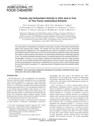
  • 4. 7776 J. Agric. Food Chem., Vol. 56, No. 17, 2008 Zaragoza et al. ´ Table 1. Chemical Composition (g/100 g) of F. vesiculosus Extracts 1 and 2a extract 1 extract 2 water 5.3 ( 0.0 9.1 ( 0.0*** ashes 13.7 ( 0.1 13.0 ( 0.1** proteins 3.6 ( 0.0 5.4 ( 0.0*** total lipids 1.2 ( 0.0 7.5 ( 0.6*** carbohydrates 17.8 ( 0.5 11.1 ( 0.4*** uronic acids 9.9 ( 0.4 10.1 ( 0.6 total fiber 20.1 ( 1.5 1.3 ( 0.1*** iodine (mg/kg) 113 ( 1 77 ( 2*** a Results are expressed as means ( SEM. Differences between extracts by the Student’s t test are given by the following: **p < 0.01; ***p < 0.001; n ) 3 replicates. in the presence of 2 mM CaCl2 and 2 mM paraoxon. Paraoxon degradation to p-nitrophenol was monitored by the absorbance increase at 412 nm (extinction coefficient 18290 L/mol/cm) for 5 min. One unit of PON-1 hydrolyzed 1 nmol of paraoxon/min. To evaluate the O2•- scavenging activity, we used the same procedure (15) as described in the noncellular system, albeit with serial dilutions of plasma. An oxygen radical absorbance capacity (ORAC) assay was used to elucidate the antioxidant behavior of plasma against the peroxyl radicals with R-phycoerythrin serving as a redox-sensitive fluorescent indicator and ABAP as a chemical peroxyl radical generator (24). Trolox was used as a positive control. R-Phycoerythrin fluores- cence decline was measured as the difference between 5 and 30 min at 37 °C (excitation wavelength, 530 nm; emission wavelength, 585 nm). The thiobarbituric acid-reactive substances (TBARS) in plasma as indicative of malondialdehyde presence were determined by the method of Yagi (25) with one minor modification: Butylated hydroxytoluene Figure 1. (A) LC-DAD-MS chromatogram of a F. vesiculosus extract. and ethylenediaminetetraacetic acid were added to the reaction mixture (B) Ions found under the peak I (RT ) 26 min) using cone voltage values as antioxidants at a final concentration of 0.01% and 1.3 µM, higher than 30 V. The ions with (m/z)- 125 correspond to phloroglucinol, respectively. while ions with (m/z)- 83, 69, and 57 were its main ions product. The NO released in plasma was oxidized to nitrites. NO was Phloroglucinol (RT ) 5.6 min) was not found. (C) LC-MS/MS analysis in measured in deproteinized samples (100 µL) by 20% trichloroacetic parent scan mode. The ions found correspond to the parents of ion acid (1:1 v/v) at 4 °C and centrifuged at 18600g for 15 min. Supernatants (50 µL) were incubated in 5 mL of a solution (10 mM products with (m/z)- 125. KI and 10 mM H2SO4) converting nitrites to NO, which was evaluated by a 2.0 mm Iso-NOP sensor. The maximal signal was recorded and Expert Committee on Food Additives is 1000 µg/day. A new compared to a NaNO2 standard. European Union regulation (2006/352) sets to 150 µg/day the Enzymatic Protection in Rat Erythrocytes. Tests were carried out in maximum iodine allowed in food supplements. Thus, the iodine erythrocytes from 24 h-fasted male rats daily treated by oral route with content in the extracts should be taken into account in order F. Vesiculosus extracts (200 mg/kg body weight) at the end of the 4 not to exceed the limits allowed when administered to humans. week toxicity study and with phloroglucinol (43 mg/kg body weight) Total polyphenols (g/100 g extract) expressed as gallic acid in the same conditions as F. Vesiculosus-treated rats. and phloroglucinol equivalents were higher in extract 1 (28.8 To evaluate the Cu-Zn SOD activity, 750 µL of hemolysed ( 1.7 and 27.7 ( 1.5, respectively) than in extract 2 (18.0 ( erythrocytes was used according to a technique based on the inhibition of pyrogallol auto-oxidation (26). The catalase activity was measured 1.6 and 16.3 ( 0.3, respectively) (Table 2). The molecular in 20 µL of hemolysed erythrocytes at room temperature, and the masses of the polyphenolic fractions of the two extracts decomposition of H2O2 at 240 nm was evaluated (27). The rate constant demonstrate that they were similar with about 40% > 300 kDa, (k ) 107 L/mol s) for the first 30 s was calculated. 43% between 50 and 300 kDa, 7% between 30 and 50 kDa, Statistical Analysis. The statistical analysis was conducted by using 10% between 10 and 30 kDa, and traces with molecular masses the SPSS version 12 statistical analysis packages. Results were <10 and <3 kDa. The LC-DAD-MS(MS) chromatograms of expressed as the means ( standard errors of the mean (SEM). Data the F. Vesiculosus extract 2 are reported in Figure 1. The UV were evaluated using either the unpaired Student’s t test or one-factor spectrum of peak I (Figure 1A) suggests a structure of a analysis of variance. The DMS test for multiple comparisons was used phloroglucinol derivative, while the mass spectrum at high cone to detect differences among groups (p < 0.05). voltage values yields ions with (m/z)- 125, 83, 69, and 57 (Figure 1B). The ions with (m/z)- 83, 69, and 57 represent RESULTS AND DISCUSSION ions product of phloroglucinol, suggesting that peak I may Characterization of the F. Wesiculosus Extracts. Although contain a phloroglucinol moieties. Figure 1C reports the LC- extract 1 contained less proteins and lipids than extract 2, it MS/MS chromatogram obtained when acquiring the parents of was richer in carbohydrates and total fiber. The presence of total ions product with (m/z)- 125. It should be emphasized that peak fiber in the extracts is inversely related to the percentage of I was not present in the fraction lower than 10 kDa and that ethanol used in the extraction procedure (Table 1). The iodine phloroglucinol was not found in the F. Vesiculosus extracts. content (Table 1) was greater in extract 1 than in extract 2. Thus, the results seem to indicate that the polyphenolic fractions The European Scientific Committee on Food suggests an upper are high molecular weight forms of polymeric phloroglucinol. intake of 600 µg of iodine/day whether the limit set by the Joint Although there were differences in the condensed forms,
  • 5. Toxicity and Antioxidant Activity of Fucus vesiculosus J. Agric. Food Chem., Vol. 56, No. 17, 2008 7777 Table 2. Polyphenols and Fucoxanthin in F. vesiculosus Extracts 1 and 2a serve as ingredients for the formation of highly pro-oxidant radical species such as hydroxyl radical and peroxynitrite (a extract 1 extract 2 potent oxidant generated by the mol:mol reaction of O2•- and total polyphenols g gallic ac. equiv/100 g 28.8 ( 1.7 18.0 ( 1.6** NO) (29) that lead to severe damage to host cells and tissues. g phloroglucinol equiv/100 g 27.7 ( 1.5 16.3 ( 0.3** The extracts had a similar effect on reducing O2•- production fucoxanthin mg/100 g 0.26 ( 0.02 1.24 ( 0.06*** when RAW 264.7 macrophages were stimulated with PMA a Results are expressed as means ( SEM. Differences between extracts by (Table 4). Indeed, extract 1 proved to be more effective than the Student’s t test are given by the following: **p < 0.01; ***p < 0.001; n ) 3 extract 2 when LPS was used (Table 4). With regard to replicates. decreasing NO production, extract 1 was more effective than extract 2 (Table 4). A reduction of NO production by RAW structures, and molecular weights, the chemical properties were 264.7 macrophages by phloroglucinol derivatives was also similar. Thus, it was difficult, and also not particularly useful, observed by Ishii et al. (30). Furthermore, our group has to evaluate single phlorotannins in the two extracts. Moreover, previously demonstrated that NO inhibits the O2•- production monomeric forms of polyphenols were not present. by inflammatory exudate-derived polymorphonuclear leukocytes Although fucoxanthin levels were low in both extracts, these (31) by inactivating the NAD(P)H oxidase. Thus, it is likely levels were higher in extract 2 (1.24 ( 0.06 mg/100 g) (Table that the final O2•- measurement may be the result of NAD(P)H 2). The extracts also contained astaxanthin and violaxanthin in inactivation by NO and the O2•- scavenging activity, which is low concentrations (<0.05 mg/100 g). supported by the reduction of IC50 in the X/XO assay. Reduced Antioxidant Activity of the Extracts in Noncellular Sys- O2•- and NO production can indicate that the generation of tems. The two extracts from F. Vesiculosus showed a good either hydroxyl radicals or peroxynitrite will be reduced. The activity in a broad spectrum of the antioxidant chemical tests. capacity of the two extracts to protect against oxidative damage Both extracts had important reducing power (Table 3); thus, caused by free radicals may be of great interest in the prevention they are likely to contain molecules that act as electron donors, of the acute and chronic inflammatory processes that occur in converting them into more stable products, thereby terminating many diseases and are concomitant with the activation of the radical chain reaction. Although this approach is being used polymorphonuclear leukocytes and macrophages. However, in the evaluation of antioxidant activity of dietary polyphenols, these cells also have an antimicrobial function because of their caution is necessary to extrapolate the results to the physiological phagocytic nature and capacity to release reactive species into environment, as this assay is performed in acidic pH value. the phagosome. The measurement of O2•- and NO production Extract 1 proved to be a more potent antioxidant in terms of by RAW 264.7 macrophages indicated that extract 1 exhibited a variety of radical-generating systems such as O2•- (381%), higher antioxidant activity than extract 2, which may be related DPPH radical (218%), peroxyl radicals (71%), and also in the to the 60% higher polyphenols or polyphenols with higher ABTS radical cation (80%) than did extract 2 (Table 3). The antioxidant activity. O2•- scavenging activity is relevant because although this radical As observed with extracts from seaweed species other than does not directly initiate lipid oxidation, but it does generate F. Vesiculosus obtained by 50% ethanol, the greater total hydroxyl radicals by the Fenton reaction. No assays on the polyphenol content correlates with greater O2•- scavenging antioxidant activity of F. Vesiculosus have been performed to activity in vitro (9). However, evidence of antioxidant activity date. However, assays using other brown seaweeds rich in in vitro cannot be easily extrapolated into an in vivo setting phloroglucinol or in phloroglucinol derivatives showed great due to the fact that once ingested polyphenols and carotenoids reducing power and DPPH radical scavenging activity (7) and undergo extensive modification during first-pass metabolism and O2•- (8, 9) and DPPH radical scavenging activities (8), also because of the concentrations tested (32). respectively. Azo compounds such as ABAP generate peroxyl Toxicity Studies. The LD50 by the oral route in rats for radicals at a reproducible and constant rate. Phenols are the most extracts 1 and 2 (Table 5) ranged between 1000 and 2000 and common antioxidant compounds to readily scavenge peroxyl >2000 mg/kg, respectively, while with female mice, they ranged radicals by donating hydrogen atoms as well as by stabilizing between 1000 and 2000 and >750 mg/kg, respectively. As the resulting antioxidant radical by electron delocalization and/ expected, administration of the two extracts by an intraperitoneal or intramolecular hydrogen bonding or by further oxidation (28). route resulted in higher toxicity than by the oral route. Moreover, The higher activity of extract 1 is likely to be related to the the extracts were more toxic in mice than in rats (Table 5). greater content of total polyphenols. In addition, polyphenols The Irwin test did not show important alterations even by the and lipophilic antioxidants such as carotenoids can be measured intraperitoneal route. with the ABTS radical cation test. Fucoxanthin has also been described to be a scavenger of DPPH (10, 11) and ABTS Treated female rats ingested approximately less food (13% radicals (11). The higher levels of fucoxanthin present in extract reduction along the 4 week toxicity study) than their respective 2 should be partly responsible for the antioxidant activity (10). control; thus, their weight gain was also reduced. The hema- The capacity of the two extracts from F. Vesiculosus to tological values did not reveal any pathological modification scavenge O2•-, peroxyl radicals, or artificial radicals provides and were found to range within the normal limits for rats. We support for antioxidant efficacy in vitro. However, the cell-based only detected a decrease in the white cell count in those groups in vitro results, as well as those obtained ex vivo after treated with extract 2 (about 50%) and in the group treated with administering the extracts, are considered more predictive than the high dose of extract 1 (about 30%). However, the differential the present results based on chemically generated oxidants. white cell percentage was maintained. No differences were Antioxidant Activity of the Extracts in a Cellular System. detected in any of the coagulation parameters assayed. Activated inflammatory cells, such as macrophages, contain We have also observed an increase of R-amylase in rats NAD(P)H-oxidase. The NAD(P)H-oxidase and the inducible treated either with the high dose of extract 1 (44%) or with NO synthase are strongly induced upon exposure to bacterial both doses of extract 2 (about 36%), although these values were endotoxin and inflammatory cytokines. These enzymes mediate within the normal limits for rats. In addition, we observed an the massive production of O2•- and NO, respectively, which increase in Na+ urine excretion when extracts 1 and 2 (68 and
  • 6. 7778 J. Agric. Food Chem., Vol. 56, No. 17, 2008 Zaragoza et al. ´ Table 3. Reducing Power and Antioxidant Activities of F. vesiculosus Extracts 1 and 2 by Noncellular Systemsa extract 1 extract 2 phloroglucinol reducing power (mmols quercetin equiv/g extract) 1.5 ( 0.1 b 1.0 ( 0.0 a 8.5 ( 0.1 c O2•- scavenging activity (IC50, µg/mL) 182 ( 7 b 694 ( 35 c 86 ( 4 a DPPH (IC50, µg/mL) 11.9 ( 0.1 a 26 ( 1 c 18.0 ( 0.9 b TRAP (mmols trolox equiv/g extract) 2.4 ( 0.1 a 1.4 ( 0.1 a 28.7 ( 1.8 b ABTS scavenging activity (mmols trolox equiv/g extract) 1.8 ( 0.1 b 1.0 ( 0.1 a 17.0 ( 0.2 c a Significant differences were evaluated by the DMS test. Mean values within a row with unlike superscript letters were significantly different, p < 0.05. Results are expressed as means ( SEM; n ) 3 replicates. Table 4. O2•- and NO Production by F. vesiculosus Extracts 1 and 2 in plasma following the daily 4 week treatment at 200 mg/kg, RAW 264.7 Macrophagesa probably due to their low systemic uptake (32), and, thus, small reached concentrations. inducible agent extract 1 extract 2 Nonenzymatic Antioxidant Protection in Rat Plasma. O2•- (IC50, µg/mL) PMA 38 ( 4 31 ( 4 LPS 39 ( 5 68 ( 6** Knowledge-based antioxidant efficacy must drive from in vivo/ NO (IC50, µg/mL) PMA 37 ( 5 not inhibition ex vivo data, and its activity can be through a direct scavenging LPS 95 ( 10 >100 of free radicals. The increased reducing power (29%), PON-1 activity (33%), and O2•- scavenging activity (25%) in the plasma a Results are expressed as means ( SEM. Differences between extracts by of rats treated with the extract 2 (Table 7) indicate that some Student’s t test are given by the following: **p < 0.01; n ) 4 replicates. compounds were absorbed and their presence in the parent or Table 5. Approximate LD50 Values of F. vesiculosus Extracts 1 and 2 metabolized forms will exhibit antioxidant activity in the organism although the -SH groups were maintained at levels LD50 (mg/kg body weight) similar to those of the control. An increase in the reducing power animal route sex extract 1 extract 2 has also been observed in rats a short time after the administra- tion of a very high single dose (2000 mg/kg containing 600 rat oral female 1000-2000 >2000 male 1000-2000 >2000 mg/kg polyphenols) of brown seaweeds extract (VNP) (7). The i.p. female 250 >500 fact that plasma nitrite concentration remained constant while male 1000-2000 500 plasma exhibited a higher O2•- scavenging activity may indicate mice oral female 1000-2000 <750 that less peroxynitrite is being produced. This observation is male 1000 500 corroborated by the significantly reduced levels of TBARS i.p. female 150-200 250-500 male 150-200 250-500 (17%) (Table 7), which most likely reflects not only plasma’s increased ability to scavenge free radicals but also PON-1’s greater hydrolytic activity. PON-1 protects low-density lipo- 90%, respectively) were administered at the highest dose. proteins from oxidative modification by reactive oxygen species However, we have not observed a decrease in plasma Na+ and thus significantly contributes to the atheroprotective effect concentration. This may be explained by an effect of extracts of high-density lipoproteins. It hydrolyzes phospholipid hydro- on Na+ release from the metabolism of organic molecules such peroxides and cholesterol ester hydroperoxides (esterase activity) as uronic acids, as NaCl was not present in the extracts. Fecal and reduces lipid hydroperoxides to the respective hydroper- blood yielded negative results throughout the 4 week of daily oxides as well as degrades hydrogen peroxide (peroxidase oral doses of F. Vesiculosus extracts, indicating an absence of activity) (23). Thus, antioxidants in extract 2 maintain ex vivo lesions in the gastrointestinal tract. their reducing capacity and O2•- scavenging activity, in addition In terms of organ weight (Table 6), we found an increase in to other activities. It is likely that the increased activity of extract the relative liver weight in male rats treated with the high dose 2 ex vivo is caused by a distinct phlorotannin content and to of extract 2 (25%) and in the kidneys of male rats treated with the presence of fucoxanthin, as carotenoids react with peroxyl the high dose of extract 1 (21%). Increased liver weight stemmed radicals by forming an adduct. not from fat accumulation, since this was not observed in the Enzymatic Antioxidant Protection in Rat Erythrocytes. anatomopathological study, nor was it due to alcohol, as the The Cu-Zn SOD activity was increased (about 32%) in rats latter was absent from extracts. Moreover, the absence of any treated with the extract 2 and phloroglucinol (Figure 2A). The changes in liver and kidney functional parameters (transami- catalase activity (Figure 2B), however, remained in both groups nases, alkaline phosphatase, and bilirubin and urea and creati- of extract-treated rats at levels similar to those of the control nine, respectively) supports a lack of toxicity in these organs. group. An increased Cu-Zn SOD activity can be seen as an In general, the anatomopathological changes observed were additional protective mechanism against oxidative stress if slight or moderate and neither matched the severity of acute catalase activity is sufficiently high enough to counteract the alterations nor were dose-dependent, and they were slightly H2O2 generated by the SOD. Depending upon the phenolic greater in rats treated with extract 2. Small alterations in both compound involved (its structure, specificity to the tissue, and extract treated rats groups were observed in lungs that may be the degree of systemic bioavailability), its effect on rat anti- related to a hypersensitivity reaction stemming from factors other oxidant enzymes is likely to be different (33). than extract administration. The overall results obtained from Our results indicate that the oxidative stress observed in the 4 week toxicity study indicate that even at the daily dose of plasma and erythrocytes is reduced when rats are treated with 750 mg/kg body weight for 4 weeks, no relevant signs of toxicity the extract 2 for a period of 4 weeks, although extract 1 occurred with the two F. Vesiculosus extracts studied. contained 60% more total polyphenols than extract 2, which is Phloroglucinol and Fucoxanthin Evaluation in Rat Plasma. probably a reflection of absorption and the rate of metabolism Phloroglucinol and fucoxanthin studied by HPLC-MS/MS, in and also of the levels of fucoxanthin, although compounds were their either parent or metabolized forms, were not found in rat not detected. One possibility is that the antioxidant effects ex
  • 7. Toxicity and Antioxidant Activity of Fucus vesiculosus J. Agric. Food Chem., Vol. 56, No. 17, 2008 7779 Table 6. Body and Relative Organ Weights of Female Rats after a Daily Dose of F. vesiculosus Extracts 1 and 2 during a 4 Week Perioda body weight heart stomach liver spleen lungs kidneys ovaries/testes females control 211 ( 6 a 0.44 ( 0.02 a 1.07 ( 0.08 a 3.41 ( 0.25 a 0.30 ( 0.01 a 0.68 ( 0.06 a 0.54 ( 0.03 a 0.15 ( 0.03 a extract 1, 200 mg/kg 201 ( 9 a 0.43 ( 0.03 a 0.94 ( 0.07 a 3.79 ( 0.07 a 0.34 ( 0.02 a 0.88 ( 0.23 a 0.51 ( 0.02 a 0.13 ( 0.01 a extract 1, 750 mg/kg 190 ( 5 a 0.44 ( 0.03 a 1.01 ( 0.03 a 3.79 ( 0.38 a 0.34 ( 0.02 a 0.70 ( 0.10 a 0.49 ( 0.03 a 0.13 ( 0.01 a extract 2, 200 mg/kg 209 ( 4 a 0.41 ( 0.01 a 1.03 ( 0.05 a 3.41 ( 0.05 a 0.29 ( 0.01 a 1.06 ( 0.21 a 0.51 ( 0.02 a 0.15 ( 0.02 a extract 2, 750 mg/kg 196 ( 8 a 0.43 ( 0.02 a 1.05 ( 0.05 a 3.53 ( 0.22 a 0.34 ( 0.02 a 0.67 ( 0.13 a 0.55 ( 0.02 a 0.17 ( 0.04 a males control 302 ( 10 a 0.40 ( 0.01 a 0.84 ( 0.04 a 3.16 ( 0.13 a 0.24 ( 0.01 a 0.63 ( 0.14 a 0.48 ( 0.02 a 0.73 ( 0.08 a extract 1, 200 mg/kg 299 ( 8 a 0.42 ( 0.01 a 0.91 ( 0.07 a 3.63 ( 0.20 ab 0.31 ( 0.05 a 0.59 ( 0.02 a 0.52 ( 0.02 ab 0.75 ( 0.05 a extract 1, 750 mg/kg 285 ( 6 a 0.41 ( 0.00 a 0.93 ( 0.05 a 3,28 ( 0.19 a 0.28 ( 0.01 a 0.64 ( 0.03 a 0.59 ( 0.02 b 0.85 ( 0.05 a extract 2, 200 mg/kg 295 ( 6 a 0.43 ( 0.02 a 0.98 ( 0.04 a 3.42 ( 0.10 a 0.26 ( 0.01 a 0.60 ( 0.03 a 0.55 ( 0.02 ab 0.76 ( 0.02 a extract 2, 750 mg/kg 270 ( 5 a 0.44 ( 0.02 a 0.97 ( 0.03 a 3.97 ( 0.19 b 0.31 ( 0.02 a 0.82 ( 0.11 a 0.57 ( 0.04 ab 0.90 ( 0.07 a a Significant differences were evaluated by DMS test. Mean values within a column with unlike superscript letters were significantly different, p < 0.05. Results are expressed as means ( SEM; n ) 7 rats. Table 7. Oxidative Stress Parameters in Plasma of Male Rats after a Daily Dose (200 mg/kg) of F. vesiculosus Extracts 1 and 2 during a 4 Week Perioda control extract 1 extract 2 phloroglucinol reducing power (mM quercetin equiv) 139 ( 12 a 148 ( 12 a 180 ( 13 b 182 ( 13 b -SH groups (mM glutathione equiv) 0.23 ( 0.02 a 0.26 ( 0.02 a 0.26 ( 0.02 a 0.24 ( 0.02 a PON-1 activity (U/mL) 179 ( 7 a 214 ( 12 bc 238 ( 17 c 200 ( 11 ab O2•- scavenging activity (IC50, µg/mL) 45 ( 4 b 39 ( 4 ab 34 ( 3 a 42 ( 4 ab ORAC (mM trolox equiv) 2.2 ( 0.1 a 2.7 ( 0.2 a 2.7 ( 0.2 a 2.8 ( 0.2 a NO concentration (µM) 2.9 ( 0.2 a 3.0 ( 0.2 a 2.9 ( 0.2 a 3.1 ( 0.2 a TBARS (µM) 8.2 ( 0.5 b 7.9 ( 0.4 ab 6.8 ( 0.5 a 7.2 ( 0.6 ab a Significant differences were evaluated by DMS test. Mean values within a row with unlike superscript letters were significantly different, p < 0.05. Results are expressed as means ( SEM; n ) 7 rats. vivo are mediated by metabolites with also biological activity. This paper confirms that antioxidant efficacy can be demon- It is also likely that a synergic effect among polyphenols (34) strated in one system but fails to protect in others. These or carotenoids (35), in their either parent or metabolized forms, observations reflect the distinct mechanism of action between takes place. the two seaweed extracts. In conclusion, here, we provide evidence that the two extracts obtained from F. Vesiculosus lack toxicity and exhibit important antioxidant activity against chemically generated oxidants and in vitro. However, only extract 2 shows an antioxidant activity ex vivo through preventing oxidant formation, scavenging O2•- and reducing active intermediates. Our findings are relevant, as they elucidate the potential future application of the F. Vesiculosus extract 2 (Healsea) both as functional foods and as food supplements. Although extrapolation of findings from animal experiments to humans is difficult, it is conceivable that F. Vesiculosus extracts will maintain the antioxidant activity in humans. ABBREVIATIONS USED O2•-, superoxide anion; NO, nitric oxide; DAD, diode array detection; X/XO, xanthine/xanthine oxidase; DPPH, 2,2-di- phenyl-1-picrylhydrazyl; ABAP, 2,2′-azo-bis-2-amidinopropane; ABTS, 2′-azinobis(3-ethylbenzothiazoline-6-sulfonic acid); SOD, superoxide dismutase; PMA, phorbol 12-myristate 13-acetate; LPS, lipopolysaccharide; L-NIO, N5-(1-iminoethyl)-L-ornithine dihydrochloride; IC50, inhibitory concentration 50; TRAP, total radical antioxidant parameter; LD50, lethal dose 50; PON, paraoxonase; ORAC, oxygen radical absorbance capacity; TBARS, thiobarbituric acid-reactive substances. ACKNOWLEDGMENT Figure 2. Enzymatic antioxidant activities in erythrocytes from male rats We thank Robin Rycroft for editing the manuscript. after a daily dose (200 mg/kg) of F. vesiculosus extracts 1 and extract 2 LITERATURE CITED during a 4 week period. (A) Cu-Zn SOD. (B) Catalase. Data are expressed as means ( SEM. Significant differences were evaluated by (1) Rice-Evans, C. A.; Miller, N. J.; Paganga, G. Structure-antioxidant DMS test, p < 0.05. Data with different letters are significantly different; activity relationships of flavonoids and phenolic acids. Free n ) 7 rats. Radical Biol. Med. 1996, 20, 933–956.
  • 8. 7780 J. Agric. Food Chem., Vol. 56, No. 17, 2008 Zaragoza et al. ´ (2) Krinsky, N. I.; Kyung-Jin, Y. Carotenoid-radical interactions. (20) Irwin, S. Comprehensive observational assessment: Ia. A system- Biochem. Biophys. Res. Commun. 2003, 305, 754–760. atic, quantitative procedure for assessing the behavioral and (3) Kuo, S. M.; Leavitt, P. S.; Lin, C. P. Dietary flavonoids interact physiologic state of the mouse. Psychopharmacology (Berlin) with trace metals and affect metallothionein level in human 1968, 13, 222–257. intestinal cells. Biol. Trace Elem. Res. 1998, 62, 135–153. (21) Needhan, C. D.; Simpson, R. G. The benzidine test for occult (4) Robak, J.; Gryglewski, R. J. Flavonoids are scavengers of blood in faces. Q. J. Med. 1952, 21, 123–133. superoxide anions. Biochem. Pharmacol. 1998, 37, 837–841. (22) Ellman, G.; Lysko, H. A precise method for the determination of (5) Cook, N. C.; Samman, S. Flavonoids. Chemistry, metabolism, whole blood and plasma sulfhydryl groups. Anal. Biochem. 1979, cardioprotective effects, and dietary source. J. Nutr. Biochem. 93, 98–102. 1996, 7, 66–76. (23) Aviram, M.; Hardak, E.; Vaya, J.; Mahmood, S.; Milo, S.; (6) Ragan, M. A.; Glombitza, K. W. Phlorotannins, brown algal Hoffman, A.; Billicke, S.; Draganov, D.; Rosenblat, M. Human polyphenols. Progr. Phycol. Res. 1986, 4, 129–241. serum paraoxonases (PON1) Q and R selectively decrease lipid (7) Kang, K.; Park, Y.; Hawng, H. J.; Kim, S. H.; Lee, J. G.; Shin, peroxides in human coronary and carotid atherosclerotic lesions: H. C. Antioxidative properties of brown algae polyphenolics and PON1 esterase and peroxidase-like activities. Circulation 2000, 101, 2510–2517. their perspectives as chemopreventive agents against vascular risk (24) Glazer, A. N. Phycoerythrin fluorescence-based assay for reactive factors. Arch. Pharm. Res. 2003, 26, 286–293. oxygen species. Methods Enzymol. 1990, 186, 161–168. (8) Lee, S. M.; Na, M. K.; An, R. B.; Min, B. S.; Lee, H. K. (25) Yagi, K. Assay for blood plasma or serum. Methods Enzymol. Antioxidant activity of two phloroglucinol derivatives from 1984, 105, 328–331. Dryopteris crassirhizoma. Biol. Pharm. Bull. 2003, 26, 1354– (26) Marklund, S. L. Pyrogallol autooxidation. In Handbook of Methods 1356. for Oxygen Radical Research; Greenwald, R. A., Ed.; CRC Press: (9) Nakai, M.; Kageyama, N.; Nakahara, K.; Miki, W. 2006. Boca Raton, FL, 1985; Vol. 24, pp 3-247. ´ Phlorotannins as radical scavengers from the extract of Sargassum (27) Aebi, H. E. Catalase in vitro. Methods Enzymol. 1984, 105, 121– ringgoldianum. Marine Biotechnol. 2006, 8, 409–414. 126. (10) Yan, X.; Chuda, Y.; Suzuki, M.; Nagata, T. Fucoxanthin as the (28) Frankel, E. N.; Meyer, A. S. The problems of using one- major antioxidant in Hijikia fusiformis, a common edible seaweed. dimensional methods to evaluate multifunctional food and biologi- Biosci., Biotechnol., Biochem. 1999, 63, 605–607. cal antioxidants. J. Sci. Food Agric. 2000, 8, 1925–1941. (11) Sachindra, N. M.; Sato, E.; Maeda, H.; Hosokawa, M.; Niwano, (29) Liochev, S. I.; Fridovich, I. Superoxide and nitric oxide: conse- Y.; Kohno, M.; Miyashita, K. Radical scavenging and singlet quences of varying rates of production and consumption: A oxygen quenching activity of marine carotenoid fucoxanthin and theoretical treatment. Free Radical Biol. Med. 2002, 33, 137– its metabolites. J. Agric. Food Chem. 2007, 55, 8516–8522. 141. (12) Singleton, V. L.; Rossi, J. A. Colorimetry of total phenolics with (30) Ishii, R.; Masakazu, H.; Koichi, S.; Munehisa, A.; Susumu, K. phosphomolybdic-phosphotungstic acid reagents. Am. J. Enol. Inhibitory effects of phloroglucinol derivates from Mallotus Vitic. 1965, 16, 144–158. japonicus on nitric oxide production by a murine macrophage- (13) Verhagen, H.; Auroma, O. I.; VanDelft, J. H. M.; Dragsted, L. O.; like cell line, RAW 264.7, activated by lipopolysaccaride and Ferguson, L. R.; Knasmuller, S.; Pool-Zobel, B. L.; Poulsen, H. E.; ¨ interferon-γ. Biochim. Biophys. Acta 2001, 1568, 74–82. Williamson, G.; Yannai, S. The 10 basic requeriments for a (31) Rodenas, J.; Mitjavila, M. T.; Carbonell, T. Nitric oxide inhibits ´ scientific paper reporting antioxidant, antimutagenic or anticar- superoxide production by inflammatory polymorphonuclear leu- cinogenic potential of test substances in in vitro experiments and kocytes. Am. J. Physiol. 1998, 274, C827–C830. in animal studies in vivo. Food Chem. Toxicol. 2003, 41, 603– (32) Kroon, P. A.; Clifford, M. N.; Crozier, A.; Day, A. J.; Donovan, 610. J. L.; Manach, C.; Williamson, G. How should we assess the (14) Oyaizu, M. Studies on products of browning reaction: antioxidative effects of exposure to dietary polyphenols in vitro. Am. J. Clin. activities of products of browning reaction prepared from glu- Nutr. 2004, 80, 15–21. cosamine. Jpn. J. Nutr. 1986, 44, 307–315. (33) Breinholt, V.; Lauridsen, S. T.; Dragsted, L. O. Differential effects (15) Ukeda, H.; Maeda, S.; Ishii, T.; Sawamura, M. Spectrophotometric of dietary flavonoids on drug metabolism and antioxidant enzymes assay for superoxide dismutase based on tetrazolium salt 3′-{1- in female rats. Xenobiotica 1999, 29, 1227–1240. [(phenylamino)-carbonyl]-3,4-tetrazolium}-bis(4-methoxy-6-ni- (34) Pignatelli, P.; Ghiselli, A.; Buchetti, B.; Carnevale, R.; Natella, tro)benzenesulfonic acid hydrate reduction by xanthine-xanthine F.; Germano, G.; Fimognari, F.; Di Santo, S.; Lenti, L.; Violi, F. ´ Polyphenols synergistically inhibit oxidative stress in subjects oxidase. Anal. Biochem. 1997, 251, 206–209. given red and white wine. Atherosclerosis 2006, 188, 77–83. (16) Gerhauser, C. Mechanism-based in vitro screening of potential ¨ (35) Stahl, W.; Junghans, A.; de Boer, B.; Driomina, E. S.; Briviba, cancer chemopreventive agents. Mutat. Res. 2003, 523-524, 163– K.; Sies, H. Carotenoid mixtures protect multilamellar liposomes 172. against oxidative damage: synergistic effects of lycopene and (17) Visioli, F.; Galli, C. Evaluation of antioxidant capacity by lutein. FEBS Lett. 1998, 427, 305–308. chemiluminescence. Anal. Biochem. 1997, 249, 244–246. (18) Re, R.; Pellegrini, N.; Proteggente, A.; Pannala, A.; Yang, M.; Received for review March 7, 2008. Revised manuscript received July Rice-Evans, C. A. Antioxidant activity applying an improved 2, 2008. Accepted July 7, 2008. We thank the Generalitat de Catalunya ABTS radical cation decolourization assay. Free Radical Biol. (2005SGR00269) and the University of Barcelona for supporting M.C.Z. Med. 1999, 26, 1231–1237. This research work was financially supported by the EU SEAHEALTH (19) Rodenas, J.; Mitjavila, M. T.; Carbonell, T. Simultaneous genera- ´ project under Grant QLK1-2002-02433. tion of nitric oxide and superoxide by inflammatory cells in rats. Free Radical Biol. Med. 1995, 18, 869–875. JF8007053