
Recommended
Recommended
More Related Content
What's hot
What's hot (18)
Similar to Neuroprotective Effects of Withania somnifera Dunn. in Hippocampal Sub-regions of Female Albino Rat
Similar to Neuroprotective Effects of Withania somnifera Dunn. in Hippocampal Sub-regions of Female Albino Rat (20)
Recently uploaded
Recently uploaded (20)
Neuroprotective Effects of Withania somnifera Dunn. in Hippocampal Sub-regions of Female Albino Rat
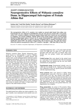
- 1. SHORT COMMUNICATION Neuroprotective Effects of Withania somnifera Dunn. in Hippocampal Sub-regions of Female Albino Rat Sushma Jain,1 Sunil Dutt Shukla,1 Kanika Sharma2 and Maheep Bhatnagar1 * 1 Department of Zoology, University College of Science, M.L. Sukhadia University, Udaipur 313001, India 2 Department of Botany, University College of Science, M.L. Sukhadia University, Udaipur 313001, India The neuroprotective effects of W. somnifera were studied on stressed adult female Swiss albino rats. Experimental rats were subjected to immobilization stress for 14 h and were treated with a root powder extract of W. somnifera available as Stresscom capsules (Dabur India Ltd). Control rats were maintained in completely, non stressed conditions. Thionin stained serial coronal sections (7mm) of brain passing through the hippocampal region of stressed rats (E1 group) demonstrated 85% degenerating cells (dark cells and pyknotic cells) in the CA2 and CA3 sub-areas. Treatment with W. somnifera root powder extract significantly reduced (80%) the number of degenerating cells in both the areas. The study thus demon- strates the antistress neuroprotective effects of W. somnifera. Copyright # 2001 John Wiley & Sons, Ltd. Keywords: W. somnifera; root powder; hippocampus; cell degeneration; immobilization stress; rat. INTRODUCTION Withania somnifera Dunn. (Family Solanaceae) com- monly known as ‘Ashwagandha’ is used as ‘Medhya Rasayana’ for the treatment of mental diseases and anxiety states, in the traditional Indian medicinal system (Chatterjee and Prakesh, 1995). It is an erect shrub 0.5–2 m high and grows throughout the dry and sub-tropical parts of India. The drug reduces the levels of acetylcho- line and catecholamine and increases the levels of serotonin and histamine in brain tissue (Handa, 1993). W.somnifera has also been shown to possess both depressant and anticonvulsant properties (Kulkarni and Verma, 1993; Kulkarni et al., 1993; Kulkarni and George, 1996). Ziauddin et al., (1996) studied its adaptogenic, antistress, anticonvulsant and cognitivedysfunction prop- erties. The antioxidative effects of the plant extract have been demonstrated (Bhattacharya et al., 1997; Panda and Kar, 1997). Cytoprotective effects of W. somnifera have also been demonstrated in cancer (Uma Devi, 1996). The present study was undertaken to investigate the antistress and neuroprotective effects of W. somnifera root extract on neuron cell bodies in hippocampal sub- regions of the adult female rat brain. MATERIALS AND METHODS Ten adult female Swiss albino rats, body weight 60 Æ 5g, age 2 months, were maintained in the animal room at a controlled temperature (26° Æ 2°C) and a light and dark cycle (12 h light and 12 h dark) for 7 days and were provided with food and water ad libitum. Rats were divided into two groups: control and experimental groups. Control group. Rats were placed in the animal room (n = 3 per cage). It was locked for 24 h. The next morning the animal room was opened and the rats were killed immediately by decapitation. Experimental group. Experimental animals were di- vided into five groups E1–E5. Animals in these groups were subjected to stress for specific periods and treated with the experimental drug. Stress protocol. Rats were placed in tightly fitting ventilated plastic boxes(10 Â 5cm) for a specific stress period (14 h) every day. The animals were unable to move in these boxes and thus became stressed. Stress was given for 30 days. The stress period was selected on the basis of a pilot study to obtain the maximum percentage cell degeneration in the CA1-Dg region. Although habituation was observed in the general behaviour, cell degeneration was complete. Drug preparation. An extract of W.somnifera Dunn. available as a commercial preparation Stresscom capsule (Dabur India Ltd), was used. The capsule consisted of a hydroalcoholic extract of Ashwagandha root powder. This extract was standardized for withanolids and withnoles. Soya lectins, bees wax and arachis oil was used as a base for medicament and packed in soft gelatin capsules. The capsule was dissolved in water and given orally, daily (20mg/kg body weight) for 30 days. Treatment was performed everyday between 9 am and 10 am. PHYTOTHERAPY RESEARCH Phytother. Res. 15, 544–548 (2001) DOI: 10.1002/ptr.802 Copyright # 2001 John Wiley & Sons, Ltd. * Correspondence to: Dr M. Bhatnagar, Associate Professor, Department of Zoology, University College of Science, M.L. Sukhadia University, Udaipur 313001, India. E-mail: mbhatnagar@yahoo.com Received 4 November 1998 Accepted 10 August 2000
- 2. Figure 1. (1) Photomicrograph of CA2 hippocampal region of the control animal (C) showing normal cell bodies (→) (Â40). (2) Photomicrograph demonstrating CA2 region of stress (E1) group totally dark cell (") with few pyknotic nuclei (A) (Â40). (3) Photomicrograph demonstrating CA2 region of dose (E2) group. Cells showing normal cell morphology (Â40). (4) Photomicrograph demonstrating CA2 region of dose ‡ stress (E3). Normal cells with few pyknotic nuclei (Â40). Figure 2. (1) Photomicrograph showing CA3 hippocampal sub-region of control (C). Normal cell bodies (→) present (Â40). (2) Photomicrograph demonstrating CA3 region of stress (E1) group showing dark cells (") with pyknotic nuclei (A) (Â40). (3) Photomicrograph showing CA3 region of dose ‡ stress (E3) group. All are normal cell bodies (2) (Â40). (4) Photomicrograph showing CA3 of dose ‡ stress (E3) group demonstrating normal cell (→) (Â40). NEUROPROTECTIVE EFFECTS OF WITHANIA SOMNIFERA 545 Copyright # 2001 John Wiley & Sons, Ltd. Phytother. Res. 15, 544–548 (2001)
- 3. Treatment Schedule. Group E1: Rats were subjected to 14h stress every day for 30 days and killed on day 31. Group E2: Normal rats were given the drug daily for 30 days. Group E3: Rats were given the drug daily for 30 days and from day 31 onwards were subjected to stress for 30 days. Group E4: Rats were subjected to stress for 30 days and from day 31 onwards given the drug for 30 days. Group E5: Rats were given the drug and stress daily for 30 days. Parameters of study. Daily body weight, food and water consumption was recorded. After completion of the experiments, the rats were killed by decapitation, and then the brain was dissected out. The ascorbic acid level (n = 4) was measured (Natelson, 1971), in the whole fore brain homogenate to confirm the stress effects. The remaining brain tissue (n = 6) was rinsed in distilled water and fixed for 12 h in 10% neutral chilled formalin. After fixation the brains were dehydrated in alcohol series, embedded in paraffin wax and 7mm thick serial coronal sections were cut. Sections were stained with thionin stain (Clark and Sperry, 1945) for demon- stration of nerve cells. RESULTS Hippocampal sub-regions CA1-Dg were studied in the control (C) and experimental (E1–E5) groups. Identification of the hippocampal sub-regions was based on a rat brain atlas by Paxinos and Watson (1986). Photomicrographs of C, E1, E2 and E3 groups are included only. CA2 hippocampal sub-regions In the control group (Fig. 1.1) the cell bodies were 5– 6 mm in size, and multipolar in shape. All the cells in the region demonstrated a large and distinct nucleus, a centrally located nucleolus and darkly stained cytoplasm. In group E1 (Fig. 1.2) the thionin stained preparation showed degenerating cells, (85%) darkly stained cells and pyknotic nuclei in CA2 (Fig. 3). Cell bodies were 3–5mm in size and irregular in shape. A few apoptotic bodies were observed. The cells were compactly arranged in a tier of 3–5 cells. In the E2 group (Fig. 1.3) the cells were large (5–6mm), with a large centrally located nucleus. The cytoplasm was darkly stained. No degenerative characteristics were observed. In the E3 group (Fig. 1.4) the cell bodies were large, the nucleus and nucleolus were distinct in most of the cell bodies. The number of dark cell bodies, pyknotic nuclei or apoptotic bodies were reduced significantly. In the E4 and E5 groups darkly stained cells, pyknotic nuclei and vacuolated spaces were observed, similar to the E1 group. Figure 3. (1) Photomicrograph showing cellular condensation (Â100). (2) Photomicrograph showing pyknotic cell (Â100). (3) Photomicrograph showing cellular vacualization and swelling (Â100). Photomicrograph showing dark cell (Â100). 546 S. JAIN ET AL. Copyright # 2001 John Wiley & Sons, Ltd. Phytother. Res. 15, 544–548 (2001)
- 4. CA3 hippocampal sub region In the control group (C, Fig. 2.1), the cells were large (7–8mm) and multipolar in shape. All the cells demon- strated a large, distinct nucleus, centrally located nucleolus nissel material in the cytoplasm was intensely stained. Cell bodies were arranged compactly in 4–6 tiers of cells. In group E1 (Fig. 2.2) the thionin stained preparation showed degenerating cells, darkly stained cells and pyknotic nuclei (85%). Pyknotic cell bodies were comparatively small (4–5 mm) and irregular in shape. Cell bodies with condensed cytoplasm, apoptotic bodies and vacuolar spaces were also observed. In group E2 (Fig. 2.3) the pattern of cell distribution was similar to the controls. The cells were large with a distinct nucleus and nucleolus. All the cells were arranged compactly in the layer. Vacuolar spaces, pyknotic nuclei etc. were not observed. In group E3 (Fig. 2.4) the cell bodies were large with a distinct and centrally placed nucleus. The number of vacuolar spaces, pyknotic nuclei, cells with condensed or shrunken cytoplasm were drastically reduced. In the E4 and E5 groups darkly stained cells, pyknotic nuclei and vacuolated spaces were observed, similar to the E1 group. No significant change in body weight was observed in the rats of the control or experimental group in the present study. The study of ascorbic acid level in the fore brain homogenate of the control and experimental groups showed a significant reduction in ascorbic acid level after stress commensurate with the drug treatment (Fig. 4). DISCUSSION The results of the present study demonstrates that Aswagandha (W. Somnifera Dunn.) produces neuropro- tective effects and reduces the stress induced changes in neuron cell bodies in the CA2 and CA3 sub-area of the hippocampus. Stress caused a significant degeneration in the cell bodies of these areas showing as dark cells, pyknotic cells, cells with condensed nuclei and cyto- plasm. Vacuolar spaces in the cell cytoplasm were observed in thionin stained serial sections. In sections demonstrating histochemical localization of Acpase, passing through the region. After treatment with Aswagandha root powder extract, a significant reduction in the number of these degenerated cells was also observed. Dark cells, cells with shrunken nucleus and cytoplasm, and vacuolar spaces were significantly reduced in both CA2 and CA3. Interestingly animals of group E3 (drug treatment for atleast 30 days given prior to subjecting to stress) showed more cytoprotective effects than the post or simultaneous treatment. Although the mechanism of degeneration and cyto- protective effects of the drug can not be explained on basis of these experiments it is possible that an increase in corticosteroid level during stress might be associated with the cell degeneration. Several authors have reported the neurotoxicological effects of corticosterone on hippocampal cells (Sapolasky et al. 1985, 1991; Gould et al., 1990). A significant decrease in the ascorbic acid level in the brain homogenate of the stressed group and restoration to the normal level after treatment with the herbal drug substantiate our observation. The cytoprotective properties of W. somnifera and several other plants have been studied earlier. Al-Harbi et al. (1997) showed anti-ulcer and gastric cytoprotective effects of Commifora mol mol. Zhu et al. (1997) showed such effects using Cyperus rotundus in rats. Dhuley and Naik (1997) demonstrated the protective effects of a Rhinax herbal formulation on CCl4 induced liver injury. Uma Devi et al. (1993) demonstrated cytoprotective andradiosenitization effects of W.somnifera. The present study is the first to report the cytoprotec- tive antistress effects of W.somnifera on hippocampal cell bodies. REFERENCES Al-Harbi MM, Queshi S, Raza MM, Ahmed MM, Afzal M, Shah AH. 1997. Gastric-antiulcer and cytoprotective effects of Commifora mol mol (oleo-gum resin) in rats. J Ethno- pharmacol 55: 141±150. Bhattacharya SS, Satyan KS, Ghosal S. 1997. Antioxidant activity of glycowithanolides from Withania somnifera. Indian J Exp Biol 35: 236±239. Chatterjee A, Prakesh SC. 1995. The Treatise of Indian Medicinal Plants, Vol. 4, CSIR: New Delhi; 209±210. Clark G, Sperry G. 1945. A simpli®ed nissel stain with thionin. Stain Tech 23: 23±24. Duley JN, Naik SP. 1997. Protective effects of Rhinax an herbal formulation against CCl4 induced liver injury and survival in rats. J Ethnopharmacol 56: 159±164. Gould E, Woolley CS, McEwen BS. 1990. Short term glucocorticoid manipulations after neuronal morphology and survival in the adult dentate gyrus. Neuroscience 37: 367±375. Figure 4. Ascorbic acid content (mg/mL) in rat forebrain homogenate. NEUROPROTECTIVE EFFECTS OF WITHANIA SOMNIFERA 547 Copyright # 2001 John Wiley & Sons, Ltd. Phytother. Res. 15, 544–548 (2001)
- 5. Handa SS. 1995. Plants and plant products in Indian health. In Decade of the Brain, India/US Research in Mental Health and Neuroscience. Koslow, Murthy Coelho (eds). CSIR: New Delhi; 163±171. Kulkarni SK, Verma AV. 1993. Ashwagandha and Brahmi nootropic and deaddiction pro®le of psychotropic indi- genous plants. Drugs Today 29: 257±263. Kulkarni SK, Sharma A, Verma A, Ticky MK. 1993. GABA receptor mediated anticonvulsant action of Withania somnifera root extract. Indian Drugs 30: 305±312. Kulkarni SK, George B. 1996. Anticonvulsant action of Withania somnifera (Ashwagandha) root extract against pentylehetetrazol - induced kindling in mice. Phytother Res 10: 447±449. Natelson S. 1971. Technique of Clinical Chemistry, 3rd edn. Charles C. Thomas: Spring®eld. Panda S, Kar A. 1997. Evidence for free radical scavenging activity of Ashwagandha root powder in mice. Indian J Physiol Pharmacol 41: 424±426. Paxinos G, Watson C. 1986. The Rat Brain in Stereotaxic Co- ordinates, 2nd edn. Academic Press: Florida. Sapolasky RM, Kery LC, McEwen BS. 1985. Prolonged glucocorticoid exposure reduces hippocampal neuron number: Implication for aging. J Neurosci 5: 1222±1227. Sapolasky RM, Satin-Behrens BA, Armanini MP. 1991. Long term adrenalectomy causes loss of dentate gyrus and pyramidal neurons in the adult hippocampus. Exp Neurol 114: 246±249. Uma Devi P, Sharada AC, Soloman FE. 1993. Antitumor and radiosensitizing effects of Withania somnifera (Ashwa- gandha) on a transplantable mouse tumor, sarcoma-180. Indian J Exp Biol 3: 607±611. Uma Devi P. 1996. Withania somnifera Dunn. (Ashwagand- ha). Potential plant source of a promising drug for cancer chemotherapy and radiosensitization. Indian J Exp Biol 34: 927±932. Zhu M, Luk HH, Fung HS, Luk CT. 1997. Cytoprotective effects of Cyperus rotundus against ethanol induced gastric ulceration in rats. Phytother Res 11: 392±394. Ziauddin N, Phansalkar N, Patki P, Diwanay S, Patwardhan B. 1996. Studies on the immunomodulatory effects of Ashwagandha. J Ethnopharmacol 50: 69±76. 548 S. JAIN ET AL. Copyright # 2001 John Wiley & Sons, Ltd. Phytother. Res. 15, 544–548 (2001)