Bone- Alveolar Process- portion of the maxilla/mandible that supports the tooth external plate- dense cortical bone covered by periosteum with nerves, blood vessels, and cells that can differentiate into osteoblasts cancellous trabeculae located between 2 compact layers inner socket wall called lamina dura or bundle bone (insertion of Sharpey’s fibers) absence of lamina dura may be present on radiograph of periodontal diseased site Alveolar bone is constantly changing throughout life and is constantly remodeling in response to occlusal forces osteoblasts deposit bone matrix osteoclasts involved in bone resorption. Periodontal Ligament- Ligament that surrounds tooth has supportive, nutritive, regenerative, and sensory functions Provides principle anchoring mechanism of tooth to alveolar bone Contains blood supply to provide nutrients to alveolar bone Contains undifferentiated stem cells which allow for repair and regeneration of cementum, PDL, and alveolar bone Contains sensory nerve network which provides sensory tactile feedback regarding the position and pressure on the tooth, Fibers insert on alveolar bone on one end and cementum of tooth on other Width of ligament space may be evident on radiograph, width ranges from 0.1-0.25mm and is narrowest on midpoint of root. Cementum Contains structural and biochemical properties similar to alveolar bone Organic matrix composed primarily of type I and III collagen deposited on root surface by formative cells (cementoblasts).
Mobile Marketing Trends for Your Business Strategy
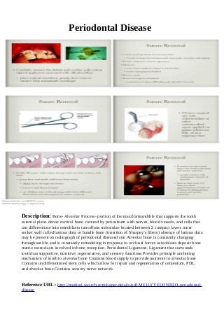
Periodontal Disease
1. Periodontal Disease
Description: Bone- Alveolar Process- portion of the maxilla/mandible that supports the tooth
external plate- dense cortical bone covered by periosteum with nerves, blood vessels, and cells that
can differentiate into osteoblasts cancellous trabeculae located between 2 compact layers inner
socket wall called lamina dura or bundle bone (insertion of Sharpey’s fibers) absence of lamina dura
may be present on radiograph of periodontal diseased site Alveolar bone is constantly changing
throughout life and is constantly remodeling in response to occlusal forces osteoblasts deposit bone
matrix osteoclasts involved in bone resorption. Periodontal Ligament- Ligament that surrounds
tooth has supportive, nutritive, regenerative, and sensory functions Provides principle anchoring
mechanism of tooth to alveolar bone Contains blood supply to provide nutrients to alveolar bone
Contains undifferentiated stem cells which allow for repair and regeneration of cementum, PDL,
and alveolar bone Contains sensory nerve network.
Reference URL : http://medical.wesrch.com/paper-details/pdf-ME1LYYEUONDZQ-periodontal-
disease