SlideShare una empresa de Scribd logo
1 de 47
CHAPTER 3 LOWER RESPIRATORY
TRACT INFECTIONS
             Acute Bronchitis (“Chest
             Cold”)

Photomicrograph     Tracheobronchial infections
of Bordetella       without pneumonia comprise a
(Haemophilus)       spectrum of disorders with
pertussis bacteria  different clinical implications:
using Gram stain    acute bronchitis, chronic
technique. CDC      bronchitis, and bronchiectasis.
                    Acute bronchitis or “acute simple
                    bronchitis” in otherwise-healthy
                    persons is extremely common,
                    usually of viral etiology, and a
          BLOOD common reason for overuse of
LYMPHOCYTOSIS antibiotics. The term “acute
IN A PATIENT        infectious bronchitis” is
WITH                sometimes used to distinguish this
PERTUSSIS. The entity from other causes of cough,
lymphocytes in this and the term “tracheobronchitis”
blood smear from is sometimes used for accuracy
an 18-month-old since the trachea is also inflamed.
child with a        However, “chest cold” is probably
Bordetella          the best term for daily practice
pertussis infection   since it implies that antibiotics are
have lobulated        seldom necessary. Bordetella
nuclei.               pertussis, the agent of whooping
Lymphocytosis is      cough, is now recognized as a
characteristic of     cause of acute bronchitis in adults.
this disorder and     Infection by either Mycoplasma
the lymphocyte        pneumoniae or Chlamydia
morphology is         pneumoniae accounts for many of
often atypical. The   the stubborn cases in which
cytology of the       symptoms fail to resolve or recur
cells could be        soon after treatment has been
mistaken for          discontinued.
neoplastic
lymphocytes.          Infection of the tracheobronchial
(Wright-Giemsa        mucosa causes local
stain) © The Johns    inflammation, increased secretion
Hopkins Autopsy       of mucus, and damage to ciliated
Resource (JHAR).      cells. Symptoms result both from
Image Archive.        the inflammatory response and
                      also from the interruption of the
                      mucociliary blanket that normally
                      cleanses the lower respiratory
                      tract. Most cases of acute
                      bronchitis (95% by some
                      estimates) are caused by viruses.
All of the common viruses
affecting the upper respiratory
tract have been implicated:
rhinoviruses, coronavirus,
respiratory syncytial virus,
adenoviruses, coxsackieviruses,
influenza viruses A and B, and
parainfluenza virus. In 2 studies in
which attempts were made to
establish a precise diagnosis, the
etiology was established in only
16% and 29% of cases with
viruses being the most common
causes.
Mycoplasma pneumoniae and
Chlamydia pneumoniae probably
play minor roles in this illness, at
least in most populations.
However, M. pneumoniae
probably causes more cases of
bronchitis than pneumonia, and C.
pneumoniae may be an important
cause of acute bronchitis in
college-aged students. Whether S.
pneumoniae, H. influenzae, and
M. catarrahalis cause chest cold
in otherwise-healthy persons is
unclear, but there is little support
for the concept of quot;acute bacterial
bronchitisquot; as a community-
acquired disease. Recently it has
been emphasized that Bordetella
pertussis can infect adults, even
when vaccinated, providing a
reservoir for causing whooping
cough among infants. Bordetella
parapertussis causes a protracted
illness similar to whooping cough
but without systemic toxicity.
Whether these observations can be
generalized to other populations is
undetermined.

The onset is typically preceded by
a prodrome of at least 24 hours
with symptoms of coryza and
pharyngitis. A dry cough,
signifying early inflammation of
the upper airway, often evolves
into a cough productive of
moderate amounts of
mucopurulent sputum. Fever,
headache, myalgias, and
retrosternal chest pain or
discomfort may be present. Fever
is most common when an
influenza virus or Mycoplasma
pneumoniae is the causative agent.
The patient rarely looks toxic.
Tracheal tenderness is often
present. Auscultation may reveal a
few coarse crackles with
occasional wheezes in the chest,
but there are no signs of
consolidation.


           Transmission electron
photomicrograph of a hamster trachea
ring infected with M. pneumoniae.
Note the orientation of the
mycoplasmas through their
specialized tip-like organelle, which
permits close association with the
respiratory epithelium. M,
                     mycoplasma; m, microvillus; C, cilia.
                     Both images used with permission.
                     From Baseman and Tully, Emerging
                     Infection Diseases 3

                     Acute Infectious
                     Exacerbations of Chronic
                     Bronchitis

           Figure  Chronic bronchitis is defined by
Chlamydial         the American Thoracic Society
inclusions ©       (ATS) as excessive sputum
Bristol Biomedical production with cough, present on
Archive. Used with most days for at least 3 months a
permission         year and not less than 2 successive
                   years, without an underlying
                   etiology such as tuberculosis or
           Figure  bronchiectasis. This common
Chlamydial         disorder, affecting up to 25% of
inclusions in an   the adult population, can lead to
endothelial cell © full-blown chronic obstructive
Bristol Biomedical pulmonary disease (COPD), the
Archive. Used with fourth-leading cause of death in
permission         the United States. The extent to
which acute exacerbations are due
to treatable infections remains
controversial.

Chronic bronchitis is caused
mainly by cigarette smoking. Air
pollution, cold and damp climates,
heredity, frequent lower
respiratory tract infections, and
immunodeficiency disorders (such
as common variable
hypogammaglobulinemia or
isolated IgA deficiency) play a
role in some patients. The
essential feature is anatomic
change in the larger airways,
including an increased number of
mucus-producing goblet cells and
mucosal gland hypertrophy in the
bronchial walls. Increased
bronchial secretions and impaired
ability to handle them lead to
chronic cough and disabling
complications.
Current opinion holds that most
acute exacerbations of chronic
bronchitis are caused by viruses or
by non-infectious agents. Viruses
have been found in as few as 7%
to as many as 64% of cases in
which they were sought. By
conservative estimate viruses
cause about one-third of cases, the
more common ones being
influenza viruses A and B,
parainfluenza virus,
coronaviruses, and rhinoviruses.
Cultures of sputum often show
non-typable strains of
Haemophilus influenzae,
Streptococcus pneumoniae, and/or
Moraxella catarrhalis. However,
the extent to which these bacteria
explain exacerbations in a given
patient is hard to determine since
they often colonize the damaged
lower respiratory tract on a more-
or-less permanent basis. Evidence
suggests that repeated episodes of
bacterial infections—especially
when caused by H. influenzae—
contribute to deterioration of
pulmonary function. S. aureus and
aerobic gram-negative rods
occasionally cause exacerbations
of chronic bronchitis. The
pathogens associated with
quot;atypical pneumoniaquot; such as M.
pneumoniae, C. pneumoniae, and
L. pneumophila probably cause
fewer than 10% of exacerbations.
Evidence to date suggests that
Chlamydia pneumoniae is more
strongly associated with the
underlying chronic bronchitis than
with its acute exacerbations.
However, Chlamydia pneumoniae
can cause a stubborn respiratory
illness lasting several weeks or
longer and tending to relapse after
each course of antibiotics.
Bronchiectasis

                    Bronchiectasis is an acquired
          Figure
                    disorder characterized
This PA x-ray
                    anatomically by abnormal
depicts
                    dilatation of bronchi and
atelectatic, and
                    bronchioles and clinically by
bronchiectatic
                    chronic productive cough and
changes in this
child’s right       frequent lower respiratory tract
                    infections. Its prevalence fell
upper pulmonary
                    dramatically after the introduction
lung field.
                    of broad-spectrum antibiotics and
Atelectasis (a
                    widespread immunization against
collapse of the
                    measles and pertussis. Although
lung) can be due
                    bronchiectasis is now uncommon,
to bronchiectasis
                    it often goes undiagnosed until
(an enlargement
                    far-advanced. Newer imaging
of the bronchial
                    studies now enable earlier
tubes) and a
                    diagnosis, and our understanding
decrease in the
                    of its causes continues to improve.
effectiveness of
their ciliated
                    Cigarette smoking, the major
mucosal lining,
                    cause of chronic bronchitis, plays
which renders
                    little role in bronchiectasis except
the lungs unable
to clear          for predisposing to recurrent
themselves of     infections. The basic problem in
clogging mucous   bronchiectasis is permanent
build-up.         structural damage to the walls of
CDC/ Dr.          bronchi and bronchioles brought
Thomas Hooten     about by the concerted action of
                  (1) infection and (2) impairment
                  of the pulmonary toilet, airway
                  obstruction, and/or a defect in host
                  defenses.

                  In the past, bronchiectasis was
                  associated especially with
                  frequent or severe lower
                  respiratory infections during
                  childhood. Bronchiectasis
                  continues to be associated with
                  such infections - especially
                  necrotizing pneumonias in which
                  treatment is delayed - but the list
                  of known causes has expanded.
                  Bronchiectasis can be the earliest
                  clue to cystic fibrosis presenting
                  during adolescence or early
                  adulthood. Staphylococcus
aureus, Pseudomonas aeruginosa,
and Pseudomonas cepacia are
often isolated from these patients.
Mycobacterium avium-
intracelluare complex (MAC)
infection is not infrequently
associated with bronchiectasis,
especially in older women and/or
thin women. Allergic
bronchopulmonary aspergillosis
often leads to bronchiectasis,
which might be prevented by early
recognition of this syndrome.
Immunodeficiency disorders, both
congenital
(hypogammaglobulinemia) and
acquired (AIDS) predispose to
bronchiectasis. The dyskinetic
cilia syndromes are sufficiently
common (about 1 in every 20,000
to 60,000 persons) that a case is
likely to occur in every medium-
sized city.
Patients with advanced
bronchiectasis experience daily
cough productive of large
amounts of mucopurulent, thick,
tenacious sputum. However, most
patients produce lesser amounts of
sputum, at least during the early
stages, and cough may be
nonproductive (quot;dry
bronchiectasisquot;) or even absent.
Dyspnea and hemoptysis are
common. Patients often give a
history of repeated respiratory
infections and sometimes give a
history of recurrent pleuritic chest
pain. Hard crackles are heard
locally over the lung fields in
about 70% of patients. Rhonchi
and widespread expiratory
wheezes are also common.
Clubbing is present in only about
3% of patients. Plain chest x-rays
(PA and lateral views) are usually
abnormal.
Acute Community-acquired
                     Pneumonia: Overview

         Figure      Pneumonia accounts for an
This AP chest x-     estimated 45,000 deaths in the
ray shows            United States each year. It is the
pneumonia of the     6th most common cause of death
left lower lobe      and the most common infectious
with early           cause of death. Since it is not a
consolidation,       reportable disease, the precise
the etiology of      incidence is unknown. Estimates
which was            suggest that 4 million cases occur
unknown.             each year, prompting 10 million
Pneumonia can        physician visits and 600,000 to 1.2
be caused by a       million hospitalizations and
variety of agents    adding $23 billion to health care
including            costs. Data suggest a 28-fold
bacteria, viruses,   increased cost for managing the
and                  disease on an inpatient basis
mycoplasmas,         ($7,517 versus $264 for outpatient
among others.        therapy). However, the mortality
Pneumonia            rate is 1% or less for patients
remains an           managed as outpatients versus
important cause      14% to 25% for those admitted to
of morbidity and     the hospital. Physicians often
mortality in the     overestimate the short-term
United States as     mortality risk, but erring toward
both a primary,      hospitalization is understandable
and secondary        given the potentially fatal nature
infection.           of the disease.
CDC/Dr.              Microorganisms can enter the
Thomas Hooten        lungs by aspiration, inhalation, or
                     by way of the bloodstream
                     (hematogenous pneumonia).
         Figure      Aspiration of bacteria that have
This                 colonized the oropharynx is by far
anteroposterior      the most common mechanism.
x-ray reveals a      Most humans aspirate small
bilaterally          amounts of oropharyngeal
progressive          secretions on a nightly basis.
plague infection     Microorganisms that are not
involving both       removed by the mucociliary
lung fields.         blanket are taken up and killed by
The first signs of   pulmonary alveolar macrophages,
plague are fever,    the last line of defense. This
headache,            process, called pulmonary
weakness, and        clearance, is impaired by viral
rapidly              respiratory infections, tobacco
developing           smoke, chronic lung disease,
pneumonia with       alcohol, and many other factors
shortness of        associated with debilitating
breath, chest       diseases. One or more chronic
pain, cough, and    diseases are present in the
sometimes           majority of adult patients with
bloody or watery    pneumonia (58% to 89% of
sputum,             patients in various studies).
eventually          Alcoholism predisposes to
progressing for 2   aspiration, but cigarette smoking
- 4 days into       is the main avoidable risk factor
respiratory         for community-acquired
failure and         pneumonia in adults.
shock.
CDC/Dr. Jack        Inhalation of aerosolized particles
Poland              is an important route of entry for
                    many viruses including the
                    influenza viruses and, most
                    recently, the Hantaviruses.
                    Bacteria that cause pneumonia by
                    airborne transmission include M.
                    tuberculosis, Yersinia pestis
                    (plague), Bacillus anthracis
                    (anthrax), and probably
                    Legionella pneumophila
                    (Legionnaire’s disease) and
                    Francisella tularensis (tularemia).
Spore-producing fungi such as
Histoplasma capsulatum,
Blastomyces dermatitidis, and
Coccidioides immitis also cause
inhalation pneumonia.

Hematogenous pneumonia
classically develops from septic
pulmonary emboli, frequently
resulting in patchy or nodular
bilateral pulmonary infiltrates
sometimes accompanied by
pleural effusions. In inner-city
populations, a familiar scenario
consists of bilateral pneumonia
associated with S. aureus
endocarditis on the tricuspid or
pulmonic valves of injecting drug
users. Another scenario consists of
emboli from septic
thrombophlebitis: for example, of
the pelvic veins (pelvic
inflammatory disease, septic
abortion), internal jugular vein
(the Lemierre syndrome) or any
large vein where a catheter has
been inserted. Hematogenous
seeding of the lungs possibly
explains some pneumonias caused
by gram-negative bacteria and by
unusually virulent organisms such
as F. tularensis (tularemia).

The microbial cause of
community-acquired pneumonia
is usually difficult to determine. In
prospective studies of patients
requiring hospitalization, a cause
is found in only 40% to 70% of
cases. In primary care practice, a
far greater fraction of cases are
never diagnosed. Most of these
cases respond to empiric therapy.
Published data concerning the
causes of pneumonia vary from
one region to another, but some
generalizations are possible.
Mycoplasma pneumoniae has
been determined to be the most
common cause in some
communities, when presumptive
diagnoses were taken into
account, followed by
Streptococcus pneumoniae and
Chlamydia pneumoniae. Adults
with compromised host defenses
are likely to have pneumococcal
pneumonia, but can also have
pneumonia due to H. influenzae,
Moraxella catarrhalis, S. aureus,
or aerobic gram-negative rods.
There is wide agreement that S.
pneumoniae is the most common
cause of community-acquired
pneumonia requiring
hospitalization. An emerging and
controversial area concerns the
frequency of pneumonia caused
by more than one microorganism.
In one study, a second pathogen
was found in about 10% of
patients with pneumonia due to a
conventional bacterial pathogen,
but in 55% of patients in whom an
quot;atypicalquot; pathogen was found.
In 1938, the term quot;atypical
pneumoniaquot; was introduced to
describe quot;an unusual form of
tracheobronchopneumonia with
severe constitutional symptomsquot;.
It later became customary to
distinguish between quot;classical
bacterial pneumoniaquot; and
quot;atypical pneumoniaquot;. This
distinction was challenged during
the 1990s when researchers found
it difficult if not impossible to
differentiate these illnesses on
clinical grounds. Some authorities
now suggest abandoning the term
quot;atypical pneumoniaquot;. Others
keep the term since it enriches our
appreciation of the disease, forces
us to consider unusual etiologies,
and reminds us that the quot;atypical
pneumoniasquot; do not respond to β-
lactam antibiotics. For these latter
reasons and for the sake of clarity,
we keep the terms here even while
agreeing that in some cases it may
be impossible to distinguish
between quot;classic bacterial
pneumoniaquot; and quot;atypical
pneumoniaquot; in actual clinical
practice.

quot;Typical pneumoniaquot; is an
alveolar disease whereas quot;atypical
pneumoniasquot; affects mainly the
tracheobronchial mucosa and
interstitium of the lung; hence, the
different clinical manifestations.

Classical bacterial pneumonia
begins with sudden onset of fever,
chills, pleuritic chest pain, and
productive cough. In the absence
of impaired consciousness or
inebriation, patients usually seek
medical care within 6 hours of the
onset of symptoms. Chills occur
in about 50% and chest pain in
about 30% of patients. Most
patients are febrile although some,
especially the elderly, may have
normal or subnormal
temperatures. The respiratory rate
is usually increased. Physical
examination often reveals signs of
consolidation such as dullness to
percussion, pectoriloquy, and
egophony (e to a change). Lobar
consolidation is present on chest
x-ray in about one-third of
patients. The white blood count is
usually elevated with a shift-to-
the-left. However, leukopenia
rather than leukocytosis may be
present and portends a poor
prognosis.

Atypical pneumonia, on the other
hand, usually begins gradually.
The insidious onset is often
brought out by asking, quot;When was
the last time you were in your
usual good health?quot; Constitutional
symptoms are usually more
prominent than the pulmonary
symptoms. Chest pain is
experienced as substernal
discomfort. Cough is non-
productive or productive of only
scanty amounts of sputum.
Relative bradycardia is frequently
present. The trachea may be
tender but the lung fields are
essentially clear to auscultation,
prompting one to be surprised by
the extent of infiltrate present on
chest x-ray. The white blood
count is often normal or near
normal. Modest elevation of liver
enzymes (specifically, the
aminotransferases - AST (SGOT)
and ALT (SGPT) - is often
present. Atypical pneumonia, in
summary, seldom presents as an
acute, life-threatening medical
problem but forces the physician
to expand the differential
diagnosis.

Pneumococcal Pneumonia
Streptococcus pneumoniae
remains the major cause of severe
community-acquired pneumonia
and, worldwide, a leading cause of
death. It accounts for about two-
thirds of cases of bacteremic
pneumonia, and is the most
common cause of pneumonia
leading to hospitalization in all
age groups. Some authorities
believe that S. pneumoniae may
cause up to one-half of all
community-acquired pneumonias.
There is concern that the
incidence of pneumococcal
disease may be increasing at the
same time that drug resistance is
becoming much more common.
Primary care clinicians should
strive to make pneumococcal
vaccination an imperative for
patients at increased risk.

S. pneumoniae is a common
colonizer of the nasopharynx.
Invasive pneumococcal disease
occurs most often after a new
serotype has been acquired,
typically after an incubation of
one to three days. Viral illness
increases the incidence of disease
presumably by interfering with
normal host defenses. Risk factors
for invasive pneumococcal disease
include extremes of age,
alcoholism, HIV disease, end-
stage renal disease, sickle cell
disease, diabetes mellitus,
dementia, malnutrition,
malignancies, diseases affecting B
lymphocyte function (notably,
multiple myeloma and
hypogammaglobulinemia), and
immunosuppressive disorders.
Patients with asplenia are
susceptible to fulminant
pneumococcal disease. The
pneumococcus does not invade
cells as readily as do some of the
other streptococci. However once
in the lungs, it passes easily from
one alveolus to another through
the pores of hence the basis Cohn
until stopped by the dense
connective tissue of a fissure for
lobar consolidation.

As classically described by
previous generations of clinicians,
S. pneumoniae causes a lobar
pneumonia with by the sudden
onset of fever with a single, hard-
shaking chill, cough productive of
rusty-colored mucopurulent
sputum, and pleuritic chest pain.
The patient presents with systemic
toxicity including tachypnea.
Physical examination reveals
crepitant râles, tubular breath
sounds, and signs of lobar
consolidation (dullness to
percussion, egophony with e to a
change, and pectoriloquy). Today,
however, pneumococcal disease is
often a more subtle illness. Patchy
                     infiltrates and bronchopneumonia
                     are relatively common. It is often
                     difficult to say precisely what
                     represents pneumococcal
                     pneumonia and what does not
                     unless blood cultures are positive.

                     Bacterial Pneumonia due to
                     Agents other than S.
                     pneumoniae
          Figure
Haemophilus
                     Among the numerous bacteria
influenzae as
                     other than S. pneumoniae that
seen using a
                     sometimes cause acute
Gram-stain
                     community-acquired pneumonia,
technique.
                     the most common are H.
During the flu
                     influenzae, S. aureus,
outbreak of 1918
                     Streptococcus pyogenes,
H. influenzae
                     miscellaneous aerobic gram-
was termed
                     negative rods, and anaerobic
Pfeiffer's
                     quot;mouth floraquot; bacteria. Patients
Bacillus, where it
                     with these pneumonias often have
was found in the
                     significant underlying disease,
sputum of many
                     severe pneumonia, or both.
influenza
patients, and       Therefore, hospitalization is
thought to be the   usually indicated.
cause of
influenza.          H. influenzae is a frequent cause
CDC                 of pneumonia in elderly patients
                    and in patients with serious
                    underlying diseases including
                    chronic obstructive lung disease.
                    The pneumonia usually has a
                    patchy or segmental distribution
                    characteristic of
                    bronchopneumonia as opposed to
                    lobar pneumonia. A sputum
                    Gram’s stain showing small,
                    pleomorphic gram-negative
                    coccobacilli can be virtually
                    diagnostic.

                    Staphylococcus aureus
                    pneumonia, when community-
                    acquired, tends to be an acute,
                    fulminant about 1% of cases,
                    except during influenza
                    epidemics. Influenza virus
                    infection markedly predisposes to
staphylococcal colonization of the
respiratory mucosa.
Staphylococcal pneumonia tends
to be a necrotizing process with
abscess formation. The chest x-ray
sometimes shows air pockets
known as pneumatoceles,
especially in children.

Streptococcus pyogenes (group A
streptococcal) pneumonia is also
uncommon except during
influenza epidemics. This
pneumonia is often accompanied
by the rapid development of large
empyemas. Chest tube drainage is
often necessary, resulting in
prolonged hospitalization.

Klebsiella pneumoniae is a
relatively common cause of
pneumonia in patients suffering
from alcoholism. The pneumonia
often assumes a lobar distribution.
Classically, this pneumonia
affects the upper lobes and causes
a “bulging fissure” on chest x-ray.
E. coli and other aerobic gram-
negative rods are relatively
common causes of pneumonia in
the frail elderly. Pseudomonas
aeruginosa, although a common
cause of nosocomial pneumonia,
is rarely associated with
community-acquired pneumonia
in patients without underlying
lung disease or severe debility.

Pneumonia due to quot;mouth floraquot;
bacteria - by which is meant a
combination of anaerobic and
aerobic bacteria with the
anaerobes usually predominating -
occurs most frequently in patients
suffering from alcoholism and
poor oral hygiene and results from
aspiration. The sputum is usually
copious and often foul smelling.
quot;Mouth floraquot; pneumonia in an
edentulous patient should prompt
suspicion of underlying lung
cancer. A foul odor to the breath
is present in many but not all of
these patients. This form of
pneumonia is often associated
with lung abscess and with
empyema due to bronchopleural
fistula.

Mycoplasma pneumoniae
pneumonia

Formerly known as the quot;Eaton
agentquot;, Mycoplasma pneumoniae
is the most commonly identified
cause of atypical pneumonia
although its precise incidence is
unknown. Various investigators
have determined this
microorganism to be the cause of
13% to 27% of community-
acquired pneumonias. It can also
cause hospital-acquired
pneumonias, and it has caused as
many as 50% of pneumonias
during epidemics in closed
populations. Mycoplasma
pneumoniae pneumonia becomes
less common after age 40, but
older persons may experience
more severe manifestations.

M. pneumoniae is a cell-wall-
deficient organism with particular
affinity for the respiratory tract
epithelium. Many of the disease
manifestations are now thought to
be immune-mediated. Close,
prolonged contact promotes
transmission by respiratory
secretions. There is currently
interest in the extent to which M.
pneumoniae accompanies other
agents as a co-pathogen. In one
study, an additional pathogen was
found in about two-thirds of
patients with M. pneumoniae
pneumonia who required
hospitalization; S. pneumoniae
was most commonly found, but
Legionella species and Chlamydia
pneumoniae were also identified.

It is estimated that of persons
infected with M. pneumoniae,
about 20% are symptomatic, about
70% develop a mild respiratory
illness (pharyngitis and/or
tracheobronchitis), and fewer than
10% develop pneumonia. The
disease occurs in all age groups
including toddlers and the elderly
but peaks between ages 5 to 15
years.

After an incubation period of
about 3 weeks, symptoms begin
gradually with fever, headache,
malaise, chills, sore throat,
substernal productive, and cough.
The cough is initially non-
productive, paroxysmal, and
worse at night. It commonly
becomes productive later in the
illness. Physical examination is
usually unimpressive. Bullous
myringitis (inflammation of the
tympanic membrane with bullae)
is uncommon, occurring at most
in about 5% of patients, but has a
high positive predictive value for
M. pneumoniae infection. More
commonly there is mild
tenderness over the paranasal
sinuses, mild erythema of the
posterior pharyngeal mucosa, soft
cervical lymphadenopathy, and
tracheal tenderness. Scattered
râles and wheezes may be present
but are usually unimpressive.

The white blood count is normal
in 75% or more of cases.
Thrombocytosis can occur as an
acute-phase response. Liver
enzymes, notably the
aminotransferases (AST and
ALT), are often mildly elevated.
The chest x-ray commonly shows
infiltrates that are much more
extensive than one would have
suspected from physical
examination. The most common
pattern is a peribronchial
pneumonia in which thickened
bronchial shadows are surrounded
by streaky interstitial infiltrates
and patchy atelectasis. Other
patterns include nodular infiltrates
and hilar lymphadenopathy. The
lower lobes are most commonly
involved, and pleural effusions -
which can be especially severe in
patients with sickle cell disease -
occur in up to 20% of patients
when carefully sought.

Extrapulmonary manifestations of
M. pneumoniae pneumonia
sometimes dominate the clinical
picture and include hemolytic
anemia, rashes including the life-
threatening Stevens-Johnson
syndrome, central nervous system
complications (about 0.1% of
patients, especially children),
cardiac complications, and
polyarthritis.

Chlamydia pneumoniae
pneumonia

Chlamydia pneumoniae, described
in 1986 as the TWAR agent, has
been determined by some
researchers to be the third or
fourth most common cause of
community-acquired pneumonia,
explaining perhaps 10% to 14% of
cases (up to 28% in some series).
Pneumonia is recognized most
frequently among the elderly, in
whom it can be severe.

Chlamydia pneumoniae is
classified as a bacterium on the
basis of its cell wall and growth
properties. Unlike most bacteria,
however, it grows only as an
intracellular parasite. Serologic
studies suggest that most humans
gain experience with C.
pneumoniae at some point in their
lives, although immunity is short-
lived. About 50% of all persons
have antibodies by age 20, and up
to 75% of elderly persons are
seropositive. It is also thought that
most infections (up to 90%) are
asymptomatic. Transmission is
probably person-to-person by
respiratory secretions.

After an incubation period of
several weeks, most patients
experience gradual onset of non-
specific upper and lower
respiratory symptoms including
those of sinusitis, otitis, and
pharyngitis. Sore throat with
hoarseness is often prominent
among the initial symptoms and
tends to be the dominant symptom
in college-aged persons.
Symptoms of pneumonia tend to
develop slowly. Often, patients
have experienced symptoms for
several weeks before seeking
medical care. The history
sometimes suggests a biphasic
illness, as follows: (1) upper
respiratory infection with sore
throat that resolved, then (2) lower
respiratory infection with cough.

The severity is age-dependent.
Children under age 5 seldom have
evidence of significant disease.
University students often present
with a 10-day history of sore
throat or hoarseness with minimal
fever. The mean age of patients
with pneumonia is about 56 years.
Ronchi and râles are present on
physical examination more
frequently than in M. pneumoniae
pneumonia, even among patients
who do not complain of cough.
The white blood count is usually
normal. Chest x-ray may show
one or more infiltrates, the most
common finding being a single,
patchy, subsegmental infiltrate.

Wheezing is sometimes present.
Accumulating evidence suggests
that C. pneumoniae sometimes
precipitates adult-onset asthma.
Reported extrapulmonary
manifestations of C. pneumoniae
infection include
meningoencephalitis, cerebellar
dysfunction, Guillain-Barré
syndrome, reactive arthritis, and
myocarditis. The possibility that
C. pneumoniae might cause
coronary artery disease has
received much attention. High
antibodies to C. pneumoniae have
been observed in patients with
chronic obstructive lung disease,
sarcoidosis, and lung cancer but
an etiologic link is unclear.

Chlamydia psittaci
(Psittacosis; Ornithosis)

About 100 to 200 cases of
psittacosis are reported in the U.S.
each year, but the true incidence is
thought to be much higher.
Mortality can be high if the
diagnosis is not suspected.

Chlamydia psittaci infects many
and perhaps all species of birds,
which may remain asymptomatic
or show symptoms and signs of
illness such as anorexia, dyspnea,
and ruffled feathers. Strains of C.
psittaci that are most virulent for
humans tend to be those
associated with psittacine birds
(from the Latin psittacus, or
parrot), such as parrots, parakeets,
macaws, cockatoos, and
budgerigars, and also with
turkeys. Humans who develop
psittacosis are commonly bird
fanciers or work in poultry farms
(notably, turkey farms), abattoirs,
processing plans, pet shops, or
veterinarians’ offices. The
organism is usually acquired by
inhalation, but human-to-human
transmission occurs on rare
occasions.

After an incubation period of 5 to
15 days, patients develop
symptoms and signs of illness
ranging in severity from mild to
life threatening. Atypical
pneumonia, the most
characteristic form of the disease,
is manifest by headache, fever,
and non-productive cough. Chest
x-ray is usually abnormal (75%)
of cases, most commonly showing
consolidation of one lower lobe.
The radiographic findings are
usually much more striking than
the findings on auscultation of the
chest. Psittacosis can also present
as a typhoidal illness (fever,
malaise, relative bradycardia,
                     hepatosplenomegaly), a non-
                     specific flu-like quot;viral syndromequot;,
                     a mononucleosis-like syndrome,
                     or as fever of unknown origin.

                     Legionnaire’s disease
          Figure     First identified in 1976 during an
This micrograph      outbreak at an American Legion
of a biopsied        Convention in Philadelphia,
lung tissue          Legionnaire’s disease is now
specimen stained     recognized as a relatively
with the CDC's       common cause of both
modified             community-acquired and hospital-
Dieterle silver      acquired pneumonia. The
impregnation         incidence exhibits wide
procedure,           geographic variation - from less
revealed small,      than 1% to more than 16% of
blunt,               community-acquired pneumonias
pleomorphic          - reflecting to a large extent the
intracellular, and   degree of contamination of water
extracellular        reservoirs by the causative
bacilli, which       organisms. Unlike pneumonias
stain brown to       due to M. pneumoniae and C.
black against a       pneumoniae, cases that can be
pale yellow           treated on an outpatient basis tend
background;           to be the exception rather than the
Mag. 500x.            rule.
Dieterle’s stain is
a preparation         Legionella species are gram-
used when             negative bacteria that stain poorly
                      and survive intracellularly. More
Legionella
                      than 40 species, and more than 60
pneumophila
bacteria are          serogroups, of Legionella are now
suspected.            recognized. Legionella
Uranyl nitrate is     pneumophila is the most
applied in order      commonly encountered species,
to first sensitize    causing at least 80% of clinical
the slide mount,      infections.
which is
subsequently          Legionellosis seems to be a
treated with gum      disease of human progress
mastic, followed      brought about by devices that
by an incubation      maintain water at warm
period, after         temperatures and produce
which the slide is    aerosols. In water, the organisms
soaked in silver      multiply within amebas; in
nitrate. Lastly,      humans they multiply within
the slide is          alveolar macrophages. The
“developed” in a   disease is spread by water rather
hydroquinone,      than by person-to-person contact.
sodium sulfite,    Contamination of water sources
acetone,           has been associated with
formaldehyde,      numerous outbreaks in settings
pyridine, and      ranging from inner-city hospitals
gum mastic bath.   to luxury cruise liners.
The Legionella
                Clinically severe cases of
pneumophila
                Legionnaire’s disease tend to
bacteria, if
present, will   occur in persons with
stain black. CDCcompromised host defenses, most
                often in the setting of chronic
                obstructive pulmonary disease,
         Figure immunosuppression, or advanced
                age. A mild form of Legionellosis,
This silver-
                known as Pontiac fever, is a self-
stained
micrograph of a limited disease presenting as
                fever, malaise, headaches, and
lung tissue
                chills without pneumonia. Pontiac
specimen
                fever resolves within a few days
revealed the
                without antibiotic therapy.
presence of
                Legionnaire’s disease, the more
Legionella
                familiar and more severe form of
pneumophila
                Legionellosis, affects persons of
bacteria. The
specimen was       all ages and presents with
taken from a       symptoms that overlap those of
victim of the      quot;classic bacterialquot; and quot;atypicalquot;
1976               pneumonia.
Legionnaires’
disease outbreak  After an incubation period of 2 to
in Philadelphia.  10 days, patients experience the
CDC               onset of fever, headache, anorexia,
                  malaise, and myalgia. At this
                  point, respiratory symptoms are
                  usually not prominent, the cough
          Figure
                  being only minimally productive.
Legionella
                  Some patients have chest pain
pneumophila
                  and, if the sputum is blood-tinged,
multiplying
                  pulmonary embolism is often
inside a cultured
                  suspected. Gastrointestinal
human lung
                  symptoms with nausea, vomiting,
fibroblast
                  diarrhea, and abdominal pain can
CDC
                  also dominate the clinical picture.
                  Alternatively, neurologic
          Figure symptoms can be the presenting
Anteroposterior complaint, variably manifested as
                  headache, lethargy, and change in
x-ray reveals
                  mental status.
bilateral
pulmonary
infiltrates in a   Fever is usually present. Relative
patient with       bradycardia is found more often in
Legionnaires'      older patients and in those with
disease CDC        severe pneumonia. Examination
                   of the chest usually shows râles
                   and, later in the illness, signs of
                   consolidation. The peripheral
                   blood commonly shows
                   leukocytosis and
                   thrombocytopenia. Hyponatremia
                   (serum sodium < 130 mEq/L) is
                   more common in Legionnaire’s
                   disease than in most other
                   pneumonias. Hypophosphatemia
                   also occurs. There is frequently
                   evidence of liver and renal
                   dysfunction. Hematuria and
                   proteinuria are common. There is
                   no characteristic feature on chest
                   x-ray. The most common pattern
                   is a patchy infiltrate involving one
                   lobe, which progresses to
                   consolidation. Infiltrates can
                   assume a diffuse or interstitial
                   pattern, and pleural effusions are
common.

Más contenido relacionado

La actualidad más candente (20)

Pneumonia
PneumoniaPneumonia
Pneumonia
 
The respiratory system - Pneumonia
The respiratory system - PneumoniaThe respiratory system - Pneumonia
The respiratory system - Pneumonia
 
Pneumonia
PneumoniaPneumonia
Pneumonia
 
Pneumonia - Classification & Etiology
Pneumonia - Classification & EtiologyPneumonia - Classification & Etiology
Pneumonia - Classification & Etiology
 
Pneumonia
PneumoniaPneumonia
Pneumonia
 
Pneumonia ppt
Pneumonia pptPneumonia ppt
Pneumonia ppt
 
bacterial pneumonia
bacterial pneumoniabacterial pneumonia
bacterial pneumonia
 
Lower respiratory tract infection Pneumonia
Lower respiratory tract infection PneumoniaLower respiratory tract infection Pneumonia
Lower respiratory tract infection Pneumonia
 
pneumonia
pneumoniapneumonia
pneumonia
 
Pneumonia
PneumoniaPneumonia
Pneumonia
 
Pneumonia in children
Pneumonia in childrenPneumonia in children
Pneumonia in children
 
Pneumonia Made Easy (Everything about it)
Pneumonia Made Easy (Everything about it)Pneumonia Made Easy (Everything about it)
Pneumonia Made Easy (Everything about it)
 
Pneumonia
PneumoniaPneumonia
Pneumonia
 
Pneumonia
Pneumonia Pneumonia
Pneumonia
 
Pneumonia
PneumoniaPneumonia
Pneumonia
 
Pneumonia
PneumoniaPneumonia
Pneumonia
 
med
medmed
med
 
Pneumonia
PneumoniaPneumonia
Pneumonia
 
Atypical pneumonia
Atypical pneumoniaAtypical pneumonia
Atypical pneumonia
 
classification of pnemonia
classification of pnemoniaclassification of pnemonia
classification of pnemonia
 

Destacado

Lower respiratory tract infection
Lower respiratory tract infectionLower respiratory tract infection
Lower respiratory tract infectionSuzana Arbutina
 
Lower respiratory tract infections ppt
Lower respiratory tract infections pptLower respiratory tract infections ppt
Lower respiratory tract infections pptManali Solanki
 
Lower respiratory tract infection (LRTI) in
Lower respiratory tract infection (LRTI) inLower respiratory tract infection (LRTI) in
Lower respiratory tract infection (LRTI) inOsama Felemban
 
Urinary Tract Infection
Urinary Tract InfectionUrinary Tract Infection
Urinary Tract InfectionDeep Deep
 
83608028 acute-bronchitis-case-study
83608028 acute-bronchitis-case-study83608028 acute-bronchitis-case-study
83608028 acute-bronchitis-case-studyhomeworkping3
 
Prinsip-prinsip Pemasaran Bab 7 - Strategi Pemasaran yang Digerakkan oleh Pel...
Prinsip-prinsip Pemasaran Bab 7 - Strategi Pemasaran yang Digerakkan oleh Pel...Prinsip-prinsip Pemasaran Bab 7 - Strategi Pemasaran yang Digerakkan oleh Pel...
Prinsip-prinsip Pemasaran Bab 7 - Strategi Pemasaran yang Digerakkan oleh Pel...Mirza Syah
 
Acute bronchitis in children
Acute bronchitis in childrenAcute bronchitis in children
Acute bronchitis in childrenFabio Grubba
 
a case presentation on Acute bronchitis
 a case presentation  on Acute bronchitis  a case presentation  on Acute bronchitis
a case presentation on Acute bronchitis Anvy Anvia
 
Bronchitis
BronchitisBronchitis
Bronchitisfitango
 
bronchitis (pathology and treatment)by srota dawn
bronchitis (pathology and treatment)by srota  dawnbronchitis (pathology and treatment)by srota  dawn
bronchitis (pathology and treatment)by srota dawnSrota Dawn
 
2009外文讲义1
2009外文讲义12009外文讲义1
2009外文讲义1Deep Deep
 
张结 3 1 1
张结 3 1 1张结 3 1 1
张结 3 1 1Deep Deep
 
Thyroid Disease2
Thyroid Disease2Thyroid Disease2
Thyroid Disease2Deep Deep
 
Spontaneous Esophageal Rupture 修改后
Spontaneous Esophageal Rupture  修改后Spontaneous Esophageal Rupture  修改后
Spontaneous Esophageal Rupture 修改后Deep Deep
 
Certain Gastrointestinal Disorders Can Be Life Threatening And Require Emerge...
Certain Gastrointestinal Disorders Can Be Life Threatening And Require Emerge...Certain Gastrointestinal Disorders Can Be Life Threatening And Require Emerge...
Certain Gastrointestinal Disorders Can Be Life Threatening And Require Emerge...Deep Deep
 

Destacado (20)

Lower respiratory tract infection
Lower respiratory tract infectionLower respiratory tract infection
Lower respiratory tract infection
 
Lower respiratory tract infections ppt
Lower respiratory tract infections pptLower respiratory tract infections ppt
Lower respiratory tract infections ppt
 
Lower respiratory tract infection (LRTI) in
Lower respiratory tract infection (LRTI) inLower respiratory tract infection (LRTI) in
Lower respiratory tract infection (LRTI) in
 
Urinary Tract Infection
Urinary Tract InfectionUrinary Tract Infection
Urinary Tract Infection
 
Bronchitis
BronchitisBronchitis
Bronchitis
 
83608028 acute-bronchitis-case-study
83608028 acute-bronchitis-case-study83608028 acute-bronchitis-case-study
83608028 acute-bronchitis-case-study
 
Moraxella catarrhalis
Moraxella catarrhalisMoraxella catarrhalis
Moraxella catarrhalis
 
Prinsip-prinsip Pemasaran Bab 7 - Strategi Pemasaran yang Digerakkan oleh Pel...
Prinsip-prinsip Pemasaran Bab 7 - Strategi Pemasaran yang Digerakkan oleh Pel...Prinsip-prinsip Pemasaran Bab 7 - Strategi Pemasaran yang Digerakkan oleh Pel...
Prinsip-prinsip Pemasaran Bab 7 - Strategi Pemasaran yang Digerakkan oleh Pel...
 
Acute bronchitis in children
Acute bronchitis in childrenAcute bronchitis in children
Acute bronchitis in children
 
a case presentation on Acute bronchitis
 a case presentation  on Acute bronchitis  a case presentation  on Acute bronchitis
a case presentation on Acute bronchitis
 
Bronchitis
BronchitisBronchitis
Bronchitis
 
Moraxella
MoraxellaMoraxella
Moraxella
 
bronchitis (pathology and treatment)by srota dawn
bronchitis (pathology and treatment)by srota  dawnbronchitis (pathology and treatment)by srota  dawn
bronchitis (pathology and treatment)by srota dawn
 
Bronchitis lecture in children
Bronchitis lecture in childrenBronchitis lecture in children
Bronchitis lecture in children
 
2009外文讲义1
2009外文讲义12009外文讲义1
2009外文讲义1
 
Dffy
DffyDffy
Dffy
 
张结 3 1 1
张结 3 1 1张结 3 1 1
张结 3 1 1
 
Thyroid Disease2
Thyroid Disease2Thyroid Disease2
Thyroid Disease2
 
Spontaneous Esophageal Rupture 修改后
Spontaneous Esophageal Rupture  修改后Spontaneous Esophageal Rupture  修改后
Spontaneous Esophageal Rupture 修改后
 
Certain Gastrointestinal Disorders Can Be Life Threatening And Require Emerge...
Certain Gastrointestinal Disorders Can Be Life Threatening And Require Emerge...Certain Gastrointestinal Disorders Can Be Life Threatening And Require Emerge...
Certain Gastrointestinal Disorders Can Be Life Threatening And Require Emerge...
 

Similar a Chapter 3 Lower Respiratory Tract Infections

Similar a Chapter 3 Lower Respiratory Tract Infections (20)

Bronchiectasis
BronchiectasisBronchiectasis
Bronchiectasis
 
Lower respiratory Disorders.pdf
Lower respiratory  Disorders.pdfLower respiratory  Disorders.pdf
Lower respiratory Disorders.pdf
 
Understanding the bronchiectasis prognosis
Understanding the bronchiectasis prognosisUnderstanding the bronchiectasis prognosis
Understanding the bronchiectasis prognosis
 
Pneumonia
PneumoniaPneumonia
Pneumonia
 
Nursing management Lower respiratort problems.pptx
Nursing management Lower respiratort problems.pptxNursing management Lower respiratort problems.pptx
Nursing management Lower respiratort problems.pptx
 
BRONCHO PNEUMONIA PRESESNTATION.pptx
BRONCHO PNEUMONIA PRESESNTATION.pptxBRONCHO PNEUMONIA PRESESNTATION.pptx
BRONCHO PNEUMONIA PRESESNTATION.pptx
 
Amr El Said
Amr  El SaidAmr  El Said
Amr El Said
 
SUPPRATIVE LUNG DISEASES.pptx
SUPPRATIVE LUNG DISEASES.pptxSUPPRATIVE LUNG DISEASES.pptx
SUPPRATIVE LUNG DISEASES.pptx
 
Urti
UrtiUrti
Urti
 
New PPT Presentation.pptx
New PPT Presentation.pptxNew PPT Presentation.pptx
New PPT Presentation.pptx
 
Common suppurative diseases of lung- Bronchiectasis...!
Common suppurative diseases of lung- Bronchiectasis...!Common suppurative diseases of lung- Bronchiectasis...!
Common suppurative diseases of lung- Bronchiectasis...!
 
Pneumonia
PneumoniaPneumonia
Pneumonia
 
pneumonia.pptx
pneumonia.pptxpneumonia.pptx
pneumonia.pptx
 
Lung pathology 1
Lung pathology 1Lung pathology 1
Lung pathology 1
 
Bronchiectasis
BronchiectasisBronchiectasis
Bronchiectasis
 
Bacterial infections of respiratory tract_.ppt
Bacterial infections of respiratory tract_.pptBacterial infections of respiratory tract_.ppt
Bacterial infections of respiratory tract_.ppt
 
RTIs
RTIsRTIs
RTIs
 
Pneumonia ppt
Pneumonia pptPneumonia ppt
Pneumonia ppt
 
Pneumonia ppt
Pneumonia pptPneumonia ppt
Pneumonia ppt
 
Upper Respiratory Infections
Upper Respiratory InfectionsUpper Respiratory Infections
Upper Respiratory Infections
 

Más de Deep Deep

Poison2 1285794193199-phpapp01
Poison2 1285794193199-phpapp01Poison2 1285794193199-phpapp01
Poison2 1285794193199-phpapp01Deep Deep
 
Mayo Clinic Notes On Tof
Mayo Clinic Notes On TofMayo Clinic Notes On Tof
Mayo Clinic Notes On TofDeep Deep
 
Orthopaedics Surgery Tutor.Com
Orthopaedics Surgery Tutor.ComOrthopaedics Surgery Tutor.Com
Orthopaedics Surgery Tutor.ComDeep Deep
 
Introduction
IntroductionIntroduction
IntroductionDeep Deep
 
What Is Jaundice
What Is JaundiceWhat Is Jaundice
What Is JaundiceDeep Deep
 
Stress Ulcer Prophylaxis Introduction
Stress Ulcer Prophylaxis IntroductionStress Ulcer Prophylaxis Introduction
Stress Ulcer Prophylaxis IntroductionDeep Deep
 
Infectious Disease
Infectious DiseaseInfectious Disease
Infectious DiseaseDeep Deep
 
SEXUALLY TRANSMITTED DISEASES
SEXUALLY TRANSMITTED DISEASESSEXUALLY TRANSMITTED DISEASES
SEXUALLY TRANSMITTED DISEASESDeep Deep
 
Infections Of The Skin And Its Appendages
Infections Of The Skin And Its AppendagesInfections Of The Skin And Its Appendages
Infections Of The Skin And Its AppendagesDeep Deep
 
Infectious Disease
Infectious DiseaseInfectious Disease
Infectious DiseaseDeep Deep
 
Infectious Disease Git
Infectious Disease GitInfectious Disease Git
Infectious Disease GitDeep Deep
 
Sexually Transmitted Diseases ...
Sexually Transmitted Diseases                                                ...Sexually Transmitted Diseases                                                ...
Sexually Transmitted Diseases ...Deep Deep
 
金教案2 3
金教案2 3金教案2 3
金教案2 3Deep Deep
 
张结教案4
张结教案4张结教案4
张结教案4Deep Deep
 
张结 1 1 2003
张结 1 1 2003张结 1 1 2003
张结 1 1 2003Deep Deep
 
Part3.Extraction Of Teeth
Part3.Extraction Of TeethPart3.Extraction Of Teeth
Part3.Extraction Of TeethDeep Deep
 
Part2.Anesthesia
Part2.AnesthesiaPart2.Anesthesia
Part2.AnesthesiaDeep Deep
 
Part1.Anatomy
Part1.AnatomyPart1.Anatomy
Part1.AnatomyDeep Deep
 
讲课2 Blood Component
讲课2  Blood Component讲课2  Blood Component
讲课2 Blood ComponentDeep Deep
 

Más de Deep Deep (20)

Poison2 1285794193199-phpapp01
Poison2 1285794193199-phpapp01Poison2 1285794193199-phpapp01
Poison2 1285794193199-phpapp01
 
Mayo Clinic Notes On Tof
Mayo Clinic Notes On TofMayo Clinic Notes On Tof
Mayo Clinic Notes On Tof
 
Orthopaedics Surgery Tutor.Com
Orthopaedics Surgery Tutor.ComOrthopaedics Surgery Tutor.Com
Orthopaedics Surgery Tutor.Com
 
Introduction
IntroductionIntroduction
Introduction
 
What Is Jaundice
What Is JaundiceWhat Is Jaundice
What Is Jaundice
 
Stress Ulcer Prophylaxis Introduction
Stress Ulcer Prophylaxis IntroductionStress Ulcer Prophylaxis Introduction
Stress Ulcer Prophylaxis Introduction
 
Infectious Disease
Infectious DiseaseInfectious Disease
Infectious Disease
 
SEXUALLY TRANSMITTED DISEASES
SEXUALLY TRANSMITTED DISEASESSEXUALLY TRANSMITTED DISEASES
SEXUALLY TRANSMITTED DISEASES
 
Infections Of The Skin And Its Appendages
Infections Of The Skin And Its AppendagesInfections Of The Skin And Its Appendages
Infections Of The Skin And Its Appendages
 
Infectious Disease
Infectious DiseaseInfectious Disease
Infectious Disease
 
Infectious Disease Git
Infectious Disease GitInfectious Disease Git
Infectious Disease Git
 
Sexually Transmitted Diseases ...
Sexually Transmitted Diseases                                                ...Sexually Transmitted Diseases                                                ...
Sexually Transmitted Diseases ...
 
金教案2 3
金教案2 3金教案2 3
金教案2 3
 
张结教案4
张结教案4张结教案4
张结教案4
 
张结 1 1 2003
张结 1 1 2003张结 1 1 2003
张结 1 1 2003
 
张结 1 1
张结 1 1张结 1 1
张结 1 1
 
Part3.Extraction Of Teeth
Part3.Extraction Of TeethPart3.Extraction Of Teeth
Part3.Extraction Of Teeth
 
Part2.Anesthesia
Part2.AnesthesiaPart2.Anesthesia
Part2.Anesthesia
 
Part1.Anatomy
Part1.AnatomyPart1.Anatomy
Part1.Anatomy
 
讲课2 Blood Component
讲课2  Blood Component讲课2  Blood Component
讲课2 Blood Component
 

Último

VIP Call Girls Indore Kirti 💚😋 9256729539 🚀 Indore Escorts
VIP Call Girls Indore Kirti 💚😋  9256729539 🚀 Indore EscortsVIP Call Girls Indore Kirti 💚😋  9256729539 🚀 Indore Escorts
VIP Call Girls Indore Kirti 💚😋 9256729539 🚀 Indore Escortsaditipandeya
 
💎VVIP Kolkata Call Girls Parganas🩱7001035870🩱Independent Girl ( Ac Rooms Avai...
💎VVIP Kolkata Call Girls Parganas🩱7001035870🩱Independent Girl ( Ac Rooms Avai...💎VVIP Kolkata Call Girls Parganas🩱7001035870🩱Independent Girl ( Ac Rooms Avai...
💎VVIP Kolkata Call Girls Parganas🩱7001035870🩱Independent Girl ( Ac Rooms Avai...Taniya Sharma
 
High Profile Call Girls Coimbatore Saanvi☎️ 8250192130 Independent Escort Se...
High Profile Call Girls Coimbatore Saanvi☎️  8250192130 Independent Escort Se...High Profile Call Girls Coimbatore Saanvi☎️  8250192130 Independent Escort Se...
High Profile Call Girls Coimbatore Saanvi☎️ 8250192130 Independent Escort Se...narwatsonia7
 
Russian Escorts Girls Nehru Place ZINATHI 🔝9711199012 ☪ 24/7 Call Girls Delhi
Russian Escorts Girls  Nehru Place ZINATHI 🔝9711199012 ☪ 24/7 Call Girls DelhiRussian Escorts Girls  Nehru Place ZINATHI 🔝9711199012 ☪ 24/7 Call Girls Delhi
Russian Escorts Girls Nehru Place ZINATHI 🔝9711199012 ☪ 24/7 Call Girls DelhiAlinaDevecerski
 
Call Girls Colaba Mumbai ❤️ 9920874524 👈 Cash on Delivery
Call Girls Colaba Mumbai ❤️ 9920874524 👈 Cash on DeliveryCall Girls Colaba Mumbai ❤️ 9920874524 👈 Cash on Delivery
Call Girls Colaba Mumbai ❤️ 9920874524 👈 Cash on Deliverynehamumbai
 
Call Girls Nagpur Just Call 9907093804 Top Class Call Girl Service Available
Call Girls Nagpur Just Call 9907093804 Top Class Call Girl Service AvailableCall Girls Nagpur Just Call 9907093804 Top Class Call Girl Service Available
Call Girls Nagpur Just Call 9907093804 Top Class Call Girl Service AvailableDipal Arora
 
Vip Call Girls Anna Salai Chennai 👉 8250192130 ❣️💯 Top Class Girls Available
Vip Call Girls Anna Salai Chennai 👉 8250192130 ❣️💯 Top Class Girls AvailableVip Call Girls Anna Salai Chennai 👉 8250192130 ❣️💯 Top Class Girls Available
Vip Call Girls Anna Salai Chennai 👉 8250192130 ❣️💯 Top Class Girls AvailableNehru place Escorts
 
Top Rated Bangalore Call Girls Mg Road ⟟ 8250192130 ⟟ Call Me For Genuine Sex...
Top Rated Bangalore Call Girls Mg Road ⟟ 8250192130 ⟟ Call Me For Genuine Sex...Top Rated Bangalore Call Girls Mg Road ⟟ 8250192130 ⟟ Call Me For Genuine Sex...
Top Rated Bangalore Call Girls Mg Road ⟟ 8250192130 ⟟ Call Me For Genuine Sex...narwatsonia7
 
Call Girls Darjeeling Just Call 9907093804 Top Class Call Girl Service Available
Call Girls Darjeeling Just Call 9907093804 Top Class Call Girl Service AvailableCall Girls Darjeeling Just Call 9907093804 Top Class Call Girl Service Available
Call Girls Darjeeling Just Call 9907093804 Top Class Call Girl Service AvailableDipal Arora
 
Lucknow Call girls - 8800925952 - 24x7 service with hotel room
Lucknow Call girls - 8800925952 - 24x7 service with hotel roomLucknow Call girls - 8800925952 - 24x7 service with hotel room
Lucknow Call girls - 8800925952 - 24x7 service with hotel roomdiscovermytutordmt
 
Call Girls Ooty Just Call 9907093804 Top Class Call Girl Service Available
Call Girls Ooty Just Call 9907093804 Top Class Call Girl Service AvailableCall Girls Ooty Just Call 9907093804 Top Class Call Girl Service Available
Call Girls Ooty Just Call 9907093804 Top Class Call Girl Service AvailableDipal Arora
 
Best Rate (Hyderabad) Call Girls Jahanuma ⟟ 8250192130 ⟟ High Class Call Girl...
Best Rate (Hyderabad) Call Girls Jahanuma ⟟ 8250192130 ⟟ High Class Call Girl...Best Rate (Hyderabad) Call Girls Jahanuma ⟟ 8250192130 ⟟ High Class Call Girl...
Best Rate (Hyderabad) Call Girls Jahanuma ⟟ 8250192130 ⟟ High Class Call Girl...astropune
 
Bangalore Call Girls Nelamangala Number 7001035870 Meetin With Bangalore Esc...
Bangalore Call Girls Nelamangala Number 7001035870  Meetin With Bangalore Esc...Bangalore Call Girls Nelamangala Number 7001035870  Meetin With Bangalore Esc...
Bangalore Call Girls Nelamangala Number 7001035870 Meetin With Bangalore Esc...narwatsonia7
 
Night 7k to 12k Chennai City Center Call Girls 👉👉 7427069034⭐⭐ 100% Genuine E...
Night 7k to 12k Chennai City Center Call Girls 👉👉 7427069034⭐⭐ 100% Genuine E...Night 7k to 12k Chennai City Center Call Girls 👉👉 7427069034⭐⭐ 100% Genuine E...
Night 7k to 12k Chennai City Center Call Girls 👉👉 7427069034⭐⭐ 100% Genuine E...hotbabesbook
 
Call Girl Number in Panvel Mumbai📲 9833363713 💞 Full Night Enjoy
Call Girl Number in Panvel Mumbai📲 9833363713 💞 Full Night EnjoyCall Girl Number in Panvel Mumbai📲 9833363713 💞 Full Night Enjoy
Call Girl Number in Panvel Mumbai📲 9833363713 💞 Full Night Enjoybabeytanya
 
VIP Mumbai Call Girls Hiranandani Gardens Just Call 9920874524 with A/C Room ...
VIP Mumbai Call Girls Hiranandani Gardens Just Call 9920874524 with A/C Room ...VIP Mumbai Call Girls Hiranandani Gardens Just Call 9920874524 with A/C Room ...
VIP Mumbai Call Girls Hiranandani Gardens Just Call 9920874524 with A/C Room ...Garima Khatri
 
Top Rated Bangalore Call Girls Richmond Circle ⟟ 8250192130 ⟟ Call Me For Gen...
Top Rated Bangalore Call Girls Richmond Circle ⟟ 8250192130 ⟟ Call Me For Gen...Top Rated Bangalore Call Girls Richmond Circle ⟟ 8250192130 ⟟ Call Me For Gen...
Top Rated Bangalore Call Girls Richmond Circle ⟟ 8250192130 ⟟ Call Me For Gen...narwatsonia7
 
Bangalore Call Girl Whatsapp Number 100% Complete Your Sexual Needs
Bangalore Call Girl Whatsapp Number 100% Complete Your Sexual NeedsBangalore Call Girl Whatsapp Number 100% Complete Your Sexual Needs
Bangalore Call Girl Whatsapp Number 100% Complete Your Sexual NeedsGfnyt
 
Premium Call Girls Cottonpet Whatsapp 7001035870 Independent Escort Service
Premium Call Girls Cottonpet Whatsapp 7001035870 Independent Escort ServicePremium Call Girls Cottonpet Whatsapp 7001035870 Independent Escort Service
Premium Call Girls Cottonpet Whatsapp 7001035870 Independent Escort Servicevidya singh
 

Último (20)

VIP Call Girls Indore Kirti 💚😋 9256729539 🚀 Indore Escorts
VIP Call Girls Indore Kirti 💚😋  9256729539 🚀 Indore EscortsVIP Call Girls Indore Kirti 💚😋  9256729539 🚀 Indore Escorts
VIP Call Girls Indore Kirti 💚😋 9256729539 🚀 Indore Escorts
 
💎VVIP Kolkata Call Girls Parganas🩱7001035870🩱Independent Girl ( Ac Rooms Avai...
💎VVIP Kolkata Call Girls Parganas🩱7001035870🩱Independent Girl ( Ac Rooms Avai...💎VVIP Kolkata Call Girls Parganas🩱7001035870🩱Independent Girl ( Ac Rooms Avai...
💎VVIP Kolkata Call Girls Parganas🩱7001035870🩱Independent Girl ( Ac Rooms Avai...
 
High Profile Call Girls Coimbatore Saanvi☎️ 8250192130 Independent Escort Se...
High Profile Call Girls Coimbatore Saanvi☎️  8250192130 Independent Escort Se...High Profile Call Girls Coimbatore Saanvi☎️  8250192130 Independent Escort Se...
High Profile Call Girls Coimbatore Saanvi☎️ 8250192130 Independent Escort Se...
 
Russian Escorts Girls Nehru Place ZINATHI 🔝9711199012 ☪ 24/7 Call Girls Delhi
Russian Escorts Girls  Nehru Place ZINATHI 🔝9711199012 ☪ 24/7 Call Girls DelhiRussian Escorts Girls  Nehru Place ZINATHI 🔝9711199012 ☪ 24/7 Call Girls Delhi
Russian Escorts Girls Nehru Place ZINATHI 🔝9711199012 ☪ 24/7 Call Girls Delhi
 
Call Girls Colaba Mumbai ❤️ 9920874524 👈 Cash on Delivery
Call Girls Colaba Mumbai ❤️ 9920874524 👈 Cash on DeliveryCall Girls Colaba Mumbai ❤️ 9920874524 👈 Cash on Delivery
Call Girls Colaba Mumbai ❤️ 9920874524 👈 Cash on Delivery
 
Call Girls Nagpur Just Call 9907093804 Top Class Call Girl Service Available
Call Girls Nagpur Just Call 9907093804 Top Class Call Girl Service AvailableCall Girls Nagpur Just Call 9907093804 Top Class Call Girl Service Available
Call Girls Nagpur Just Call 9907093804 Top Class Call Girl Service Available
 
Vip Call Girls Anna Salai Chennai 👉 8250192130 ❣️💯 Top Class Girls Available
Vip Call Girls Anna Salai Chennai 👉 8250192130 ❣️💯 Top Class Girls AvailableVip Call Girls Anna Salai Chennai 👉 8250192130 ❣️💯 Top Class Girls Available
Vip Call Girls Anna Salai Chennai 👉 8250192130 ❣️💯 Top Class Girls Available
 
Russian Call Girls in Delhi Tanvi ➡️ 9711199012 💋📞 Independent Escort Service...
Russian Call Girls in Delhi Tanvi ➡️ 9711199012 💋📞 Independent Escort Service...Russian Call Girls in Delhi Tanvi ➡️ 9711199012 💋📞 Independent Escort Service...
Russian Call Girls in Delhi Tanvi ➡️ 9711199012 💋📞 Independent Escort Service...
 
Top Rated Bangalore Call Girls Mg Road ⟟ 8250192130 ⟟ Call Me For Genuine Sex...
Top Rated Bangalore Call Girls Mg Road ⟟ 8250192130 ⟟ Call Me For Genuine Sex...Top Rated Bangalore Call Girls Mg Road ⟟ 8250192130 ⟟ Call Me For Genuine Sex...
Top Rated Bangalore Call Girls Mg Road ⟟ 8250192130 ⟟ Call Me For Genuine Sex...
 
Call Girls Darjeeling Just Call 9907093804 Top Class Call Girl Service Available
Call Girls Darjeeling Just Call 9907093804 Top Class Call Girl Service AvailableCall Girls Darjeeling Just Call 9907093804 Top Class Call Girl Service Available
Call Girls Darjeeling Just Call 9907093804 Top Class Call Girl Service Available
 
Lucknow Call girls - 8800925952 - 24x7 service with hotel room
Lucknow Call girls - 8800925952 - 24x7 service with hotel roomLucknow Call girls - 8800925952 - 24x7 service with hotel room
Lucknow Call girls - 8800925952 - 24x7 service with hotel room
 
Call Girls Ooty Just Call 9907093804 Top Class Call Girl Service Available
Call Girls Ooty Just Call 9907093804 Top Class Call Girl Service AvailableCall Girls Ooty Just Call 9907093804 Top Class Call Girl Service Available
Call Girls Ooty Just Call 9907093804 Top Class Call Girl Service Available
 
Best Rate (Hyderabad) Call Girls Jahanuma ⟟ 8250192130 ⟟ High Class Call Girl...
Best Rate (Hyderabad) Call Girls Jahanuma ⟟ 8250192130 ⟟ High Class Call Girl...Best Rate (Hyderabad) Call Girls Jahanuma ⟟ 8250192130 ⟟ High Class Call Girl...
Best Rate (Hyderabad) Call Girls Jahanuma ⟟ 8250192130 ⟟ High Class Call Girl...
 
Bangalore Call Girls Nelamangala Number 7001035870 Meetin With Bangalore Esc...
Bangalore Call Girls Nelamangala Number 7001035870  Meetin With Bangalore Esc...Bangalore Call Girls Nelamangala Number 7001035870  Meetin With Bangalore Esc...
Bangalore Call Girls Nelamangala Number 7001035870 Meetin With Bangalore Esc...
 
Night 7k to 12k Chennai City Center Call Girls 👉👉 7427069034⭐⭐ 100% Genuine E...
Night 7k to 12k Chennai City Center Call Girls 👉👉 7427069034⭐⭐ 100% Genuine E...Night 7k to 12k Chennai City Center Call Girls 👉👉 7427069034⭐⭐ 100% Genuine E...
Night 7k to 12k Chennai City Center Call Girls 👉👉 7427069034⭐⭐ 100% Genuine E...
 
Call Girl Number in Panvel Mumbai📲 9833363713 💞 Full Night Enjoy
Call Girl Number in Panvel Mumbai📲 9833363713 💞 Full Night EnjoyCall Girl Number in Panvel Mumbai📲 9833363713 💞 Full Night Enjoy
Call Girl Number in Panvel Mumbai📲 9833363713 💞 Full Night Enjoy
 
VIP Mumbai Call Girls Hiranandani Gardens Just Call 9920874524 with A/C Room ...
VIP Mumbai Call Girls Hiranandani Gardens Just Call 9920874524 with A/C Room ...VIP Mumbai Call Girls Hiranandani Gardens Just Call 9920874524 with A/C Room ...
VIP Mumbai Call Girls Hiranandani Gardens Just Call 9920874524 with A/C Room ...
 
Top Rated Bangalore Call Girls Richmond Circle ⟟ 8250192130 ⟟ Call Me For Gen...
Top Rated Bangalore Call Girls Richmond Circle ⟟ 8250192130 ⟟ Call Me For Gen...Top Rated Bangalore Call Girls Richmond Circle ⟟ 8250192130 ⟟ Call Me For Gen...
Top Rated Bangalore Call Girls Richmond Circle ⟟ 8250192130 ⟟ Call Me For Gen...
 
Bangalore Call Girl Whatsapp Number 100% Complete Your Sexual Needs
Bangalore Call Girl Whatsapp Number 100% Complete Your Sexual NeedsBangalore Call Girl Whatsapp Number 100% Complete Your Sexual Needs
Bangalore Call Girl Whatsapp Number 100% Complete Your Sexual Needs
 
Premium Call Girls Cottonpet Whatsapp 7001035870 Independent Escort Service
Premium Call Girls Cottonpet Whatsapp 7001035870 Independent Escort ServicePremium Call Girls Cottonpet Whatsapp 7001035870 Independent Escort Service
Premium Call Girls Cottonpet Whatsapp 7001035870 Independent Escort Service
 

Chapter 3 Lower Respiratory Tract Infections

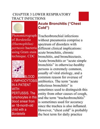
  • 1. CHAPTER 3 LOWER RESPIRATORY TRACT INFECTIONS Acute Bronchitis (“Chest Cold”) Photomicrograph Tracheobronchial infections of Bordetella without pneumonia comprise a (Haemophilus) spectrum of disorders with pertussis bacteria different clinical implications: using Gram stain acute bronchitis, chronic technique. CDC bronchitis, and bronchiectasis. Acute bronchitis or “acute simple bronchitis” in otherwise-healthy persons is extremely common, usually of viral etiology, and a BLOOD common reason for overuse of LYMPHOCYTOSIS antibiotics. The term “acute IN A PATIENT infectious bronchitis” is WITH sometimes used to distinguish this PERTUSSIS. The entity from other causes of cough, lymphocytes in this and the term “tracheobronchitis” blood smear from is sometimes used for accuracy an 18-month-old since the trachea is also inflamed. child with a However, “chest cold” is probably Bordetella the best term for daily practice
  • 2. pertussis infection since it implies that antibiotics are have lobulated seldom necessary. Bordetella nuclei. pertussis, the agent of whooping Lymphocytosis is cough, is now recognized as a characteristic of cause of acute bronchitis in adults. this disorder and Infection by either Mycoplasma the lymphocyte pneumoniae or Chlamydia morphology is pneumoniae accounts for many of often atypical. The the stubborn cases in which cytology of the symptoms fail to resolve or recur cells could be soon after treatment has been mistaken for discontinued. neoplastic lymphocytes. Infection of the tracheobronchial (Wright-Giemsa mucosa causes local stain) © The Johns inflammation, increased secretion Hopkins Autopsy of mucus, and damage to ciliated Resource (JHAR). cells. Symptoms result both from Image Archive. the inflammatory response and also from the interruption of the mucociliary blanket that normally cleanses the lower respiratory tract. Most cases of acute bronchitis (95% by some estimates) are caused by viruses.
  • 3. All of the common viruses affecting the upper respiratory tract have been implicated: rhinoviruses, coronavirus, respiratory syncytial virus, adenoviruses, coxsackieviruses, influenza viruses A and B, and parainfluenza virus. In 2 studies in which attempts were made to establish a precise diagnosis, the etiology was established in only 16% and 29% of cases with viruses being the most common causes. Mycoplasma pneumoniae and Chlamydia pneumoniae probably play minor roles in this illness, at least in most populations. However, M. pneumoniae probably causes more cases of bronchitis than pneumonia, and C. pneumoniae may be an important cause of acute bronchitis in college-aged students. Whether S.
  • 4. pneumoniae, H. influenzae, and M. catarrahalis cause chest cold in otherwise-healthy persons is unclear, but there is little support for the concept of quot;acute bacterial bronchitisquot; as a community- acquired disease. Recently it has been emphasized that Bordetella pertussis can infect adults, even when vaccinated, providing a reservoir for causing whooping cough among infants. Bordetella parapertussis causes a protracted illness similar to whooping cough but without systemic toxicity. Whether these observations can be generalized to other populations is undetermined. The onset is typically preceded by a prodrome of at least 24 hours with symptoms of coryza and pharyngitis. A dry cough, signifying early inflammation of the upper airway, often evolves
  • 5. into a cough productive of moderate amounts of mucopurulent sputum. Fever, headache, myalgias, and retrosternal chest pain or discomfort may be present. Fever is most common when an influenza virus or Mycoplasma pneumoniae is the causative agent. The patient rarely looks toxic. Tracheal tenderness is often present. Auscultation may reveal a few coarse crackles with occasional wheezes in the chest, but there are no signs of consolidation. Transmission electron photomicrograph of a hamster trachea ring infected with M. pneumoniae. Note the orientation of the mycoplasmas through their specialized tip-like organelle, which permits close association with the
  • 6. respiratory epithelium. M, mycoplasma; m, microvillus; C, cilia. Both images used with permission. From Baseman and Tully, Emerging Infection Diseases 3 Acute Infectious Exacerbations of Chronic Bronchitis Figure Chronic bronchitis is defined by Chlamydial the American Thoracic Society inclusions © (ATS) as excessive sputum Bristol Biomedical production with cough, present on Archive. Used with most days for at least 3 months a permission year and not less than 2 successive years, without an underlying etiology such as tuberculosis or Figure bronchiectasis. This common Chlamydial disorder, affecting up to 25% of inclusions in an the adult population, can lead to endothelial cell © full-blown chronic obstructive Bristol Biomedical pulmonary disease (COPD), the Archive. Used with fourth-leading cause of death in permission the United States. The extent to
  • 7. which acute exacerbations are due to treatable infections remains controversial. Chronic bronchitis is caused mainly by cigarette smoking. Air pollution, cold and damp climates, heredity, frequent lower respiratory tract infections, and immunodeficiency disorders (such as common variable hypogammaglobulinemia or isolated IgA deficiency) play a role in some patients. The essential feature is anatomic change in the larger airways, including an increased number of mucus-producing goblet cells and mucosal gland hypertrophy in the bronchial walls. Increased bronchial secretions and impaired ability to handle them lead to chronic cough and disabling complications.
  • 8. Current opinion holds that most acute exacerbations of chronic bronchitis are caused by viruses or by non-infectious agents. Viruses have been found in as few as 7% to as many as 64% of cases in which they were sought. By conservative estimate viruses cause about one-third of cases, the more common ones being influenza viruses A and B, parainfluenza virus, coronaviruses, and rhinoviruses. Cultures of sputum often show non-typable strains of Haemophilus influenzae, Streptococcus pneumoniae, and/or Moraxella catarrhalis. However, the extent to which these bacteria explain exacerbations in a given patient is hard to determine since they often colonize the damaged lower respiratory tract on a more- or-less permanent basis. Evidence suggests that repeated episodes of
  • 9. bacterial infections—especially when caused by H. influenzae— contribute to deterioration of pulmonary function. S. aureus and aerobic gram-negative rods occasionally cause exacerbations of chronic bronchitis. The pathogens associated with quot;atypical pneumoniaquot; such as M. pneumoniae, C. pneumoniae, and L. pneumophila probably cause fewer than 10% of exacerbations. Evidence to date suggests that Chlamydia pneumoniae is more strongly associated with the underlying chronic bronchitis than with its acute exacerbations. However, Chlamydia pneumoniae can cause a stubborn respiratory illness lasting several weeks or longer and tending to relapse after each course of antibiotics.
  • 10. Bronchiectasis Bronchiectasis is an acquired Figure disorder characterized This PA x-ray anatomically by abnormal depicts dilatation of bronchi and atelectatic, and bronchioles and clinically by bronchiectatic chronic productive cough and changes in this child’s right frequent lower respiratory tract infections. Its prevalence fell upper pulmonary dramatically after the introduction lung field. of broad-spectrum antibiotics and Atelectasis (a widespread immunization against collapse of the measles and pertussis. Although lung) can be due bronchiectasis is now uncommon, to bronchiectasis it often goes undiagnosed until (an enlargement far-advanced. Newer imaging of the bronchial studies now enable earlier tubes) and a diagnosis, and our understanding decrease in the of its causes continues to improve. effectiveness of their ciliated Cigarette smoking, the major mucosal lining, cause of chronic bronchitis, plays which renders little role in bronchiectasis except the lungs unable
  • 11. to clear for predisposing to recurrent themselves of infections. The basic problem in clogging mucous bronchiectasis is permanent build-up. structural damage to the walls of CDC/ Dr. bronchi and bronchioles brought Thomas Hooten about by the concerted action of (1) infection and (2) impairment of the pulmonary toilet, airway obstruction, and/or a defect in host defenses. In the past, bronchiectasis was associated especially with frequent or severe lower respiratory infections during childhood. Bronchiectasis continues to be associated with such infections - especially necrotizing pneumonias in which treatment is delayed - but the list of known causes has expanded. Bronchiectasis can be the earliest clue to cystic fibrosis presenting during adolescence or early adulthood. Staphylococcus
  • 12. aureus, Pseudomonas aeruginosa, and Pseudomonas cepacia are often isolated from these patients. Mycobacterium avium- intracelluare complex (MAC) infection is not infrequently associated with bronchiectasis, especially in older women and/or thin women. Allergic bronchopulmonary aspergillosis often leads to bronchiectasis, which might be prevented by early recognition of this syndrome. Immunodeficiency disorders, both congenital (hypogammaglobulinemia) and acquired (AIDS) predispose to bronchiectasis. The dyskinetic cilia syndromes are sufficiently common (about 1 in every 20,000 to 60,000 persons) that a case is likely to occur in every medium- sized city. Patients with advanced bronchiectasis experience daily
  • 13. cough productive of large amounts of mucopurulent, thick, tenacious sputum. However, most patients produce lesser amounts of sputum, at least during the early stages, and cough may be nonproductive (quot;dry bronchiectasisquot;) or even absent. Dyspnea and hemoptysis are common. Patients often give a history of repeated respiratory infections and sometimes give a history of recurrent pleuritic chest pain. Hard crackles are heard locally over the lung fields in about 70% of patients. Rhonchi and widespread expiratory wheezes are also common. Clubbing is present in only about 3% of patients. Plain chest x-rays (PA and lateral views) are usually abnormal.
  • 14. Acute Community-acquired Pneumonia: Overview Figure Pneumonia accounts for an This AP chest x- estimated 45,000 deaths in the ray shows United States each year. It is the pneumonia of the 6th most common cause of death left lower lobe and the most common infectious with early cause of death. Since it is not a consolidation, reportable disease, the precise the etiology of incidence is unknown. Estimates which was suggest that 4 million cases occur unknown. each year, prompting 10 million Pneumonia can physician visits and 600,000 to 1.2 be caused by a million hospitalizations and variety of agents adding $23 billion to health care including costs. Data suggest a 28-fold bacteria, viruses, increased cost for managing the and disease on an inpatient basis mycoplasmas, ($7,517 versus $264 for outpatient among others. therapy). However, the mortality Pneumonia rate is 1% or less for patients remains an managed as outpatients versus important cause 14% to 25% for those admitted to of morbidity and the hospital. Physicians often
  • 15. mortality in the overestimate the short-term United States as mortality risk, but erring toward both a primary, hospitalization is understandable and secondary given the potentially fatal nature infection. of the disease. CDC/Dr. Microorganisms can enter the Thomas Hooten lungs by aspiration, inhalation, or by way of the bloodstream (hematogenous pneumonia). Figure Aspiration of bacteria that have This colonized the oropharynx is by far anteroposterior the most common mechanism. x-ray reveals a Most humans aspirate small bilaterally amounts of oropharyngeal progressive secretions on a nightly basis. plague infection Microorganisms that are not involving both removed by the mucociliary lung fields. blanket are taken up and killed by The first signs of pulmonary alveolar macrophages, plague are fever, the last line of defense. This headache, process, called pulmonary weakness, and clearance, is impaired by viral rapidly respiratory infections, tobacco developing smoke, chronic lung disease, pneumonia with alcohol, and many other factors
  • 16. shortness of associated with debilitating breath, chest diseases. One or more chronic pain, cough, and diseases are present in the sometimes majority of adult patients with bloody or watery pneumonia (58% to 89% of sputum, patients in various studies). eventually Alcoholism predisposes to progressing for 2 aspiration, but cigarette smoking - 4 days into is the main avoidable risk factor respiratory for community-acquired failure and pneumonia in adults. shock. CDC/Dr. Jack Inhalation of aerosolized particles Poland is an important route of entry for many viruses including the influenza viruses and, most recently, the Hantaviruses. Bacteria that cause pneumonia by airborne transmission include M. tuberculosis, Yersinia pestis (plague), Bacillus anthracis (anthrax), and probably Legionella pneumophila (Legionnaire’s disease) and Francisella tularensis (tularemia).
  • 17. Spore-producing fungi such as Histoplasma capsulatum, Blastomyces dermatitidis, and Coccidioides immitis also cause inhalation pneumonia. Hematogenous pneumonia classically develops from septic pulmonary emboli, frequently resulting in patchy or nodular bilateral pulmonary infiltrates sometimes accompanied by pleural effusions. In inner-city populations, a familiar scenario consists of bilateral pneumonia associated with S. aureus endocarditis on the tricuspid or pulmonic valves of injecting drug users. Another scenario consists of emboli from septic thrombophlebitis: for example, of the pelvic veins (pelvic inflammatory disease, septic abortion), internal jugular vein (the Lemierre syndrome) or any
  • 18. large vein where a catheter has been inserted. Hematogenous seeding of the lungs possibly explains some pneumonias caused by gram-negative bacteria and by unusually virulent organisms such as F. tularensis (tularemia). The microbial cause of community-acquired pneumonia is usually difficult to determine. In prospective studies of patients requiring hospitalization, a cause is found in only 40% to 70% of cases. In primary care practice, a far greater fraction of cases are never diagnosed. Most of these cases respond to empiric therapy. Published data concerning the causes of pneumonia vary from one region to another, but some generalizations are possible. Mycoplasma pneumoniae has been determined to be the most common cause in some
  • 19. communities, when presumptive diagnoses were taken into account, followed by Streptococcus pneumoniae and Chlamydia pneumoniae. Adults with compromised host defenses are likely to have pneumococcal pneumonia, but can also have pneumonia due to H. influenzae, Moraxella catarrhalis, S. aureus, or aerobic gram-negative rods. There is wide agreement that S. pneumoniae is the most common cause of community-acquired pneumonia requiring hospitalization. An emerging and controversial area concerns the frequency of pneumonia caused by more than one microorganism. In one study, a second pathogen was found in about 10% of patients with pneumonia due to a conventional bacterial pathogen, but in 55% of patients in whom an quot;atypicalquot; pathogen was found.
  • 20. In 1938, the term quot;atypical pneumoniaquot; was introduced to describe quot;an unusual form of tracheobronchopneumonia with severe constitutional symptomsquot;. It later became customary to distinguish between quot;classical bacterial pneumoniaquot; and quot;atypical pneumoniaquot;. This distinction was challenged during the 1990s when researchers found it difficult if not impossible to differentiate these illnesses on clinical grounds. Some authorities now suggest abandoning the term quot;atypical pneumoniaquot;. Others keep the term since it enriches our appreciation of the disease, forces us to consider unusual etiologies, and reminds us that the quot;atypical pneumoniasquot; do not respond to β- lactam antibiotics. For these latter reasons and for the sake of clarity, we keep the terms here even while
  • 21. agreeing that in some cases it may be impossible to distinguish between quot;classic bacterial pneumoniaquot; and quot;atypical pneumoniaquot; in actual clinical practice. quot;Typical pneumoniaquot; is an alveolar disease whereas quot;atypical pneumoniasquot; affects mainly the tracheobronchial mucosa and interstitium of the lung; hence, the different clinical manifestations. Classical bacterial pneumonia begins with sudden onset of fever, chills, pleuritic chest pain, and productive cough. In the absence of impaired consciousness or inebriation, patients usually seek medical care within 6 hours of the onset of symptoms. Chills occur in about 50% and chest pain in about 30% of patients. Most patients are febrile although some,
  • 22. especially the elderly, may have normal or subnormal temperatures. The respiratory rate is usually increased. Physical examination often reveals signs of consolidation such as dullness to percussion, pectoriloquy, and egophony (e to a change). Lobar consolidation is present on chest x-ray in about one-third of patients. The white blood count is usually elevated with a shift-to- the-left. However, leukopenia rather than leukocytosis may be present and portends a poor prognosis. Atypical pneumonia, on the other hand, usually begins gradually. The insidious onset is often brought out by asking, quot;When was the last time you were in your usual good health?quot; Constitutional symptoms are usually more prominent than the pulmonary
  • 23. symptoms. Chest pain is experienced as substernal discomfort. Cough is non- productive or productive of only scanty amounts of sputum. Relative bradycardia is frequently present. The trachea may be tender but the lung fields are essentially clear to auscultation, prompting one to be surprised by the extent of infiltrate present on chest x-ray. The white blood count is often normal or near normal. Modest elevation of liver enzymes (specifically, the aminotransferases - AST (SGOT) and ALT (SGPT) - is often present. Atypical pneumonia, in summary, seldom presents as an acute, life-threatening medical problem but forces the physician to expand the differential diagnosis. Pneumococcal Pneumonia
  • 24. Streptococcus pneumoniae remains the major cause of severe community-acquired pneumonia and, worldwide, a leading cause of death. It accounts for about two- thirds of cases of bacteremic pneumonia, and is the most common cause of pneumonia leading to hospitalization in all age groups. Some authorities believe that S. pneumoniae may cause up to one-half of all community-acquired pneumonias. There is concern that the incidence of pneumococcal disease may be increasing at the same time that drug resistance is becoming much more common. Primary care clinicians should strive to make pneumococcal vaccination an imperative for patients at increased risk. S. pneumoniae is a common
  • 25. colonizer of the nasopharynx. Invasive pneumococcal disease occurs most often after a new serotype has been acquired, typically after an incubation of one to three days. Viral illness increases the incidence of disease presumably by interfering with normal host defenses. Risk factors for invasive pneumococcal disease include extremes of age, alcoholism, HIV disease, end- stage renal disease, sickle cell disease, diabetes mellitus, dementia, malnutrition, malignancies, diseases affecting B lymphocyte function (notably, multiple myeloma and hypogammaglobulinemia), and immunosuppressive disorders. Patients with asplenia are susceptible to fulminant pneumococcal disease. The pneumococcus does not invade cells as readily as do some of the
  • 26. other streptococci. However once in the lungs, it passes easily from one alveolus to another through the pores of hence the basis Cohn until stopped by the dense connective tissue of a fissure for lobar consolidation. As classically described by previous generations of clinicians, S. pneumoniae causes a lobar pneumonia with by the sudden onset of fever with a single, hard- shaking chill, cough productive of rusty-colored mucopurulent sputum, and pleuritic chest pain. The patient presents with systemic toxicity including tachypnea. Physical examination reveals crepitant râles, tubular breath sounds, and signs of lobar consolidation (dullness to percussion, egophony with e to a change, and pectoriloquy). Today, however, pneumococcal disease is
  • 27. often a more subtle illness. Patchy infiltrates and bronchopneumonia are relatively common. It is often difficult to say precisely what represents pneumococcal pneumonia and what does not unless blood cultures are positive. Bacterial Pneumonia due to Agents other than S. pneumoniae Figure Haemophilus Among the numerous bacteria influenzae as other than S. pneumoniae that seen using a sometimes cause acute Gram-stain community-acquired pneumonia, technique. the most common are H. During the flu influenzae, S. aureus, outbreak of 1918 Streptococcus pyogenes, H. influenzae miscellaneous aerobic gram- was termed negative rods, and anaerobic Pfeiffer's quot;mouth floraquot; bacteria. Patients Bacillus, where it with these pneumonias often have was found in the significant underlying disease, sputum of many severe pneumonia, or both. influenza
  • 28. patients, and Therefore, hospitalization is thought to be the usually indicated. cause of influenza. H. influenzae is a frequent cause CDC of pneumonia in elderly patients and in patients with serious underlying diseases including chronic obstructive lung disease. The pneumonia usually has a patchy or segmental distribution characteristic of bronchopneumonia as opposed to lobar pneumonia. A sputum Gram’s stain showing small, pleomorphic gram-negative coccobacilli can be virtually diagnostic. Staphylococcus aureus pneumonia, when community- acquired, tends to be an acute, fulminant about 1% of cases, except during influenza epidemics. Influenza virus infection markedly predisposes to
  • 29. staphylococcal colonization of the respiratory mucosa. Staphylococcal pneumonia tends to be a necrotizing process with abscess formation. The chest x-ray sometimes shows air pockets known as pneumatoceles, especially in children. Streptococcus pyogenes (group A streptococcal) pneumonia is also uncommon except during influenza epidemics. This pneumonia is often accompanied by the rapid development of large empyemas. Chest tube drainage is often necessary, resulting in prolonged hospitalization. Klebsiella pneumoniae is a relatively common cause of pneumonia in patients suffering from alcoholism. The pneumonia often assumes a lobar distribution. Classically, this pneumonia
  • 30. affects the upper lobes and causes a “bulging fissure” on chest x-ray. E. coli and other aerobic gram- negative rods are relatively common causes of pneumonia in the frail elderly. Pseudomonas aeruginosa, although a common cause of nosocomial pneumonia, is rarely associated with community-acquired pneumonia in patients without underlying lung disease or severe debility. Pneumonia due to quot;mouth floraquot; bacteria - by which is meant a combination of anaerobic and aerobic bacteria with the anaerobes usually predominating - occurs most frequently in patients suffering from alcoholism and poor oral hygiene and results from aspiration. The sputum is usually copious and often foul smelling. quot;Mouth floraquot; pneumonia in an edentulous patient should prompt
  • 31. suspicion of underlying lung cancer. A foul odor to the breath is present in many but not all of these patients. This form of pneumonia is often associated with lung abscess and with empyema due to bronchopleural fistula. Mycoplasma pneumoniae pneumonia Formerly known as the quot;Eaton agentquot;, Mycoplasma pneumoniae is the most commonly identified cause of atypical pneumonia although its precise incidence is unknown. Various investigators have determined this microorganism to be the cause of 13% to 27% of community- acquired pneumonias. It can also cause hospital-acquired pneumonias, and it has caused as many as 50% of pneumonias
  • 32. during epidemics in closed populations. Mycoplasma pneumoniae pneumonia becomes less common after age 40, but older persons may experience more severe manifestations. M. pneumoniae is a cell-wall- deficient organism with particular affinity for the respiratory tract epithelium. Many of the disease manifestations are now thought to be immune-mediated. Close, prolonged contact promotes transmission by respiratory secretions. There is currently interest in the extent to which M. pneumoniae accompanies other agents as a co-pathogen. In one study, an additional pathogen was found in about two-thirds of patients with M. pneumoniae pneumonia who required hospitalization; S. pneumoniae was most commonly found, but
  • 33. Legionella species and Chlamydia pneumoniae were also identified. It is estimated that of persons infected with M. pneumoniae, about 20% are symptomatic, about 70% develop a mild respiratory illness (pharyngitis and/or tracheobronchitis), and fewer than 10% develop pneumonia. The disease occurs in all age groups including toddlers and the elderly but peaks between ages 5 to 15 years. After an incubation period of about 3 weeks, symptoms begin gradually with fever, headache, malaise, chills, sore throat, substernal productive, and cough. The cough is initially non- productive, paroxysmal, and worse at night. It commonly becomes productive later in the illness. Physical examination is
  • 34. usually unimpressive. Bullous myringitis (inflammation of the tympanic membrane with bullae) is uncommon, occurring at most in about 5% of patients, but has a high positive predictive value for M. pneumoniae infection. More commonly there is mild tenderness over the paranasal sinuses, mild erythema of the posterior pharyngeal mucosa, soft cervical lymphadenopathy, and tracheal tenderness. Scattered râles and wheezes may be present but are usually unimpressive. The white blood count is normal in 75% or more of cases. Thrombocytosis can occur as an acute-phase response. Liver enzymes, notably the aminotransferases (AST and ALT), are often mildly elevated. The chest x-ray commonly shows infiltrates that are much more
  • 35. extensive than one would have suspected from physical examination. The most common pattern is a peribronchial pneumonia in which thickened bronchial shadows are surrounded by streaky interstitial infiltrates and patchy atelectasis. Other patterns include nodular infiltrates and hilar lymphadenopathy. The lower lobes are most commonly involved, and pleural effusions - which can be especially severe in patients with sickle cell disease - occur in up to 20% of patients when carefully sought. Extrapulmonary manifestations of M. pneumoniae pneumonia sometimes dominate the clinical picture and include hemolytic anemia, rashes including the life- threatening Stevens-Johnson syndrome, central nervous system complications (about 0.1% of
  • 36. patients, especially children), cardiac complications, and polyarthritis. Chlamydia pneumoniae pneumonia Chlamydia pneumoniae, described in 1986 as the TWAR agent, has been determined by some researchers to be the third or fourth most common cause of community-acquired pneumonia, explaining perhaps 10% to 14% of cases (up to 28% in some series). Pneumonia is recognized most frequently among the elderly, in whom it can be severe. Chlamydia pneumoniae is classified as a bacterium on the basis of its cell wall and growth properties. Unlike most bacteria, however, it grows only as an intracellular parasite. Serologic
  • 37. studies suggest that most humans gain experience with C. pneumoniae at some point in their lives, although immunity is short- lived. About 50% of all persons have antibodies by age 20, and up to 75% of elderly persons are seropositive. It is also thought that most infections (up to 90%) are asymptomatic. Transmission is probably person-to-person by respiratory secretions. After an incubation period of several weeks, most patients experience gradual onset of non- specific upper and lower respiratory symptoms including those of sinusitis, otitis, and pharyngitis. Sore throat with hoarseness is often prominent among the initial symptoms and tends to be the dominant symptom in college-aged persons. Symptoms of pneumonia tend to
  • 38. develop slowly. Often, patients have experienced symptoms for several weeks before seeking medical care. The history sometimes suggests a biphasic illness, as follows: (1) upper respiratory infection with sore throat that resolved, then (2) lower respiratory infection with cough. The severity is age-dependent. Children under age 5 seldom have evidence of significant disease. University students often present with a 10-day history of sore throat or hoarseness with minimal fever. The mean age of patients with pneumonia is about 56 years. Ronchi and râles are present on physical examination more frequently than in M. pneumoniae pneumonia, even among patients who do not complain of cough. The white blood count is usually normal. Chest x-ray may show
  • 39. one or more infiltrates, the most common finding being a single, patchy, subsegmental infiltrate. Wheezing is sometimes present. Accumulating evidence suggests that C. pneumoniae sometimes precipitates adult-onset asthma. Reported extrapulmonary manifestations of C. pneumoniae infection include meningoencephalitis, cerebellar dysfunction, Guillain-Barré syndrome, reactive arthritis, and myocarditis. The possibility that C. pneumoniae might cause coronary artery disease has received much attention. High antibodies to C. pneumoniae have been observed in patients with chronic obstructive lung disease, sarcoidosis, and lung cancer but an etiologic link is unclear. Chlamydia psittaci
  • 40. (Psittacosis; Ornithosis) About 100 to 200 cases of psittacosis are reported in the U.S. each year, but the true incidence is thought to be much higher. Mortality can be high if the diagnosis is not suspected. Chlamydia psittaci infects many and perhaps all species of birds, which may remain asymptomatic or show symptoms and signs of illness such as anorexia, dyspnea, and ruffled feathers. Strains of C. psittaci that are most virulent for humans tend to be those associated with psittacine birds (from the Latin psittacus, or parrot), such as parrots, parakeets, macaws, cockatoos, and budgerigars, and also with turkeys. Humans who develop psittacosis are commonly bird fanciers or work in poultry farms
  • 41. (notably, turkey farms), abattoirs, processing plans, pet shops, or veterinarians’ offices. The organism is usually acquired by inhalation, but human-to-human transmission occurs on rare occasions. After an incubation period of 5 to 15 days, patients develop symptoms and signs of illness ranging in severity from mild to life threatening. Atypical pneumonia, the most characteristic form of the disease, is manifest by headache, fever, and non-productive cough. Chest x-ray is usually abnormal (75%) of cases, most commonly showing consolidation of one lower lobe. The radiographic findings are usually much more striking than the findings on auscultation of the chest. Psittacosis can also present as a typhoidal illness (fever,
  • 42. malaise, relative bradycardia, hepatosplenomegaly), a non- specific flu-like quot;viral syndromequot;, a mononucleosis-like syndrome, or as fever of unknown origin. Legionnaire’s disease Figure First identified in 1976 during an This micrograph outbreak at an American Legion of a biopsied Convention in Philadelphia, lung tissue Legionnaire’s disease is now specimen stained recognized as a relatively with the CDC's common cause of both modified community-acquired and hospital- Dieterle silver acquired pneumonia. The impregnation incidence exhibits wide procedure, geographic variation - from less revealed small, than 1% to more than 16% of blunt, community-acquired pneumonias pleomorphic - reflecting to a large extent the intracellular, and degree of contamination of water extracellular reservoirs by the causative bacilli, which organisms. Unlike pneumonias stain brown to due to M. pneumoniae and C.
  • 43. black against a pneumoniae, cases that can be pale yellow treated on an outpatient basis tend background; to be the exception rather than the Mag. 500x. rule. Dieterle’s stain is a preparation Legionella species are gram- used when negative bacteria that stain poorly and survive intracellularly. More Legionella than 40 species, and more than 60 pneumophila bacteria are serogroups, of Legionella are now suspected. recognized. Legionella Uranyl nitrate is pneumophila is the most applied in order commonly encountered species, to first sensitize causing at least 80% of clinical the slide mount, infections. which is subsequently Legionellosis seems to be a treated with gum disease of human progress mastic, followed brought about by devices that by an incubation maintain water at warm period, after temperatures and produce which the slide is aerosols. In water, the organisms soaked in silver multiply within amebas; in nitrate. Lastly, humans they multiply within the slide is alveolar macrophages. The
  • 44. “developed” in a disease is spread by water rather hydroquinone, than by person-to-person contact. sodium sulfite, Contamination of water sources acetone, has been associated with formaldehyde, numerous outbreaks in settings pyridine, and ranging from inner-city hospitals gum mastic bath. to luxury cruise liners. The Legionella Clinically severe cases of pneumophila Legionnaire’s disease tend to bacteria, if present, will occur in persons with stain black. CDCcompromised host defenses, most often in the setting of chronic obstructive pulmonary disease, Figure immunosuppression, or advanced age. A mild form of Legionellosis, This silver- known as Pontiac fever, is a self- stained micrograph of a limited disease presenting as fever, malaise, headaches, and lung tissue chills without pneumonia. Pontiac specimen fever resolves within a few days revealed the without antibiotic therapy. presence of Legionnaire’s disease, the more Legionella familiar and more severe form of pneumophila Legionellosis, affects persons of bacteria. The
  • 45. specimen was all ages and presents with taken from a symptoms that overlap those of victim of the quot;classic bacterialquot; and quot;atypicalquot; 1976 pneumonia. Legionnaires’ disease outbreak After an incubation period of 2 to in Philadelphia. 10 days, patients experience the CDC onset of fever, headache, anorexia, malaise, and myalgia. At this point, respiratory symptoms are usually not prominent, the cough Figure being only minimally productive. Legionella Some patients have chest pain pneumophila and, if the sputum is blood-tinged, multiplying pulmonary embolism is often inside a cultured suspected. Gastrointestinal human lung symptoms with nausea, vomiting, fibroblast diarrhea, and abdominal pain can CDC also dominate the clinical picture. Alternatively, neurologic Figure symptoms can be the presenting Anteroposterior complaint, variably manifested as headache, lethargy, and change in x-ray reveals mental status. bilateral pulmonary
  • 46. infiltrates in a Fever is usually present. Relative patient with bradycardia is found more often in Legionnaires' older patients and in those with disease CDC severe pneumonia. Examination of the chest usually shows râles and, later in the illness, signs of consolidation. The peripheral blood commonly shows leukocytosis and thrombocytopenia. Hyponatremia (serum sodium < 130 mEq/L) is more common in Legionnaire’s disease than in most other pneumonias. Hypophosphatemia also occurs. There is frequently evidence of liver and renal dysfunction. Hematuria and proteinuria are common. There is no characteristic feature on chest x-ray. The most common pattern is a patchy infiltrate involving one lobe, which progresses to consolidation. Infiltrates can assume a diffuse or interstitial pattern, and pleural effusions are