Infection of oral mucosa
•Descargar como PPTX, PDF•
58 recomendaciones•17,027 vistas


Recomendados
Más contenido relacionado
La actualidad más candente
La actualidad más candente (20)
Destacado
Similar a Infection of oral mucosa
Similar a Infection of oral mucosa (20)
Infection of oral mucosa
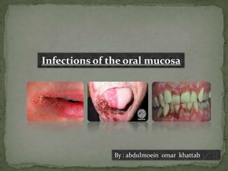
- 1. Infections of the oral mucosa By : abdulmoein omar khattab
- 2. Infections of oral mucosa Herpetic stomatitis (HSV). Herpangina, Hand,foot,and mouth disease Viral infections (Coxsackie viruses). Chickenpox and herpes zoster(shingles): (HZV). Influenza (influenza virus ). Infectious mononucleosis (glandular fever), Hairy leukoplakia (EBV). Measles ( paramyxovirus). Cytomegalovirus (CMV). Mumps( paramyxovirus). Measles (Rubeola). Human papilloma virus (HPV). Necrotizing ulcerative gingivitis (NUG) (acute ulcerative gingivitis, acute necrotizing ulcerative gingivitis (AUG, ANUG)). Bacterial infections Noma (cancrum oris). Actinomycosis. Syphilis (primary, secondary, tertiary, congenital). Tuberculosis. Leprosy. Gonorrhoea. Pseudomembranous candidosis (acute and chronic). Erythematous candidosis(acuteandchronic). Fungal infections Candida-associated denture stomatitis (chronic atrophic candidosis). Candida-associated and other forms of angular cheilitis.
- 3. *Introduction The common oral mucosal infections are caused by viruses, bacteria, and fungi. Helminthic and protozoal infections may involve oral tissues 1-Viral infections: The main viral infections of the oral mucosa are: Human immunodeficiency virus (HIV), mumps, and influenza , herpes simplex virus (HSV) and human papillomavirus (HPV) , coxsackievirus infection, measles (rubeola), and rubella, Epstein-barr virus , versilla-zoster virus, cytomegalovirus .( Mumps primarily affects salivary glands ,but may be associated with a non-specific stomatitis because of the reduced salivary flow. An acute non-specific stomatitis may also be seen as an early feature in patients with influenza.) *(HPV infections have received particular attention in recent years, as high-risk strains have been linked to some cases of oral squamous cell carcinoma. Nonetheless, many other viral infections can affect the oral cavity in humans, either as localized or systemic infections.)
- 4. *Herpetic stomatitis: The herpes simplex viruses (HSV) are DNA viruses and are the most frequent cause of a viral infection of the mouth. There are two types of HSV with serological, biological, and clinical differences. Type 1 is traditionally associated with infections of the skin and oral mucous membranes, and type 2 virus with infections of the genitalia. However, in sexually active subjects either type may be associated with oral and/or genital infection. HSV have also been implicated in recurrent erythema multiforme.
- 5. Primary infection with HSV type 1 is common and is transmitted by droplet spread or contact with the lesions. It occurs predominantly in young children and in the majority of cases is subclinical or causes a mild pharyngitis. However, in some patients it presents as primary herpetic gingivostomatitis. Following an incubation period of about 5 days the patient complains of prodromal symptoms of malaise and fever, and within a day or two the mouth becomes uncomfortable with the development of numerous small vesicles on any part of the oral mucosa and lips. There is also usually a widespread inflammation of the gingiva which is erythematous and oedematous .
- 6. The vesicles soon ulcerate .and become secondarily infected and this is accompanied by regional lymphadenitis. Fresh crops of vesicles develop over the next few days, but the symptoms begin to subside about the sixth day of fever with the oral lesions taking 10-14 days to resolve. Circumoral crusting lesions on the lips may be seen, the crusting being due to coagulation of serum which exudes from ruptured vesicles . Extraoral lesions may also be present, particularly in children. For example, vesicles may occur on the skin of the chin as aresult of drooling of saliva and on the fingers as a result of sucking. Infections involving the nailbed, herpetic whitlow, may be quite painful and infection may be transmitted to the eyes by rubbing. Histological examination of an intact herpetic vesicle shows an intraepithelial blister .The vesicle results from distension and rupture of the virally infected epithelial cells by intracellular
- 7. oedema and the coalescence of disrupted cells. The rupture of infected cells releases new viral particles to infect adjacent epithelial cells and the virus also gains access to the sensory axons of the trigeminal nerve. The infected cells are swollen and have eosinophilic cytoplasm and large, pale vesicular nuclei. These changes are described as ballooning degeneration. Giant cells containing many such nuclei also form as a result of fusion of the cytoplasm of infected cells .The balloon cells and multinucleate giant cells can often be identified in smears taken from an intact vesicle or from one which has recently ruptured . vesicle or from one which has recently ruptured.The lamina propria shows avariable inflammatory infiltrate, the density of which depends on the stage and severity ofthe disease, and inflammatory cells also extend into the epithelium.
- 8. About one in three of those who have had a primary infection, either clinical of subclinical, later Develop* recurrent HSV infections. This is due to reactivation of the virus which, following the primary infection, has remained latent in the sensory ganglion of the trigeminal nerve . Recurrent infection may result in asymptomatic shedding of HSV into the oral cavity or in local symptoms. Systemic symptoms are usually absent because of the immunity acquired during the primary infection. Herpes labialis is the most frequent type of recurrent infection and appears as clusters of vesicles on the lips and adjacent skin a few hours after prodromal symptoms of itching or tingling. The vesicles rupture within a short time and become crusted. They usually heal within a week. Recurrences may be brought on by a number of different stimuli, including mild febrile infections such as the common cold, ultraviolet light, mechanical trauma, menstruation, stress, and immunosuppression. Recurrent intraoral lesions occur occasionally, almost always on the hard palate or gingival.
- 9. Primary herpetic stomatitis recurrent herpetic stomatitis
- 10. *Coxsackie viruses: cause 3 conditions that can manifest in the oral cavity: hand-foot-and-mouth disease, herpangina, and acute lymphonodular pharyngitis. Herpangina: This infection is caused by various types of coxsackievirus A. Coxsackieviruses are RNA viruses. The infection is seen most commonly in children and is characterized, clinically, by the sudden onset of a mild illness with fever, anorexia, dysphagia, and sore throat. Vesicles, which rapidly break down into ulcers 1-2 mm in diameter, are seen on the tonsils, soft palate, and uvula The symptoms persist for 2-3 days only. Clinically, it may be difficult to distinguish from acute primary herpes. However, the latter is agingivostomatitis, whereas herpangina is an oropharyngitis.
- 11. Hand,foot,and mouth disease: This infection is also caused by various types of coxsackievirus A, especially type 16. It occurs predominantly in children and is transmitted in conditions of close association such as withinhouseholds. The disease is characterized by shallow, painful oral ulcers together with vesicles and ulcers on the hands and feet .It usually lasts about 7-10 days. *Differentials for hand-foot-and-mouth disease: Aphthous Stomatitis Herpetiform ulcers Primary and secondary herpetic stomatitis
- 12. *Differentials for herpangina: Primary herpetic gingivostomatitis Acute herpetic pharyngotonsillitis Aphthous Stomatitis Herpetiform stomatitis Gonococcal pharyngitis Erythema Multiforme Bacterial tonsillitis Viral pharyngitis
- 13. Foot – hand – mouth disease ( Herpangina)
- 14. Chickenpox and herpes zoster(shingles): Both chickenpox and herpes zoster are caused by the same virus, the varicella- zoster virus (VZV), which is another member of the herpesviruses.The lesions of chickenpox may be found on the oral mucosa, especially the soft palate and fauces, and may precede the characteristic skin rash. The oral lesions usually present as small ulcers.Intact vesicles are seldom seen but when examined histologically show cytopathic effects indistinguishable from those of herpes simplex. Zoster is the manifestation of recurrent infection following a primary attack of chickenpox, and in this respect is similar to recurrent herpes simplex infection except that repeated attacks of zoster are unusual.
- 15. following a primary attack of chickenpox, and in this respect is similar to recurrent herpes simplex infection except that repeated attacks of zoster are unusual. Following infection by chickenpox, the virus remains latent in the sensory ganglia probably for the remainder of the life of the host. Reactivation of the virus to cause zoster is uncommon but may occur apparently spontaneously or when the host defences are depressed. Severe infection may be seen in immunocompromised patients. The lesions are localized to the distribution of one or more sensory nerves. The characteristic unilateral vesicular eruption is frequently preceded by prodromal symptoms of pain and paraesthesia for up to two weeks. When the trigeminal nerve is involved the first division (ophthalmic) is most frequently affected. Involvement of the second or third division causes facial pain and the patient may complain of toothache, followed by the development of vesicles in the distribution of one or more branches of the trigeminal nerve. The vesicles may be entirely intraoral where they rapidly ulcerate and become secondarily infected
- 16. The disease usually runs a course of about 14 days. The most distressing complication of zoster is postherpetic neuralgia, probably caused by fibrosis in and around the sensory nerves and ganglia. Although zoster involves sensory nerves it occasionally presents with lower motor neurone-type facial paralysis as the Ramsay-Hunt syndrome. The syndrome is due to extension of primary involvement of the geniculate ganglion to affect the facial nerve.
- 17. *Infectious mononucleosis (glandular fever): This infection is caused by the Epstein-Barr virus (EBV), a member of the herpes group. It occurs predominantly in teenagers and young adults and is transmitted by contact with saliva, especially by kissing, either from an infected patient or a healthy carrier. The disease is characterized by lymph node enlargement, fever, and inflammation of the pharynx, and may be associated with prolonged periods of malaise lasting months or more. Petechial haemorrhages at the junction of the soft and hard palate and inflammation and ulceration of theoral mucosa may be seen, but the oral changes are non-specific. Cases have also been reported presenting as pericoronitis. As discussed in previous chapters, EBV is associated with Burkitt's lymphoma and has also beendetected in oral squamous cell carcinoma.
- 18. *Hairy leukoplakia Hairy leukoplakia (HL) presents as a white patch which cannot be removed. It occurs most frequently on the lateral borders of the tongue, bilaterally, but occasionally other areas of the oral mucosa are affected. The most characteristic lesions present as vertical white folds on the lateral border of the tongue with a raised, corrugated, or hairy surface. Histological examination of HL typically shows acanthotic, parakeratinized epithelium often with long, finger-like surface projections of parakeratin producing the hairy or corrugated appearance seen clinically. Candidal hyphae may be present in the parakeratin, but the candidosis is a secondary rather than a causal infection
- 19. There is an absence of associated inflammatory cells both in the epithelium and in the lamina propria. Swollen or balloon cells with prominent cell boundaries are present as a band in the prickle celllayer beneath the parakeratin (Some have small, darkly staining nuclei and perinuclear vacuoles. The swollen cells contain Epstein-Barr virus and have been described as koilocytes or koilocyte-like cells .(Strictly, the term 'koilocyte' should be confined to cells infected with HPV.) Demonstration of EBV is essential to confirm the diagnosis.
- 20. Measles( paramyxovirus): This infection occurs predominantly in children, and in developed countries is usually a mild disease with low mortality. In other countries the mortality is high. Prodromal symptoms may resemble a common cold and are accompanied by the appearance of Koplik's spots on the oral mucosa, especially the buccal mucosa opposite the molar teeth. They present as pin-point bluish-white spots against an erythematous background, and range from few in number to several hundred. They are likely to be overlooked, particularly as they start to disappear as the characteristic skin rash develops some 3-4 days later. In parts of West Africa, noma (cancrum oris), may occur as acomplication of measles in malnourished patients.
- 21. Measles
- 22. Cytomegalovirus(CMV): Cytomegalovirus (CMV) is a herpes group virus that rarely causes disease in immunocompetent individuals but which is an important pathogen in immunocompromised hosts, for example AIDS patients and organ transplant patients. However, subclinical infection is common, affecting 40-80 per cent of adults. Previously uninfected transplant patients may acquire the virus from the transplanted organ or transfused blood. The most common oral manifestation is non-specific oral ulceration. CMV infection of salivary glands is also common but usually asymptomatic .However, it has been reported that the virus is strongly associated with the xerostomia seen in AIDS patients.
- 24. *Mumps: The mumps virus is a member of the Paramyxoviridae family and of the Rubulavirus genus. This genus includes the mumps virus; Newcastle disease virus; and human parainfluenza virus types 2, 4a, and 4b. Transmission of the mumps virus occurs via direct contact, via droplets, or via fomites, and it enters through the nose or mouth. More intimate contact is necessary for the spread of mumps than for measles or varicella. During the incubation period (approximately 16-18 days), the virus proliferates in the upper respiratory tract and regional lymph nodes. Viremia follows with secondary spread to the glandular and neural tissues. Inflammation of infected tissues leads to the manifestations of parotitis and aseptic meningitis. Patients are most contagious 1-2 days before the onset of parotitis.
- 25. *Clinical history: Prodromal symptoms include a low-grade fever, malaise, lack of appetite, and headache. A day after the initial symptoms appear, reports of earache and tenderness of the parotid glands are common. One parotid gland often enlarges a few days after the other, and unilateral involvement is observed in one fourth of patients with salivary gland involvement. Once maximum parotid gland swelling has occurred, the pain, fever, and tenderness quickly diminish. The affected parotid gland returns to its normal size within 1 week. Complications of parotitis are uncommon. Recurrent acute and chronic sialadenitis are potential complications of parotitis.
- 27. Measles (Rubeola): Measles, also known as rubeola, is an acute, infectious, highly contagious disease that frequently occurs in children. In the United States, the incidence of measles has dramatically decreased as a result of the measles vaccine; however, it remains a significant problem in developing countries.
- 28. *Human papilloma virus: Human papillomavirus (HPV) is a virus from the papillomavirus family that is capable of infecting humans. Like all papillomaviruses, HPVs establish productive infections only in keratinocytes of the skin or mucous membranes. While the majority of the known types of HPV cause no symptoms in most people, some types can cause warts (verrucae), while others can – in a minority of cases – lead to cancers of the cervix, vulva, vagina, penis, oropharynx and anus. Recently, HPV has been linked with an increased risk of cardiovascular disease. In addition, HPV 16 and 18 infections are strongly associated with an increased odds ratio of developing oropharyngeal (throat) cancer.
- 29. More than 30 to 40 types of HPV are typically transmitted through sexual contact and infect the anogenital region. Some sexually transmitted HPV types may cause genital warts. Persistent infection with "high-risk" HPV types — different from the ones that cause skin warts — may progress to precancerous lesions and invasive cancer. HPV infection is a cause of nearly all cases of cervical cancer. However, most infections with these types do not cause disease.
- 30. 2-Bacterial infections *Introduction It is surprising how few specific bacterial infections of the oral mucosa occur in view of the enormous number of bacteria and the wide range of species (including pathogens) normally present in the oral cavity. This is a reflection of the normal defence mechanisms operating in the mouth. These include the barrier function of the oral epithelium, the mechanical cleansing action and the specific and non-specific antimicrobial substances in saliva, and the migration of phagocytic cells, predominantly neutrophils, into the gingival crevice and oral cavity.
- 31. *Necrotizing ulcerative gingivitis (NUG) (acute ulcerative gingivitis, acute necrotizing ulcerative gingivitis (AUG, ANUG)) This condition is now relatively uncommon in industrialized countries, although there has been a global increase associated with HIV infection. In industrialized countries the disease occurs mostly in young adults and is more common in males than females, but in developing countries it is seen almost exclusively in children, related to poverty and malnutrition. The infection presents with necrosis and crater-like, punched-out ulceration of the interdental papillae of sudden onset which may also involve the gingival margins .The ulcers are covered with a greyish-green pseudomembrane demarcated from the surrounding mucosa by a linear erythema. Other signs and symptoms include gingival bleeding, either spontaneously or on minor trauma, pain or soreness of the gums, marked halitosis, a bad taste often described as metallic, and increased salivation. Malaise, cervical lymphadenopathy, and fever may be present in advanced cases.
- 32. There is a high recurrence rate of infection unless underlying predisposing factors are adequately treated. The precise role of the various types of bacteria is unclear, but the disease is usually regarded as an endogenous, opportunistic polymicrobial infection. Apersistent form of necrotizing gingivitis is associated with HIV infection and NUG is an important factor in the development of noma.
- 33. *Noma (cancrum oris): Noma is a severe, rapidly developing gangrene of the orofacial tissues and jaws that occurs almost exclusively in developing countries, especially sub-Saharan Africa. The majority of cases are preceded by NUG, followed by rapid spread of necrosis from the original gingival lesions into the cheek and development of an area of demarcated gangrene of the orofacial tissues . Almost all cases of noma appear to develop in malnourished children whose resistance has been lowered still further by intercurrent infection such as measles or malaria. The microbiological features are similar to NUG but Fusobacterium necrophorum and Prevotella intermedia are considered to be key organisms.
- 35. Actinomycosis: Actinomycosis is a chronic, suppurative, polymicrobial infection caused by endogenous bacteria, amongst which Actinomyces species, especially Actinomyces israelii, predominate. Actinomyces are anaerobic bacteria. In actinomycosis other anaerobic bacteria act synergistically, creating conditions suitable for the growth of Actinomyces species as well as contributing to tissue injury. The soft tissues of the submandibular area and neck are most commonly involved in cervicofacial actinomycosis. The infection is characterized by multiple foci of chronic suppuration. It presents with the development of firm swellings which eventually soften and are accompanied by the formation of pus which discharges through multiple sinuses
- 36. Pain is variable and the swellings are often painless. The multiple abscesses which eventually form tend to point on to the skin rather than the mucosal surface and are accompanied by marked fibrosis of the surrounding tissues. Infection is endogenous, and either a tooth socket, most commonly a lower third molar, or an infected root canal are thought to be the portals of entry. Actinomycotic lesions develop as areas of granulomatous inflammation surrounded by abundant granulation tissue and fibrous tissue. Fresh foci of infection develop adjacent to the primary site following transport of some of the organisms by phagocytic cells, and a central area of suppurative necrosis eventually develops in most of these foci. Granules consisting of tangled meshes of Grampositive filaments of actinomyces with radiating filaments projecting on the surface are present and may be seen clinically as 'sulphur granules' in pus from actinomycotic lesions.
- 38. *Syphilis (primary, secondary, tertiary, congenital): Syphilis is an infection caused by the spirochaete Treponema pallidum. The incidence in Western countries has now declined significantly. The primary lesion (chancre) usually occurs on the genitalia, but in a minority of patients may present on the oral mucosa, usually the lips. It appears as a shallow, painless ulcer with an indurated base. The regional lymph nodes are also enlarged. Histologically, the chancre consists of ulcerated granulation tissue with a dense mononuclear inflammatory cell inflltrate chiefly composed of plasma cells. It heals spontaneously within a 3-6 week period. The signs and symptoms of secondary syphilis develop about 6 weeks after the appearance of the primary chancre, some 2-3 months after the initial exposure. A generalized skin rash is the predominant feature and may be accompanied by oral lesions of which the so- called mucous patch is the most frequent. 'Mucous patches' are flat areas of ulceration. They are usually multiple and may coalesce to produce lesions of irregular outline called 'snail-track ulcers'.
- 39. Tertiary or late-stage syphilis may develop many years after the initial exposure. Gummas (areas of necrosis associated with delayed (type IV) hypersensitivity reactions to syphilitic antigens) may occur, especially on the hard palate, leading to perforation into the nasal cavity . Histologically, a gumma consists of a central mass of coagulative necrosis surrounded by granulation tissue infiltrated by lymphocytes, plasma cells, and macrophages with occasional giant cells. Spirochaetes are very scanty or absent. Endarteritis obliterans is thought to be the cause of the atrophic glossitis which is also a feature of tertiary syphilis, the smooth surface of the tongue being broken up by fissures resulting from atrophy and fibrosis of the tongue musculature. Hyperkeratosis (syphilitic leukoplakia) frequently follows, and carcinoma of the tongue develops with far greater frequency than coincidence would permit. However, tertiary syphilis is now a rare factor in the aetiology of oral cancer .
- 40. Congenital syphilis is now rare in affluent countries but in some community groups, such as drug abusers or those with lack of prenatal care, including some of the poorer countries of the world, it is still an important cause of miscarriage, stillbirth, or neonatal infection. Congenital syphilitic infection is associated with infection of the developing tooth germs of the permanent incisors (Hutchinson's incisors) and first molars (Moon's molars or mulberry molars). The maxillary central incisors are most frequently involved and are characterized by central notching of the incisal edge and a tapering 'screwdriver' appearance. Mulberry molars, usually the first permanent molars, are characterized by hypoplastic defects of the occlusal surface and defective cusp development with rounded, globular masses of hard tissue producing their mulberry appearance. Collapse of the bridge of the nose, due to infection and destruction of the developing nasal bones, produces the characteristic saddle deformity of the bridge and the 'dished' appearance of the face.
- 41. hutchinson,s incisors assochiated to congintal type of syphilis
- 42. *Tuberculosis: Tuberculosis is an infection caused by mycobacteria, usually Mycobacterium tuberculosis, but M. bovis and other atypical mycobacteria may also cause disease in man. Globally, about 8 million people develop tuberculosis each year but oral infection is uncommon. Primary lesions of the oral mucosa may occur, but secondary infections associated with the coughing-up of infected sputum from pulmonary tuberculosis are more likely. A chronic, painless undermined ulcer covered with a greyish-yellow slough occurring most commonly on the tongue is the classical description of oral tuberculosis, but other types of ulcer and granulating gingival lesions have also been described. Patients may also present with tuberculous lymphadenitis, most frequently affecting the cervical nodes, but the intra- and extraparotid groups are occasionally involved. Diagnosis usually follows a biopsy, and the finding of typical tuberculoid granulomas, but tubercle bacilli must be demonstrated either in stained sections or by culture of the tissue before the diagnosis can be established.
- 44. *Leprosy: Leprosy is another infection caused by a mycobacterium, Mycobacterium leprae. It is rare in Northern Europe and America but endemic to certain tropical areas. Two forms of infection exist depending on the immune response of the host to the organism. In the lepromatous type there is widespread infection throughout the body, whilst in the tuberculoid type the infection remains localized. Oral lesions occur almost exclusively in the lepromatous type and have been reported in about 50 Percent of patients.They present as nodular inflammatory masses which tend to ulcerateand heal with fibrosis. The hard and soft palates, anterior gingivae in the maxilla, and the tongue are mostoften affected. Oral lesions are usually secondary to nasal involvement. Patients with lepromatous leprosy may show varying degrees of facial deformity associated with bone lesions, particularly of the nasomaxillary complex.
- 46. *Gonorrhoea: Gonorrhoea is a venereal disease caused by Neisseria gonorrhoea. Although the oral mucosa is considered to be highly resistant to gonococcal infection, tonsillar and oropharyngeal lesions have been reported in sexually active adults, particularly homosexuals. The oral manifestations vary from a generalized painful erythematous stomatitis to vesiculation and ulceration associated with burning and pain on speaking and swallowing. Lesions have been reported from all areas of the mucosa.
- 47. 3-Fungal infections: *Candida species and opportunistic infection: The fungal infections of the oral mucosa most frequently encountered are those due to species of the genus Candida. Candida albicans is the principal species associated with infection, but other species such as C. glabrata, C. tropicalis, C. krusei, and C. parapsilosis are also pathogenic for man. Most Candida species are dimorphic and exist as ovoid yeast forms or hyphae. They multiply primarily by the production of buds from ovoid yeast cells.
- 48. Candida species, principally C. albicans, are commensal organisms in the mouth of about 40 per cent of the population. Carriage rates are increased in the presence of systemic disease, and in pregnancy, tobacco smokers, and denture wearers. In children the peak carriage, about 45 per cent, occurs during the first 18 months of life. The primary oral reservoir for the organism in carriers is the dorsum of the tongue. There is an overlap in the candidal counts in saliva from carriers and from individuals showing infection, and so isolation of Candida from the mouth of an adult is not confirmatory evidence of infection and must be considered together with the clinical findings. It is often presumed that Candida has a direct aetiological relationship with a lesion if hyphae are present in smears or in histological sections of the lesion, the presence of yeasts alone not being regarded as confirmatory evidence.
- 49. Candida species are notorious opportunistic pathogens whenever the balance between the host and the organism is disturbed. Both local and systemic factors are important in the pathogenesis of candidal infections .They act by altering the homeostatic mechanisms which maintain the host-organism balance, and the defence mechanisms of the oral cavity in health. Non-specific and specific factors are involved in protection of the oral mucosa from candidal infection. Non-specific factors include shedding of epithelial cells, salivary flow, antimicrobial factors in saliva, commensal bacteria, and the phagocytic activity of neutrophils and macrophages. Specific antibodies to Candida are present in the sera of most individuals, but secretory immunity is probably more important than systemic immunity in protection of the oral mucosa.
- 50. For example, secretory IgA levels in saliva are raised in patients with oral candidosis and may inhibit adherence of the organisms to oral epithelium. There is considerable evidence that cell-mediated immune responses are impaired in patients with candidal infection, particularly in chronic candidoses. Many of the predisposing factors associated with drugs and systemic disease may act by suppressing the host's immune response, leading to varying degrees of immunocompromisation.
- 51. *Pseudomembranous candidosis (acute and chronic): Pseudomembranous candidosis (commonly referred to as thrush) is usually an acute infection but persistent infection may be seen in immunocompromised hosts, in which case it is described as chronic. The signs and symptoms of acute and chronic types are essentially the same; the duration is the distinguishing feature. Thrush also occurs in up to 5 per cent of newborn infants, when it is probably associated with immature antimicrobial defences, Candida being picked up from the birth canal. The infection is also seen in about 10 per cent of elderly debilitated patients. As for any patient with candidal infection it is important to identify underlying predisposing factors . The disease presents clinically as a thick white coating (the pseudomembrane) on the affected mucosa, said to resemble milk curds. which can be wiped away (albeit with difficulty in some cases) to leave a red, raw, and often bleeding base. Lesions may occur on any mucosal surface of the mouth and vary in size from small drop-like areas to confluent plaques covering a wide area.
- 52. The pseudomembranous plaque consists of the superficial necrotic and desquamating parakeratotic layers of the epithelium infiltrated by candidal hyphae and yeasts, and by an acute inflammatory exudate with abundant neutrophils and fibrin. Examination of appropriately stained smears of the pseudomembrane reveals the epithelial and inflammatory debris matted together by candidal hyphae and yeasts, and may be useful in some cases in establishing the diagnosis.
- 53. *Erythematous candidosis(acute and chronic): Erythematous candidosis is usually an acute infection but, as for pseudomembranous candidosis, chronic infection may occur. It is seen most commonly on the dorsum of the tongue in patients undergoing prolonged corticosteroid or antibiotic therapy, but it may develop after only a few days of topical application of an antibiotic. Antibiotic therapy alters the oral bacterial flora allowing resistant organisms, such as Candida, to flourish, and the condition is sometimes referred to as antibiotic sore tongue. It presents as a red and often painful area of oral mucosa, most commonly on the dorsum of the tongue, which may also appear depapillated (previously the condition was referred to as acute atrophic candidosis). The palate is also often involved.
- 55. *Candida-associated denture stomatitis (chronic atrophic candidosis): This common and usually symptomless condition was previously referred to as chronic atrophic candidosis. It is regarded as being a secondary candidal infection of tissues modified by the continual wearing of often ill-fitting dentures and is associated with poor denture hygiene. The condition is characterized clinically by chronic erythema and oedema of the mucosa directly covered by the denture, the affected mucosa being clearly delineated by the outline of the denture. The palate is almost invariably affected but it is very unusual to see lesions related to lower dentures, probably because they fit much less closely than upper dentures and so an environment favouring the overgrowth of Candida is not present. The condition may also occur under orthodontic appliances.
- 56. Clinically, three patterns of inflammation can be identified (Newton's classification): (1) pin-point areas of erythema - localized inflammation . (2) diffuse areas of erythema - generalized inflammation. (3) erythema associated with a granular or multinodular mucosal surface - chronic inflammatory papillary hyperplasia . The confined space between the mucosa and the upper denture, inadequate cleaning of the fitting surface, and wearing the denture throughout the night all appear to favour the overgrowth of Candida, resulting in a local imbalance of the host-parasite relationship. A high-carbohydrate diet predisposes to infection. The fitting surface of the denture is the reservoir of infection and improved denture hygiene is key to the management of this condition, in order to avoid reinfection. Candida adheres readily to acrylic and microscopic irregularities on the fitting surface also provide a suitable environment for growth and retention of the organism. Invasion of epithelium by candidal mycelia is not a feature of this condition.
- 58. *Candida-associated and other forms of angular cheilitis: Angular cheilitis is amultifactorial disease of infectious origin .It occurs predominantly in denture wearers and is seen in about 30 per cent of patients with denture stomatitis and less frequently with other types of oral candidosis. In patients with no evidence of candidal infection Staphylococcus aureus or, less frequently, beta-haemolytic streptococci may be involved. In some cases, combinations of these organisms are implicated. Clinically, angular cheilitis is characterized by soreness, erythema, and fissuring at the corners of the mouth .Deep folds of skin at the angles of the mouth, which may be associated with loss of occlusal height in old age or result from incorrectly designed or old dentures, may be contributory factors. The folds predispose to infection because of local maceration of the keratinized layers of the skin as a result of continual wetting by saliva. Nutritional deficiencies, in particular iron deficiency and deficiencies of riboflavin, folic acid, and of vitamin B12, are predisposing factors in some cases.
- 60. *Median rhomboid glossitis: This condition is characteristically located in the midline of the dorsal surface of the tongue, just anterior to the foramen caecum. The lesion is roughly rhomboidal in shape and is devoid of papillae. The surface appears reddish in colour and may be smooth, nodular, or fissured . It is usually asymptomatic. The pathogenesis of this condition is uncertain. For many years it was regarded as a developmental anomaly, and although this cannot definitely be excluded, in most cases it is associated with candidal infection. This suggests that the lesion represents a peculiarly localised form of chronic candidosis, although it is unlikely that the area will repapillate following antifungal therapy. The posterior dorsum of the tongue is the main reservoir of candida in the mouth and it is possible that local factors, such as trauma or variation in the surface anatomy, allow candidal hyphae to proliferate leading to infection of the superficial epithelial layers and development of the lesion. In some patients, usually smokers, an opposing ('kissing') lesion may be seen on the palate and the term 'multifocal candidosis' has been used to describe this. Histologically, the area is devoid of lingual papillae and the surface is covered by parakeratotic acanthotic squamous epithelium. There is neutrophil infiltration of the parakeratin associated with infection by candidal hyphae, and chronic inflammatory cell infiltration of the lamina propria, often with fibrosis.
- 62. Chronic mucocutaneous candidoses: The chronic mucocutaneous candidoses are a rare group of disorders characterized by persistent superficial candidal infections of mucosae, nails, and skin, the oral mucosa being involved in almost all cases .Oral lesions resemble those seen in chronic hyperplastic candidosis and may