Digital Imaging
•Descargar como PPTX, PDF•
52 recomendaciones•11,728 vistas

In this fast growing techo-rich world, we need to have the basics go more stronger to equip ourselves efficiently.

Recomendados
Más contenido relacionado
La actualidad más candente
La actualidad más candente (20)
Similar a Digital Imaging
Similar a Digital Imaging (20)
Más de Dipika Bumb, Indian Cancer Society, New Delhi
Más de Dipika Bumb, Indian Cancer Society, New Delhi (7)
Último
Último (20)
Digital Imaging
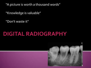
- 1. “A picture is worth a thousand words” “Knowledge is valuable” “Don’t waste it”
- 2. •INTRODUCTION •USES, ADVANATAGES, DISADVANTAGES •EQUIPMENT •ANALOG TO DIGITAL CONVERSION •DIGITAL DETECTOR SYSTEMS & CHARACTERSTICS •DIGITAL IMAGE DISPLAY •DIGITAL SUBSTRACTION RADIOGRAPHY •CONCLUSION •REFERENCES
- 3. Technological advancement-Cassetteless system Method of capturing radiographic Image Sensor Break in Electronic pieces Present and Store Computer
- 4. Detect lesions, diseases, conditions Information during root canal procedures, implants Evaluate growth & development Changes secondary to caries,trauma, periodontal diseases Progress of treatment
- 5. Superior gray scale resolution Easy reproducibility Reduced exposure to radiation Detection of defects & 3D visualization of dental structures Effective patient education tool No loss of quality due to chemical processing Lower equipment & film cost Enhancement of diagnostic image
- 6. No Darkroom Transmission of Images for Consultation Instant Viewing of Images
- 7. Initial set up costly Sensor size thicker than intraoral film Infection control difficult Receptors susceptible to rough handling and costly to replace Legal considerations
- 8. FILM BASED IMAGING DIGITAL IMAGING Density-overall degree of Brighteness-equivalent darkening Latitude- measure of range Dynamic range: number of of exposure-distinguish shades of gray i.e pixel density Film speed-faster film-less Linearity-direct radiation relationship b/n exposure & image density Contrast-diff in density- Contrast resolution-small areas of radiograph diff in density
- 9. Resolution-distinguish b/n Spatial frequency-measure of resolution-lines pairs/mm small objects that are close Background electronic noise: together small electric current that Radiographic mottle-app conveys no information but of uneven density of an serves to obscure electronic exposed film/graininess signal Sharpness- ability to define Signal to noise ratio- an edge/display density Fraction of output signal≈ boundaries diagnostic information(signal) + signal (no information-noise)
- 10. INTRAORAL EXTRAORAL Direct PhotoStimulable Phosphor Indirect Based radiography (PSP)- Storage phosphor imaging computed radiography Charged couple device (CCD) systems-solid state linear array of photoiodides
- 11. DIRECT INDIRECT
- 12. • Dental x-ray unit-radiation source Same for conventional but adapted to 1/100th second exposure time • Sensor-intraroral –no film, extraoral-PSP plates • Computer-DIGITAL IMAGE DISPLAY
- 13. Numeric format of image content & discreteness • Spatial distribution of picture elements(pixels) • Different shades of gray of each of the pixels 20 20 15 10 10 C1 0 5 1 2 3 4 5 6 7 8 9 10 0 1 2 3 4 5 6 7 8 9 10 Conventional-continuous Digital-inc /dec density spectrum
- 14. Small box or well-electrons are produced by x-ray exposure-deposited Row/column-coordinate in matrix Digital equivalent of a silver crystal Ordered arrangement
- 15. Sampling- small range of voltage values grouped together-single value Quantization- every sampled signal is assigned a value Stored in computer & represent image- computer organize pixels-gives shade of gray-correspond to no. assigned value during quantization
- 16. CONTRAST RESOLUTION SPATIAL RESOLUTION DETECTOR LATITUDE DETECTOR SENSITIVITY
- 17. • Ability – distinguish - densities • Interaction of attenuation characteristics of tissues being imaged • Ability of computer display to portray diff in density • Ability of observer to recognize differences Noise- densities captured limited by inaccuracies in image acquisition
- 18. To distinguish fine detail Limit of resolution-function of pixel size Resolution measured- units of line pair per millimeter LINE PAIR(lp) - line & its associated space- lp/mm 2 pixels required to resolve a line pair
- 19. Ability of imaging receptor to capture a range of X-ray exposure Full range of densities – gingiva to enamel PSP receptors – larger latitude CCD & CMOS-similar to film-enhanced by contrast & brightness
- 20. Sensitivity of detector to respond to small amt of radiation Factors : detector efficiency ,pixel size, noise
- 21. Charge Coupled Device ( CCD) Complementary Metal Oxide Semiconductors(CMOS) Photostimulable Phosphor Plates (PSP) Flat Panel Detectors(FPD)
- 22. Solid state detector–thin wafer of silicon chip + electronic circuit Sensitive to xrays/light Enclosed in plastic housing(protect oral environment) Electronic cable/wire system-fiberoptic cable- sensor attached to computer-ADC Length-8-35 feet
- 23. Wireless/cordless system-cable connection replaced by micowave transmittor Pixel size- 20 to70 microns-307,200 pixels CCD more sensitive to light than X-rays Layer of scintillating material (Gadolinium oxybromide)coated on the CCD directly/coupled to surface – fiber optics-inc xray absorption efficiency Linear array of Pixel = OPG & Ceph More time - complete scan
- 24. X radiation breaks covalent bonds b/n silicon atom – electron hole pairs “charge packets”-positive potential Each packet = one pixel Charge pattern = latent image “Bucket bridge” fashion-row of pixel charges to next End of row-as voltage-Charge transmitted - ADC
- 25. High Resolution = 22.5 Standard = 45 Standard High Standard & High Resolution #2 #1 #0
- 26. Silicon based semiconductors-pixel isolation from neighboring pixel-directly connected Charge transferred to transistor as small voltage Read by Frame Grabber Stored and displayed as digital gray value
- 27. Digital cameras, digital dental radiography, CPU chips 25 %more resolution, cheap, durable
- 28. Similar to CCD, no computer used CID xray sensor, cord, plug-inserted into light source on camera platform-system monitor- seconds Same as intraoral camera Color printer
- 29. Absorb & store energy from xray- stimulated by light- app wavelenghth-release energy as light (PHOSPHORESCENCE) “Europium doped” barium fluorohalide Crystal lattice- barium+iodine+chlorine+bromine Europium-imperfection in lattice F-centre
- 30. Exposed to sufficient energy source Valence electrons absorb energy-move in conduction band Electron-halogen vacencies-trapped Latent image Red light 600nm-electrons released by barium fluorohalide to conduction band When electron returns to europium ion, energy is released in green spectrum b/n 300 and 500 nm Red filter at photomultiplier tube selectively removes stimulating light, & green light converted to voltage Voltage signal quantified by ADC, stored & displayed as digital image
- 31. (Europium Activated Barium FluoroHalide) BaFX:Eu , (X= Cl, Br, or I)
- 32. PSP plates –sizes as intraoral and extraoral film PSP plate -erased before use - ghost images Latent image on PSP plate can be read by: Stationary plate scans - rotating multifaceted mirror reflecting beam of red laser light.( fast & slow scan direction) Rotating plate scans – rotation of drum past a fixed laser provides a scan Resolution of PSP systems determined by: Thickness of phosphor material Diameter of the laser beam
- 33. Plates processed quickly Susceptible to bending & scratching – permanent artifacts in receptor – obscure information of potential diagnostic value Semidark environment- plate handling Red light- not safe
- 34. Casette and PSP PSP digitizer Workstation
- 35. Provide large matrix areas - pixel sizes <100 microns Direct digital imaging-Larger areas of body-head Two types: Indirect detectors sensitive to visible light intensifying screen converts x-rays energy to light Direct detectors- photoconductor material(selenium) Similar to silicon, high atomic no-more absorption
- 36. Thin film transistor (TFT)-laptop, flatpanel computers Digitizes, processes, stores-immediate viewing-good speed Dental procedures-root canal, implants Hardcopy-printed Transmitted electronically
- 37. Spilt screen technology-multiple images on same screen Magnification-linear & angular measurements Image restoration-raw data received- corrected-before visible image on screen Image enhancement-brightness & contrast, sharpness, colour
- 38. Image analysis-extract non pictorial information-segmentation Image compression-reducing no. of digital files.lossy- irreversible Image synthesis-CT, MRI, PET-acquired data- multiple projections-new image-sectioning
- 39. Tomosynthesis-selective focusing on an arbitrary slice-through object-shift & adding basic projection Localized computed tomography(micro CT radiography, Microtomography) -invitro-study of mineral tissues -complex facial fractures Work as CT
- 40. Enhances the mineral changes that have occurred over time against a homogenous background of unchanged anatomy Subtraction of gray scales-b/n 2 images Change-light & dark areas-loss of bone(dark area), gain (light area)
- 41. Subtle changes in bone-before & after periodontal therapy Periapical region Condylar changes
- 42. Subtraction radiography. The image to the right is the result of the subtraction of the second image from the first image. Note the dark area indicating bone loss (red arrow) that was not visible on the original image
- 43. The Digital Imaging and Communications in Medicine (DICOM) Standard is a detailed specification that describes semantics and syntax for exchanging images and associated information. The standard applies to the operation of the interface which is used to transfer data in and out of an imaging device.
- 44. DICOM Display Workstation Storage, Query/Retrieve, Study Component LiteBox Query/Retrieve Results Management DICOM Acquisition Media Exchange Print Management DICOM Query/Retrieve, Archive Patient & Study
- 47. Film images->digital format->enhanced Better quality 2 parts-drum scan with reading & writing units Minicomputer with subsystems Ad-overall improved contrast, trabecular fine marrow spaces, low density=high density Disad-artifacts & noise
- 48. Microscopic imaging-single frame digital camera-attach dental microscope-high quality images Intraoral cameras-photo of single tooth, procedure, documentation & patient education barrier sheath-contamination
- 49. Fiberoptic imaging-endoscope-root canals -0.7 mm & 11.8 mm dia light fiberoptic probe- insert in root canal -disposable-optical grade plastic PSP radiography-trial file length Digital photography
- 51. Oral Radiology, Principles and interpretation, 5th edition – White & Pharoah Text book of Dental and Maxillofacial Radiology, Freny. R.Karjodkar Essentials of Dental Radiography & Radiology, III edition, Eric Whaites