

Recomendados
Más contenido relacionado
La actualidad más candente
La actualidad más candente (20)
Similar a Cementum
Similar a Cementum (20)
Más de Ali Tahir
Último
Último (20)
Cementum
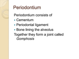
- 1. Periodontium Periodontium consists of Cementum Periodontal ligament Bone lining the alveolus Together they form a joint called Gomphosis
- 2. Cementum Hard, avascular connective tissue that covers the roots of teeth Classified into ◦ Acellular cementum ◦ Cellular cementum Development divided into ◦ Prefunctional stage ◦ Functional stage ◦ At cervical margin it is 50µm thick, progresses apicallly to 200µm.
- 4. Composition of Cementum Similar to bone 45-50% inorganic (hyroxyapatite) 50% organic (Collagens & non- collagenous proteins)
- 5. Organic components 90% is type 1 collagen Other collagens include Type III, V, VI, XII & XIV Type XII is found in ligamentous tissues and PDL Non-collagenous proteins: ◦ Alkaline phosphatase ◦ Bone sialoprotein & Dentin sialoprotein ◦ Dentin matrix protein 1 ◦ Fibronectin ◦ Osteocalcin, Osteonectin & Osteopontin ◦ Proteoglycans & Proteolipids ◦ Tenascin ◦ Growth factors including Insulin like growth factor, platelet derived growth factor
- 6. Cementum formation Cementum formation initiates at the advancing root edge HERS sends inductive signals to ecto- mesenchymal pulp cells > odontoblasts > predentin > interruption of HERS > contact of predentin with dental follicle > differentiate into cementoblasts Another theory says HERS differentiate into cementoblasts
- 8. Fate of HERS Some cells undergo apoptosis Some fragments form discrete masses surrounded by basal lamina, these are called Epithelial rests of Malassez Have a role in maintenance & regeneration of PDL Some odontogenic tumours are believed to arise from these rests If they remain attached to the forming root surface, they form enamell-pearls
- 10. Factors regulating cementogenesis Growth factors Adhesion molecules Epithelial/Enamel proteins Collagens Gla Proteins Transcription factors Signalling Molecules
- 11. Growth factors Transforming growth factor ß superfamily ◦ Promotes cell differentiation & cementogenesis during development & regeneration Platelet & Insulin derived growth factor ◦ Promote cementum formation by altering cell cycle activities Fibroblast growth factor ◦ Promote formation & regeneration of PDL tissue by inducing cell proliferation & vasculogenesis
- 12. Adhesion molecules Bone Sialoprotein/Osteopontin ◦ Promote adhesion of cells to newly forming root ◦ BSP promotes mineralization & osteopontin regulates the extent of crystal growth
- 13. Others Epithelial/Enamel Proteins: Periodontal repair & regeneration Collagens Type I & III have a role in development & regeneration of PDL Type XII maintains the PDL space Gla Proteins (∂-carboxyglutamic acid) Prevent ectopic calcifications & preserve PDL width Osteocalcin regulates crystal growth & extent of mineralization
- 14. Others Transcription factors i. Runt-related transcription factor 2(Runx-2) ii. Osterix Believed to induce cementoblast differentiation Signaling molecules Osteoprotegrin & RANKL are produced by osteoblasts & PDL fibroblasts, they mediate bone & root resorption by osteoclasts
- 15. Cementum Types Acellular ◦ Acellular extrinsic fiber cementum ◦ Acellular afibrillar cementum Cellular Intrinsic Fiber Cementum
- 16. Acellular Extrinsic Fiber Cementum (Primary Cementum) Formation: Principle tissue of attachment, covers 2/3rd of tissue or more Cementoblasts align along the newly formed unmineralized predentin (mantle dentin) These cementoblasts extend their cell processes into this predentin & deposit collagen fibrils within it Mineralization of mantle dentins starts from inside out & spreads across the cementum
- 18. Formation Initial layer of short fiber fringe > cells on root surface move away to deposit another 15-20µm of cementum (lengthens & thickens the initial FF plus non-collagenous proteins) > PDL fibroblasts deposit collagen fibrils that become stitched to the FF > cementoblasts secrete only non- collagenous proteins
- 19. AEFC Cells remain on the surface therefore called acellular cementum Light microscope shows two sets of striations ◦ Incremental deposition running parallel to root surface ◦ Inserted PDL collagen fiber bundles as short striations perpendicular to root surface No cementoid is present Innermost layer is less mineralized & outer layer shows alternative bands of more & less mineralization
- 21. Cellular Intrinsic Fiber Cementum (Secondary root is formed, a rapidly After half the cementum) forming less mineralized variety of cementum is laid called CIFC Fount in the apical third Confined to the apical & inter-radicular regions of the tooth (usually absent in incisors & canines) Because of rapid cementum deposition, an un-mineralized layer of cementum is present called cementoid & cells become entrapped in the extra-cellular matrix to form cementocytes
- 22. Cementocytes Similar to osteocytes as they reside in lacunae but their processes within the canaliculi do not form syncytium to the root surface Nourishment occurs by diffusion so the cementocytes in deeper layers may not be vital
- 23. CIFC After a rapid initial haphazard deposition, it slows down & becomes more directional In this phase cells do not become entrapped so may lead to acellular cementum Collagen laid by cementocytes is parallel to the root surface After the PDL fibers are incorporated into the cementum, they become partially mineralized > Cellular mixed fiber
- 25. How to differentiate from AEFC On light microscope ◦ Cementocytes within lacunae ◦ Laminated structure ◦ Cementoid on its surface
- 28. Cementocytes
- 29. How to differentiate EF & IF Intrinsic fibers are fine, paralled to root surface & uniformly mineralized Extrinsic fibers are larger, haphazard, mineralized variably & run perpendicular to the root surface
- 30. Acellular Afibrillar Cementum Lacks collagen No cementocytes No role in tooth attachment Appears uniform under light microscope Deposited over dentin & enamel near CEJ
- 31. Cemento-enamel junction Three types of joints are seen ◦ Cementum overlaps enamel (majority) ◦ Butt joint ◦ Gap between cementum & enamel leaving the dentin exposed
- 33. Alveolar Process Bone of the jaws containing sockets for the teeth Consists of ◦ Cortical plates (buccal & lingual) ◦ Spongiosa ◦ Alveolar bone (lining the socket) Cortical plate & alveolar bone meet at alveolar crest which is 1.5-2mm below CEJ
- 34. Alveolar bone Inner & outer components Contains many foramina b/c of which is also called cribriform plate. Because of increased radio-opacity also called lamina dura Inner alveolar bone incorporates PDL bundle fibers, therefore, also called bundle bone Bundle bone may be apposed on a lamellar bone or not
- 36. Cortical plates Surface layer of lamellar bone with haversian systems Central part of spongy bone Cortical plates are thinner in maxilla, thickest on buccal aspect of mandibular molars Spongy bone has yellow marrow rich in adipose cells, rarely has hematopoietic marrow Trabecular bone is absent in anterior teeth where cortical & alveolar bones fuse
- 38. Cementum Canaliculus GT Lacuna of cementocyte Dentin Acellular cementum Cellular cementum Hyaline layer (of Hopewell Smith) Granular layer of tomes Dentin with tubules
- 39. Aging Smooth surface becomes irregular due to calcification of ligament fiber bundles where they are attached to cementum Continues deposition of cementum occurs with age in the apical area. [Good: maintains tooth length; bad: obstructs the foramen 3. Resorption of root dentin occurs with aging which is covered by cemental repair
- 40. Cemental repair Protective function of cementoblasts after resorption of root dentin or cementum Resorption of dentin and cementum due to trauma (traumatic occlusion, tooth movement, hypereruption) Loss of cementum accompanied by loss of attachment Following reparative cementum deposition attachment is restored
- 41. Clinical Correlation Cellular cementum is similar to bone but has no nerves. Therefore it is non-sensitive to pain. Scaling produces no pain, but if cementum is removed, dentin is exposed causes sensitivity Cementum is resistant to resorption especially in younger Patients. Thus, orthodontic tooth movement causes alveolar one resorption and not tooth root loss
- 42. The End