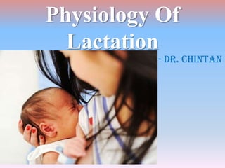
Physiology of lactation
•Descargar como PPTX, PDF•
29 recomendaciones•13,877 vistas


Recomendados
Más contenido relacionado
La actualidad más candente
La actualidad más candente (20)
Similar a Physiology of lactation
Similar a Physiology of lactation (20)
Más de DrChintansinh Parmar
Más de DrChintansinh Parmar (20)
Último
Último (20)
Physiology of lactation
- 1. Physiology Of Lactation - Dr. Chintan
- 2. Development of the Breasts The breasts begin to develop at puberty. This development is stimulated by the estrogens of the monthly female sexual cycle; Estrogens stimulate growth of the breasts’ mammary glands plus the deposition of fat to give the breasts mass. In addition, far greater growth occurs during the high estrogen state of pregnancy, and only then does the glandular tissue become completely developed for the production of milk.
- 3. Growth of the Ductal System All through pregnancy, the large quantities of estrogens secreted by the placenta cause the ductal system of the breasts to grow and branch. Simultaneously, the stroma of the breasts increases in quantity, and large quantities of fat are laid down in the stroma. Also important for growth of the ductal system are at least four other hormones: growth hormone, prolactin, the adrenal glucocorticoids, and insulin. Each of these is known to play a role in protein metabolism.
- 4. Development of the LobuleAlveolar System Final development of the breasts into milk secreting organs requires progesterone. Once the ductal system has developed, progesterone—acting synergistically with estrogen, causes additional growth of the breast lobules, with growing of alveoli and development of secretory characteristics in the cells of the alveoli. These changes are analogous to the secretory effects of progesterone on the endometrium of the uterus during the latter half of the female menstrual cycle.
- 7. Phases Of Lactation • Preparation of breast for milk secretion – mammogenesis • Synthesis & secretion of milk – lactogenesis • Expulsion of milk – galactokinesis • Maintenance of lactation - galactopoesis
- 8. Initiation of Lactation Prolactin hormone is secreted by the mother’s anterior pituitary gland, and its concentration in her blood rises steadily from the 5th week of pregnancy until birth of the baby, at which time it has risen to 10 to 20 times the normal nonpregnant level. In addition, the placenta secretes large quantities of hCS, which probably has lactogenic properties, thus supporting the prolactin from the mother’s pituitary during pregnancy. The fluid secreted during the last few days before and the first few days after parturition is called colostrum; it contains essentially the same concentrations of proteins and lactose as milk, but it has almost no fat.
- 9. Initiation of Lactation Immediately after the baby is born, the sudden loss of both estrogen and progesterone secretion from the placenta allows the lactogenic effect of prolactin from the mother’s pituitary gland to assume its natural milk promoting role, and over the next 1 to 7 days, the breasts begin to secrete copious quantities of milk instead of colostrum. growth hormone, cortisol, parathyroid hormone, and insulin. These hormones are necessary to provide the amino acids, fatty acids, glucose, and calcium required for milk formation.
- 10. Initiation of Lactation After birth of the baby, the basal level of prolactin secretion returns to the nonpregnant level over the next few weeks. Each time the mother nurses her baby, nervous signals from the nipples to the hypothalamus cause a 10- to 20-fold surge in prolactin secretion that lasts for about 1 hour. If this prolactin surge is absent or blocked as a result of hypothalamic or pituitary damage or if nursing does not continue, the breasts lose their ability to produce milk within 1 week or so. Milk production can continue for several years if the child continues to suckle, although the rate of milk formation normally decreases considerably after 7 to 9 months.
- 12. Hypothalamic Control The hypothalamus mainly stimulates production of all the other hormones, but it mainly inhibits prolactin production. Consequently, damage to the hypothalamus or blockage of the hypothalamic hypophysial portal system often increases prolactin secretion. prolactin inhibitory hormone It is almost certainly the same as the catecholamine dopamine, which is known to be secreted by the arcuate nuclei of the hypothalamus and can decrease prolactin secretion as much as 10-fold. L-Dopa decreases prolactin secretion by increasing the formation of dopamine, and bromocriptine and other dopamine agonists inhibit secretion because they stimulate dopamine receptors. Chlorpromazine and related drugs that block dopamine receptors increase prolactin secretion.
- 13. Postpartum Amenorrhea In most nursing mothers, the ovarian cycle and ovulation stop until a few weeks after cessation of nursing. The reason seems to be that the same nervous signals from the breasts to the hypothalamus that cause prolactin secretion during suckling—either because of the nervous signals themselves or because of a subsequent effect of increased prolactin — inhibit secretion of GnRH by the hypothalamus & so FSH, LH from ant. pituitary. After several months of lactation, in some mothers, especially in those who nurse their babies only some of the time, the pituitary begins to secrete sufficient gonadotropic hormones to restore the monthly sexual cycle, even though nursing continues.
- 14. Ejection (or “Let-Down”) Process Milk is secreted continuously into the alveoli of the breasts, but milk does not flow easily from the alveoli into the ductal system and, therefore, does not continually leak from the breast nipples. When the baby suckles, it receives virtually no milk for the first half minute. Sensory impulses - somatic nerves from the nipples - spinal cord – hypothalamus - oxytocin . The oxytocin via blood to the breasts, where it causes myoepithelial cells to contract, thereby expressing the milk from the alveoli into the ducts. So, within 30 seconds to 1 minute after a baby begins to suckle, milk begins to flow. This process is called milk ejection or milk let-down – opposite breast also Fondling of the baby by the mother or hearing the baby crying often gives enough of an emotional signal to the hypothalamus to cause milk ejection – inhibition by psychogenic stimuli
- 15. Milk Composition More lactose, less protein, less ash
- 16. Milk Composition At the height of lactation in the human mother, 1.5 liters of milk may be formed each day (and even more if the mother has twins). about 50 grams of fat enter the milk each day, and about 100 grams of lactose, which must be derived by conversion from the mother’s glucose. Also, 2 to 3 grams of calcium phosphate may be lost each day; multiple types of antibodies and other anti-infectious agents are secreted in milk along with the nutrients. neutrophils and macrophages - Escherichia coli bacteria, which often cause lethal diarrhea in newborns.
- 17. Advantages Baby: Balanced diet – easily digestible Protection against infection Growth factors Sterile – inexpensive Right temperature – rare chances of allergy Mother: Amenorrhea – birth control Involution of uterus – oxytocin Protection against breast engorgement & so infection Protection against obesity, cancer Emotional bonding with baby
- 18. Man will be man…
- 20. Fetal Growth
- 21. Fetal Development Circulatory – heart – 4th week – 65/min – 140/min Blood (RBC): 3rd week – yolk sac, 6th week – liver, 3rd month – spleen, afterwards – bone marrow RS – no respiration – no air - inhibition of respiration during the later months of fetal life prevents filling of the lungs with fluid and debris from the meconium CNS: spinal cord, brain stem reflex – 4th month, cerebral cortex last month, even after birth
- 22. Fetal Development GIT: 4th month – ingestion & absorption of amniotic fluid – meconium formed KIDNEY: 2nd trimester, 70 – 80 % of amniotic fluid is urine – oligohydramnios Metabolism: glucose for energy – converted to fat, Ca, phosphorus accumulation in last 4 weeks – rapid weight gain due to ossification, after 4th month – X ray visibility Iron: 3rd week iron in Hb, iron store in liver, Vitamins
- 24. Adjustments Onset of Breathing – the child ordinarily begins to breathe within seconds and has a normal respiratory rhythm within less than 1 minute after birth. Asphyxia – sensory from cooling of skin Danger of hypoxia – (1) Compression of the umbilical cord; (2) Premature separation of the placenta; (3) Excessive contraction of the uterus, which can cut off the mother’s blood flow to the placenta; (4) Excessive anesthesia of the mother (5) Head trauma or prolonged delivery
- 25. Onset of Breathing Can tolerate hypoxia for 10 minutes – adult only 4 minutes Expansion of the Lungs At birth, the walls of the alveoli are at first collapsed because of the surface tension of the viscid fluid that fills them. More than 25 mm Hg of negative inspiratory pressure in the lungs is usually required to oppose the effects of this surface tension and to open the alveoli for the first time. But once the alveoli do open, further respiration can be effected with relatively weak respiratory movements. the first inspirations of the normal neonate are extremely powerful, usually capable of creating as much as 60 mm Hg negative pressure in the Intrapleural space.
- 30. Circulatory Readjustments First, loss of the tremendous blood flow through the placenta, which approximately doubles the SVR at birth. This increases the aortic pressure as well as the pressures in the left ventricle and left atrium. Second, the PVR greatly decreases as a result of expansion of the lungs. In the unexpanded fetal lungs, the blood vessels are compressed because of the small volume of the lungs. Immediately on expansion, these vessels are no longer compressed and the resistance to blood flow decreases. Also, in fetal life, the hypoxia of the lungs causes considerable tonic vasoconstriction of the lung blood vessels, but vasodilation takes place when ventilation of the lungs eliminates the hypoxia. All these changes together reduces the pulmonary arterial pressure, right ventricular pressure, and right atrial pressure.
- 31. Closure of the Foramen Ovale The low right atrial pressure and the high left atrial pressure cause blood now to attempt to flow backward through the foramen ovale; that is, from the left atrium into the right atrium. The small valve that lies over the foramen ovale on the left side of the atrial septum closes over this opening, thereby preventing further flow through the foramen ovale. In 2/3rd of all people, the valve becomes adherent over the foramen ovale within a few months to a few years and forms a permanent closure. But even if permanent closure does not occur, the left atrial pressure throughout life normally remains 2 to 4 mm Hg greater than the right atrial pressure, and the backpressure keeps the valve closed.
- 32. Closure of the Ductus Arteriosus ↑ SVR elevates the aortic pressure while ↓ PVR reduces the pulmonary arterial pressure. So, after birth, blood begins to flow backward from the aorta into the pulmonary artery through the ductus arteriosus. After only a few hours, the muscle wall of the ductus arteriosus constricts markedly, and within 1 to 8 days - functional closure of the ductus arteriosus. Then, during the next 1 to 4 months, the ductus arteriosus ordinarily becomes anatomically occluded by growth of fibrous tissue into its lumen. The degree of contraction of the smooth muscle in the ductus wall is highly related to availability of oxygen. In one of several thousand infants – PDA. The failure of closure result from excessive ductus dilation caused by vasodilating prostaglandins in the ductus wall – indomethacin role
- 33. Closure of the Ductus Venosus In fetal life, the portal blood from the fetus’s abdomen joins the blood from the umbilical vein, and these together pass by way of the ductus venosus directly into the vena cava bypassing the liver. Immediately after birth, blood flow through the umbilical vein ceases, but most of the portal blood still flows through the ductus venosus, with only a small amount passing through the channels of the liver. Within 1 to 3 hours the muscle wall of the ductus venosus contracts strongly and closes this path of flow. As a consequence, the portal venous pressure rises from near 0 to 6 to 10 mm Hg, which is enough to force portal venous blood flow through the liver sinuses.
- 35. Functional Problems Glucose – immature liver – first 2 to 3 days – fluid imbalance – weight loss RS: TV 16 ml x RR 40 = RMV 640 ml / min, FRC more due to ↑ RR BV: 300 + 75 CO: 500 ml / min BP: 70/50 – 90/60 – 110/70 Blood: Physiological hyperbilirubinemia - jaundice anemia, physiological
- 37. Functional Problems Blood: Erythroblastosis Fetalis Kidney: Acidosis, dehydration, overhydration Liver: hypoproteinemic edema, low coagulation factors Hypothermia - ↑ BMR, BSA more as compared to body mass Vitamin D – rickets, iron deficiency, Vitamin C – scurvy (cow milk) Allergy due to newly developing immunity
- 38. Endocrine Problems/Prematurity Hermaphroditism Diabetic mother – large babies – RDS Cretin dwarfism RDS – chyne stroke breathing – Oxygen therapy – danger of blindness – retrolental fibroplasia Instable body temperature – role of incubator
- 39. Thank You…