Discovery of an Accretion Streamer and a Slow Wide-angle Outflow around FUOri...
E platyhelminthes
1. 1
Zool 628 eLabs (mkl)
A
C
B
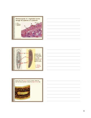
Photomicrograph of a longitudinal section
through the epidermis of a planarian
A Cilia
B Rhabdites
C Epidermis
Zool 628 eLabs (mkl)
A
B
B
A Pharynx
B Intestinal
branches
Schematic and
photomicrograph
of a whole mount
of a triclad,
showing the three
branches of the
digestive system.
Zool 628 eLabs (mkl)
Dorsal view of the pharynx (A) of a triclad. Animal has
been fed carbon particles to stain the digestive cavity (B).
A
B
2. 2
Zool 628 eLabs (mkl)
A
B B
A Pharynx
B Intestine
Pphotomicrograph of a cross section through the
pharynx region of a triclad flatworm (Dugesia).
A
Zool 628 eLabs (mkl)
Anterior end of Dugesia, showing ocelli and
auricles.
A
B
A Ocellus
B Auricle
Zool 628 eLabs (mkl)
Whole mount of a digenetic fluke.
3. 3
Zool 628 eLabs (mkl)
Female
Schistosoma
Male Schistosoma
Human Lung Fluke
A
A
AB
B
B
A Oral sucker
B Acetabulum
Zool 628 eLabs (mkl)
Whole mount of the Oriental liver fluke
(Clonorchis sinensis).
Zool 628 eLabs (mkl)
1. Mouth and oral sucker
2. Pharynx
3. Esophagus
4. Intestinal cecae
5. Genital pore
6. Ventral sucker (acetabulum)
Photomicrograph of the anterior end of the
Chinese liver fluke
4. 4
Zool 628 eLabs (mkl)
1. Uterus
2. Yolk glands
3. Intestinal ceca
4. Ovary
5. Seminal receptacle
6. Testes
7. Excretory bladder
8. Yolk ducts
Photomicrograph of the middle part of the
Chinese liver fluke
Zool 628 eLabs (mkl)
Photomicrograph of the
posterior end of the Chinese liver fluke
1. Intestinal ceca
2. Testes
3. Excretory bladder
4. Excretory pore
Zool 628 eLabs (mkl)
Trematode eggs
Miracidium
Cercaria
Photomicrographs of
trematode eggs,
miracidium larva, and
cercaria larva.
5. 5
Zool 628 eLabs (mkl)
Egg
Cercaria
Miracidium
Zool 628 eLabs (mkl)
A
B
C
A Male
B Gynecophoric canal
C Female
Copulating Schistosoma
Zool 628 eLabs (mkl)
A
B
B
A Suckers
B Hooks on rostellum
A
Scoleces of two cestodes.
6. 6
Zool 628 eLabs (mkl)
A B
C C
A Scolex
B Strobila
C Proglottid
Photomicrograph of the anterior end of a cestode.
Zool 628 eLabs (mkl)
A B
C
C
D
E
F
A Testes
B Uterus
C Ovary
D Yolk gland
E Shell gland
F Common genital pore
Structures of a
cestode proglottid
Zool 628 eLabs (mkl)
A
A Genital pore
B Ovarian capsules with
eggs or embryos
B
Photomicrograph of a
gravid proglottid
7. 7
Zool 628 eLabs (mkl)
Photomicrographs of
cysticercus larvae
Zool 628 eLabs (mkl)
A B C D
A Scolex C Mature proglottids
B Immature proglottids D Gravid proglottids
Photomicrograph of a composite
slide of a tapeworm