SlideShare una empresa de Scribd logo
1 de 12
Tutorial Notes                                         Marc Imhotep Cray,M.D.


     BASIC MEDICAL SCIENCE OF VALVULAR HEART DISEASE

Cardiac cycle
(From USMLE First Aid 2008 pg. 192)


                                                       Phases––left ventricle:
                                                       1. Isovolumetric
                                                       contraction––period between
                                                       mitral valve closure and aortic
                                                       valve opening; period of
                                                       highest O2 consumption
                                                       2. Systolic ejection––period
                                                       between aortic valve opening
                                                       and closing
                                                       3. Isovolumetric relaxation–
                                                       –period between aortic valve
                                                       closing and mitral valve
                                                       opening
                                                       4. Rapid filling––period just
                                                       after mitral valve opening
                                                       5. Reduced filling––period
                                                       just before mitral valve
                                                       closure


                    BASIC MEDICAL SCIENCE OF VALVULAR HEART DISEASE
                                           1
Tutorial Notes                                         Marc Imhotep Cray,M.D.


Ventricular Pressure-Volume
  Loop Changes in Valve
          Disease
                                     Phases––left ventricle:
                                     1. Isovolumetric contraction––period between mitral
Cardiac valve disease                valve closure and aortic valve opening; period of
significantly alters ventricular     highest O2 consumption
pressure and volume                  2. Systolic ejection––period between aortic valve
relationships during the cardiac     opening and closing
cycle. A convenient way to
analyze cardiac pressure and
                                     3. Isovolumetric relaxation––period between aortic
volume changes is by using           valve closing and mitral valve opening
ventricular pressure-volume          4. Rapid filling––period just after mitral valve
loops. The links below will          opening
illustrate the pressure-volume       5. Reduced filling––period just before mitral valve
changes that occur with the          closure
following valve defects:

      Mitral stenosis
      Aortic stenosis
      Mitral regurgitation
      Aortic regurgitation


                            BASIC MEDICAL SCIENCE OF VALVULAR HEART DISEASE
                                                   2
Tutorial Notes                                                    Marc Imhotep Cray,M.D.


                     Cardiac Valve Disease Tutorial

Aortic valve stenosis
http://en.wikipedia.org/wiki/Aortic_valve_stenosis
Aortic valve stenosis (AS) is a valvular heart disease caused by the incomplete opening of the
aortic valve.

The aortic valve controls the direction of blood flow from the left ventricle to the aorta. When in
good working order, the aortic valve does not impede the flow of blood between these two
spaces. Under some circumstances, the aortic valve becomes narrower than normal, impeding
the flow of blood. This is known as aortic valve stenosis, or aortic stenosis, often abbreviated as
AS.


An aortic valve that, due to rheumatic heart disease, has a severe stenosis (centre of image).
The pulmonary trunk is seen at the lower right (of the image). The proximal portion of right
coronary artery and its ostium can be seen at the lower left (of the image). The proximal left
main coronary artery and its ostium are seen on the right (of the image). Autopsy specimen.


                    BASIC MEDICAL SCIENCE OF VALVULAR HEART DISEASE
                                           3
Tutorial Notes                                                 Marc Imhotep Cray,M.D.


Simultaneous left ventricular and aortic pressure tracings demonstrate a pressure gradient
between the left ventricle and aorta, suggesting aortic stenosis. The left ventricle generates
higher pressures than what is transmitted to the aorta. The pressure gradient, caused by aortic
stenosis, is represented by the green shaded area. (AO = ascending aorta; LV = left ventricle;
ECG = electrocardiogram.)

Symptoms of aortic stenosis

Congestive heart failure

Syncope

Angina

BMS TUTORIAL/ Cardiovascular Physiology Concepts

Cardiovascular Physiology Concepts
Cardiac Valve Disease Pathophysiology of valve disease

CLINICAL READ/ Emedicine
Aortic Stenosis
                    BASIC MEDICAL SCIENCE OF VALVULAR HEART DISEASE
                                           4
Tutorial Notes                                                      Marc Imhotep Cray,M.D.


Aortic insufficiency
http://en.wikipedia.org/wiki/Aortic_insufficiency
Aortic insufficiency (AI), also known as aortic regurgitation (AR), is the leaking of the aortic valve
of the heart that causes blood to flow in the reverse direction during ventricular diastole, from
the aorta into the left ventricle.

Aortic insufficiency can be due to abnormalities of either the aortic valve or the aortic root (the
beginning of the aorta).


        BEFORE SURGICAL REPAIR                                   AFTER SURGICAL REPAIR


Pathophysiology

In aortic insufficiency, when the pressure in the left ventricle falls below the pressure in the
aorta, the aortic valve is not able to completely close. This causes a leaking of blood from the
aorta into the left ventricle. This means that some of the blood that was already ejected from the
heart is regurgitating back into the heart. The percentage of blood that regurgitates back
through the aortic valve due to AI is known as the regurgitant fraction.

For instance, if an individual with AI has a stroke volume of 100 ml and during ventricular
diastole 25 ml regurgitates back through the aortic valve, the regurgitant fraction is 25%. This
regurgitant flow causes a decrease in the diastolic blood pressure in the aorta, and therefore an
increase in the pulse pressure (systolic pressure - diastolic pressure).

Since some of the blood that is ejected during systole regurgitates back into the left ventricle
during diastole, there is decreased effective forward flow in AI.


                    BASIC MEDICAL SCIENCE OF VALVULAR HEART DISEASE
                                           5
Tutorial Notes                                                      Marc Imhotep Cray,M.D.


Note that while diastolic blood pressure is diminished and the pulse pressure widens, systolic
blood pressure generally remains normal or can even be slightly elevated. This is because
sympathetic nervous system and the renin-angiotensin-aldosterone axis of the kidneys
compensate for the decreased cardiac output. Catecholamines will increase the heart rate and
increase the strength of ventricular contraction, directly increasing cardiac output.
Catecholamines will also cause peripheral vasoconstriction, which causes increased systemic
vascular resistance and ensures that core organs are adequately perfused. Renin, a proteolytic
enzyme, cleaves angiotensinogen to angiotensin I, which is converted to angiotensin II, which is
also a potent vasoconstrictor. In the case of chronic aortic insufficiency with resultant cardiac
remodeling, heart failure will develop, and it is possible to see systolic pressures diminish.

Aortic insufficiency causes both volume overload (elevated preload) and pressure
overload (elevated afterload) of the heart.

The pressure overload (due to elevated pulse pressure and the systemic effects of
neuroendocrine hormones) causes left ventricular hypertrophy (LVH). There is both concentric
hypertrophy and excentric hypertrophy in AI. The concentric hypertrophy is due to the increased
left ventricular systolic pressures associated with AI, while the excentric hypertrophy is due to
volume overload caused by the regurgitant fraction.

Hemodynamics

The hemodynamic sequelae of AI are dependent on the rate of onset of AI. Acute AI and
chronic AI will have different hemodynamics and individuals will have different signs and
symptoms.

Acute aortic insufficiency

In acute AI, as may be seen with acute perforation of the aortic valve due to endocarditis, there
will be a sudden increase in the volume of blood in the left ventricle. The ventricle is unable to
deal with the sudden change in volume. In terms of the Frank-Starling curve, the end-diastolic
volume will be very high, such that further increases in volume result in less and less efficient
contraction. The filling pressure of the left ventricle will increase. This causes pressure in the left
atrium to rise, and the individual will develop pulmonary edema.

Severe acute aortic insufficiency is considered a medical emergency. There is a high mortality
rate if the individual does not undergo immediate surgery for aortic valve replacement. If the
acute AI is due to aortic valve endocarditis, there is a risk that the new valve may become
seeded with bacteria. However, this risk is small.

Acute AI usually presents as florid congestive heart failure, and will not have any of the signs
associated with chronic AI since the left ventricle had not yet developed the eccentric
hypertrophy and dilatation that allow an increased stroke volume, which in turn cause bounding
peripheral pulses. On auscultation, there may be a short diastolic murmur and a soft S1. S1 is
soft because the elevated filling pressures close the mitral valve in diastole (rather than the
mitral valve being closed at the beginning of systole).


                    BASIC MEDICAL SCIENCE OF VALVULAR HEART DISEASE
                                           6
Tutorial Notes                                                  Marc Imhotep Cray,M.D.


Chronic aortic insufficiency

If the individual survives the initial hemodynamic derailment that acute AI presents as, the left
ventricle adapts by eccentric hypertrophy and dilatation of the left ventricle, and the volume
overload is compensated for. The left ventricular filling pressures will revert to normal and the
individual will no longer have overt heart failure.

In this compensated phase, the individual may be totally asymptomatic and may have normal
exercise tolerance.

Eventually (typically after a latency period) the left ventricle will become decompensated, and
filling pressures will increase. While most individuals would complain of symptoms of congestive
heart failure to their physicians, some enter this decompensated phase asymptomatically.
Proper treatment for AI involves aortic valve replacement prior to this decompensation phase.

BMS TUTORIAL/ Cardiovascular Physiology Concepts
Cardiac Valve Disease Pathophysiology of valve disease

CLINICAL READ/eMEDICINE
Aortic Regurgitation


Mitral stenosis
http://en.wikipedia.org/wiki/Mitral_valve_stenosis
Mitral stenosis is a valvular heart disease characterized by the narrowing of the orifice of the
mitral valve of the heart.


                    BASIC MEDICAL SCIENCE OF VALVULAR HEART DISEASE
                                           7
Tutorial Notes                                                   Marc Imhotep Cray,M.D.


Mitral stenosis with marked thickening of the leaflets and left atrial hypertrophy. Superior view.
Autopsy preparation.


Rheumatic heart disease at autopsy with characteristic findings (thickened mitral valve,
thickened chordae tendineae, hypertrophied left ventricular myocardium).


                    BASIC MEDICAL SCIENCE OF VALVULAR HEART DISEASE
                                           8
Tutorial Notes                                                        Marc Imhotep Cray,M.D.


Intracardiac pressure measurements in an individual with severe mitral stenosis. Pressure
tracings in the left atrium (LA) and the left ventricle (LV) in an individual with severe mitral
stenosis. Blue areas represent the diastolic pressure gradient due to the stenotic valve.

Pathophysiology

The normal area of the mitral valve orifice is about 4 to 6 cm2. Under normal conditions, a
normal mitral valve will not impede the flow of blood from the left atrium to the left ventricle
during (ventricular) diastole, and the pressures in the left atrium and the left ventricle during
ventricular diastole will be equal. The result is that the left ventricle gets filled with blood during
early ventricular diastole, with only a small portion of extra blood contributed by contraction of
the left atrium (the "atrial kick") during late ventricular diastole.

When the mitral valve area goes below 2 cm2, the valve causes an impediment to the flow of
blood into the left ventricle, creating a pressure gradient across the mitral valve. This gradient
may be increased by increases in the heart rate or cardiac output. As the gradient across the
mitral valve increases, the amount of time necessary to fill the left ventricle with blood increases.
Eventually, the left ventricle requires the atrial kick to fill with blood. As the heart rate increases,
the amount of time that the ventricle is in diastole and can fill up with blood (called the diastolic
filling period) decreases. When the heart rate goes above a certain point, the diastolic filling
period is insufficient to fill the ventricle with blood and pressure builds up in the left atrium,
leading to pulmonary congestion.

When the mitral valve area goes less than 1 cm2, there will be an increase in the left atrial
pressures (required to push blood through the stenotic valve). Since the normal left ventricular
diastolic pressures is about 5 mmHg, a pressure gradient across the mitral valve of 20 mmHg
due to severe mitral stenosis will cause a left atrial pressure of about 25 mmHg. This left atrial
pressure is transmitted to the pulmonary vasculature and causes pulmonary hypertension.
Pulmonary capillary pressures in this level cause an imbalance between the hydrostatic
pressure and the oncotic pressure, leading to extravasation of fluid from the vascular tree and
pooling of fluid in the lungs (congestive heart failure causing pulmonary edema).

The constant pressure overload of the left atrium will cause the left atrium to increase in size. As
the left atrium increases in size, it becomes more prone to develop atrial fibrillation. When atrial
fibrillation develops, the atrial kick is lost (since it is due to the normal atrial contraction).

In individuals with severe mitral stenosis, the left ventricular filling is dependent on the atrial
kick. The loss of the atrial kick due to atrial fibrillation can cause a precipitous decrease in
cardiac output and sudden congestive heart failure.

Patients with mitral stenosis prompts a series of hemodynamic changes that frequently cause
deterioration of the patient's clinical status. A reduction in cardiac output, associated with
acceleration of heart rate and shortening of the diastolic time, frequently leads to congestive
heart failure. In addition, when AF sets in, systemic embolization becomes a real danger.

BMS TUTORIAL/ Cardiovascular Physiology Concepts

Cardiac Valve Disease Pathophysiology of valve disease

CLINICAL READ/ eMEDICINE
                    BASIC MEDICAL SCIENCE OF VALVULAR HEART DISEASE
                                           9
Tutorial Notes                                                          Marc Imhotep Cray,M.D.


Mitral Stenosis


Mitral regurgitation
http://en.wikipedia.org/wiki/Mitral_insufficiency

Mitral regurgitation (MR), mitral insufficiency or mitral incompetence is a disorder of the heart in
which the mitral valve does not close properly when the heart pumps out blood. It is the
abnormal leaking of blood from the left ventricle, through the mitral valve, and into the left
atrium, when the left ventricle contracts, i.e. there is regurgitation of blood back into the left
atrium. MR is the most common form of valvular heart disease.


An illustration of mitral annular calcification, an aging phenomenon which can cause mitral regurgitation. It
can also be associated with calcifications of aortic cusps causing stenosis and extension of calcification
into cardiac skeleton causing heart block.


Acute phase

Acute mitral regurgitation (as may occur due to the sudden rupture of a chordae tendineae or
papillary muscle) causes a sudden volume overload of both the left atrium and the left ventricle.
The left ventricle develops volume overload because with every contraction it now has to pump
out not only the volume of blood that goes into the aorta (the forward cardiac output or forward
stroke volume), but also the blood that regurgitates into the left atrium (the regurgitant volume).
The combination of the forward stroke volume and the regurgitant volume is known as the total
stroke volume of the left ventricle.

                     BASIC MEDICAL SCIENCE OF VALVULAR HEART DISEASE
                                            10
Tutorial Notes                                                     Marc Imhotep Cray,M.D.


In the acute setting, the stroke volume of the left ventricle is increased (increased ejection
fraction), this happens because of more complete emptying of heart. However, as it progresses
the LV volume increases and the contractile function deteriorates and thus leading to
dysfunctional LV and a decrease in ejection fraction.The mechanism by which the total stroke
volume is increased is known as the Frank-Starling mechanism.

The regurgitant volume causes a volume overload and a pressure overload of the left atrium.
The increased pressures in the left atrium inhibit drainage of blood from the lungs via the
pulmonary veins. This causes pulmonary congestion.

Chronic phase


Compensated

If the mitral regurgitation develops slowly over months to years or if the acute phase can be
managed with medical therapy, the individual will enter the chronic compensated phase of the
disease. In this phase, the left ventricle develops eccentric hypertrophy in order to better
manage the larger than normal stroke volume. The eccentric hypertrophy and the increased
diastolic volume combine to increase the stroke volume (to levels well above normal) so that the
forward stroke volume (forward cardiac output) approaches the normal levels.

In the left atrium, the volume overload causes enlargement of the chamber of the left atrium,
allowing the filling pressure in the left atrium to decrease. This improves the drainage from the
pulmonary veins, and signs and symptoms of pulmonary congestion will decrease.

These changes in the left ventricle and left atrium improve the low forward cardiac output state
and the pulmonary congestion that occur in the acute phase of the disease. Individuals in the
chronic compensated phase may be asymptomatic and have normal exercise tolerances.

Decompensated

An individual may be in the compensated phase of mitral regurgitation for years, but will
eventually develop left ventricular dysfunction, the hallmark for the chronic decompensated
phase of mitral regurgitation. It is currently unclear what causes an individual to enter the
decompensated phase of this disease. However, the decompensated phase is characterized by
calcium overload within the cardiac myocytes.

In this phase, the ventricular myocardium is no longer able to contract adequately to
compensate for the volume overload of mitral regurgitation, and the stroke volume of the left
ventricle will decrease. The decreased stroke volume causes a decreased forward cardiac
output and an increase in the end-systolic volume. The increased end-systolic volume translates
to increased filling pressures of the ventricular and increased pulmonary venous congestion.
The individual may again have symptoms of congestive heart failure.

The left ventricle begins to dilate during this phase. This causes a dilatation of the mitral valve
annulus, which may worsen the degree of mitral regurgitation. The dilated left ventricle causes
an increase in the wall stress of the cardiac chamber as well.

                    BASIC MEDICAL SCIENCE OF VALVULAR HEART DISEASE
                                           11
Tutorial Notes                                                    Marc Imhotep Cray,M.D.
     Demonstrations: Heart
      Sounds & Murmurs               While the ejection fraction is less in the chronic decompensated phase
http://depts.washington.edu/p        than in the acute phase or the chronic compensated phase of mitral
    hysdx/heart/demo.html            regurgitation, it may still be in the normal range (ie: > 50 percent), and
Description         Sounds           may not decrease until late in the disease course. A decreased ejection
                                     fraction in an individual with mitral regurgitation and no other cardiac
Normal                               abnormality should alert the physician that the disease may be in its
heart sounds                         decompensated phase.

Murmurs         Audio examples

Aortic
stenosis (
early)

& AS- late

Mitral
regurgitation

Pulmonic
stenosis

Aortic
insufficiency

Mitral
stenosis

Benign
murmur

Atrial septal
defect

Ventricular
septal defect

Patent
ductus
arteriosus

Extra heart
                Audio examples
sounds

Split S2


S3

                                 BASIC MEDICAL SCIENCE OF VALVULAR HEART DISEASE
                                                        12

Más contenido relacionado

La actualidad más candente

Cardiac cycle and jvp
Cardiac cycle and jvpCardiac cycle and jvp
Cardiac cycle and jvpRamesh Babu
 
Cardiac cycle (The Guyton and Hall physiology)
Cardiac cycle (The Guyton and Hall physiology)Cardiac cycle (The Guyton and Hall physiology)
Cardiac cycle (The Guyton and Hall physiology)Maryam Fida
 
The cardiac cycle new
The cardiac cycle newThe cardiac cycle new
The cardiac cycle newaratimohan
 
Cardiaccycle for share by prof. dr. muhammad abdul azeem
Cardiaccycle for share by prof. dr. muhammad abdul azeemCardiaccycle for share by prof. dr. muhammad abdul azeem
Cardiaccycle for share by prof. dr. muhammad abdul azeemDr. Muhammad Abdul Azeem
 
Cardiac cycle & pressure wave forms copy
Cardiac cycle  & pressure wave forms   copyCardiac cycle  & pressure wave forms   copy
Cardiac cycle & pressure wave forms copySuhail Mohamed P T
 
The Cardiac Cycle7
The Cardiac Cycle7The Cardiac Cycle7
The Cardiac Cycle7ratliff6275
 
Cardiac cycle-Cardiac cycle - A Small Glimpses
Cardiac cycle-Cardiac cycle -  A Small GlimpsesCardiac cycle-Cardiac cycle -  A Small Glimpses
Cardiac cycle-Cardiac cycle - A Small Glimpsespbnavs
 
The Cardiac Cycle - Mohammed Elreishi
The Cardiac Cycle - Mohammed Elreishi The Cardiac Cycle - Mohammed Elreishi
The Cardiac Cycle - Mohammed Elreishi Mohammed Elreishi
 
Cardiac cycle and its abnormality
Cardiac cycle and its abnormalityCardiac cycle and its abnormality
Cardiac cycle and its abnormalitypankaj kushwaha
 
Cardiac cycle made easy
Cardiac cycle made easy Cardiac cycle made easy
Cardiac cycle made easy Md Rahman
 
Cardiac cycle.full
Cardiac  cycle.fullCardiac  cycle.full
Cardiac cycle.fullSidra Nawaz
 

La actualidad más candente (20)

Cardiac cycle and jvp
Cardiac cycle and jvpCardiac cycle and jvp
Cardiac cycle and jvp
 
Cardiac cycle ppt (2)
Cardiac cycle ppt (2)Cardiac cycle ppt (2)
Cardiac cycle ppt (2)
 
Cardiac cycle (The Guyton and Hall physiology)
Cardiac cycle (The Guyton and Hall physiology)Cardiac cycle (The Guyton and Hall physiology)
Cardiac cycle (The Guyton and Hall physiology)
 
The cardiac cycle new
The cardiac cycle newThe cardiac cycle new
The cardiac cycle new
 
Cardiaccycle for share by prof. dr. muhammad abdul azeem
Cardiaccycle for share by prof. dr. muhammad abdul azeemCardiaccycle for share by prof. dr. muhammad abdul azeem
Cardiaccycle for share by prof. dr. muhammad abdul azeem
 
Cardiac cycle & pressure wave forms copy
Cardiac cycle  & pressure wave forms   copyCardiac cycle  & pressure wave forms   copy
Cardiac cycle & pressure wave forms copy
 
Cardiac cycle made easy
Cardiac cycle made easy Cardiac cycle made easy
Cardiac cycle made easy
 
The Cardiac Cycle7
The Cardiac Cycle7The Cardiac Cycle7
The Cardiac Cycle7
 
Cardiac cycle-Cardiac cycle - A Small Glimpses
Cardiac cycle-Cardiac cycle -  A Small GlimpsesCardiac cycle-Cardiac cycle -  A Small Glimpses
Cardiac cycle-Cardiac cycle - A Small Glimpses
 
The Cardiac Cycle - Mohammed Elreishi
The Cardiac Cycle - Mohammed Elreishi The Cardiac Cycle - Mohammed Elreishi
The Cardiac Cycle - Mohammed Elreishi
 
Cardiac cycle TAS UoL
Cardiac cycle TAS UoLCardiac cycle TAS UoL
Cardiac cycle TAS UoL
 
Cardiac cycle and its abnormality
Cardiac cycle and its abnormalityCardiac cycle and its abnormality
Cardiac cycle and its abnormality
 
Cardiac cycle
Cardiac cycleCardiac cycle
Cardiac cycle
 
Cardiac cycle
Cardiac cycleCardiac cycle
Cardiac cycle
 
Cardiac cycle made easy
Cardiac cycle made easy Cardiac cycle made easy
Cardiac cycle made easy
 
Cardiac cycle Dr. Nithil
Cardiac cycle Dr. NithilCardiac cycle Dr. Nithil
Cardiac cycle Dr. Nithil
 
Cardiac cycle.full
Cardiac  cycle.fullCardiac  cycle.full
Cardiac cycle.full
 
Cardiac cycle
Cardiac cycleCardiac cycle
Cardiac cycle
 
Cardiac cycle
Cardiac cycleCardiac cycle
Cardiac cycle
 
Cardiac cycle
Cardiac cycleCardiac cycle
Cardiac cycle
 

Destacado

Quiet times 10.24.11
Quiet times 10.24.11Quiet times 10.24.11
Quiet times 10.24.11Michael Smith
 
Terremoto del océano índico alvaro y sergio
Terremoto del océano índico alvaro y sergioTerremoto del océano índico alvaro y sergio
Terremoto del océano índico alvaro y sergiosaragalanbiogeo
 
Domingo de pentecostés c 2013 reflexión evangélica
Domingo de pentecostés c 2013 reflexión evangélicaDomingo de pentecostés c 2013 reflexión evangélica
Domingo de pentecostés c 2013 reflexión evangélicasanpablobaeza
 
Chapter 10 presentation
Chapter 10 presentationChapter 10 presentation
Chapter 10 presentationLaura Garcia
 
Probabilidad y Estadistica para Ingenieria y Ciencias. Walpole 9na Ediciión
Probabilidad y Estadistica para Ingenieria y Ciencias. Walpole 9na EdiciiónProbabilidad y Estadistica para Ingenieria y Ciencias. Walpole 9na Ediciión
Probabilidad y Estadistica para Ingenieria y Ciencias. Walpole 9na EdiciiónYefreide Navarro
 
Analisis estructural genaro delgado contreras
Analisis estructural genaro delgado contrerasAnalisis estructural genaro delgado contreras
Analisis estructural genaro delgado contrerassol huaman flores
 
物聯網好棒棒 您專屬的IoT私有雲平台
物聯網好棒棒 您專屬的IoT私有雲平台物聯網好棒棒 您專屬的IoT私有雲平台
物聯網好棒棒 您專屬的IoT私有雲平台CAVEDU Education
 

Destacado (12)

Quiet times 10.24.11
Quiet times 10.24.11Quiet times 10.24.11
Quiet times 10.24.11
 
Terremoto del océano índico alvaro y sergio
Terremoto del océano índico alvaro y sergioTerremoto del océano índico alvaro y sergio
Terremoto del océano índico alvaro y sergio
 
Domingo de pentecostés c 2013 reflexión evangélica
Domingo de pentecostés c 2013 reflexión evangélicaDomingo de pentecostés c 2013 reflexión evangélica
Domingo de pentecostés c 2013 reflexión evangélica
 
Chapter 10 presentation
Chapter 10 presentationChapter 10 presentation
Chapter 10 presentation
 
Volante xico 26 10-13
Volante xico 26 10-13Volante xico 26 10-13
Volante xico 26 10-13
 
Sistem operasi
Sistem operasiSistem operasi
Sistem operasi
 
Esfuerzo
EsfuerzoEsfuerzo
Esfuerzo
 
Presentasi BPL
Presentasi BPLPresentasi BPL
Presentasi BPL
 
Probabilidad y Estadistica para Ingenieria y Ciencias. Walpole 9na Ediciión
Probabilidad y Estadistica para Ingenieria y Ciencias. Walpole 9na EdiciiónProbabilidad y Estadistica para Ingenieria y Ciencias. Walpole 9na Ediciión
Probabilidad y Estadistica para Ingenieria y Ciencias. Walpole 9na Ediciión
 
Analisis estructural genaro delgado contreras
Analisis estructural genaro delgado contrerasAnalisis estructural genaro delgado contreras
Analisis estructural genaro delgado contreras
 
物聯網好棒棒 您專屬的IoT私有雲平台
物聯網好棒棒 您專屬的IoT私有雲平台物聯網好棒棒 您專屬的IoT私有雲平台
物聯網好棒棒 您專屬的IoT私有雲平台
 
Hit and-miss transform
Hit and-miss transformHit and-miss transform
Hit and-miss transform
 

Similar a IVMS- Basic Medical Science of Valvular Heart Disease

Matters of the heart: cardiac physiology
Matters of the heart: cardiac physiologyMatters of the heart: cardiac physiology
Matters of the heart: cardiac physiologyBien Eli Nillos
 
Cardiovascular System Pathology 2014v2 edited by @drjennings argwings
Cardiovascular System Pathology 2014v2 edited by @drjennings argwingsCardiovascular System Pathology 2014v2 edited by @drjennings argwings
Cardiovascular System Pathology 2014v2 edited by @drjennings argwingsJennings Agingu jenningsadd@gmail.com
 
Physiology part 2
Physiology part 2Physiology part 2
Physiology part 2sahar sasi
 
CVS 006 007 Development of the heart anatomy .pdf
CVS  006 007 Development of the heart anatomy .pdfCVS  006 007 Development of the heart anatomy .pdf
CVS 006 007 Development of the heart anatomy .pdfAHMED ASHOUR
 
ventricular systole (cardiac cycle).pptx
ventricular systole (cardiac cycle).pptxventricular systole (cardiac cycle).pptx
ventricular systole (cardiac cycle).pptxHalimaTarin
 
INDICATIONS FOR VALVULAR SURGERIES
INDICATIONS FOR VALVULAR SURGERIESINDICATIONS FOR VALVULAR SURGERIES
INDICATIONS FOR VALVULAR SURGERIESRevathy Ambikadevi
 
Cardiac cycle,e.c.g,heart sounds
Cardiac cycle,e.c.g,heart soundsCardiac cycle,e.c.g,heart sounds
Cardiac cycle,e.c.g,heart soundsDr. Jaishree Tiwari
 
Cardiac cycle Physiology
Cardiac cycle PhysiologyCardiac cycle Physiology
Cardiac cycle PhysiologyRaghu Veer
 
Cardiac cycle & sound
Cardiac cycle  & soundCardiac cycle  & sound
Cardiac cycle & soundKanthlal SK
 
Lec # 5, cardiovascular system
Lec # 5, cardiovascular systemLec # 5, cardiovascular system
Lec # 5, cardiovascular systemAli Sheikh
 
Ventricular PV loop 2019
Ventricular PV loop 2019Ventricular PV loop 2019
Ventricular PV loop 2019DIPAK PATADE
 
mitral stenosis AHA guidlines 2014
mitral stenosis AHA guidlines 2014mitral stenosis AHA guidlines 2014
mitral stenosis AHA guidlines 2014Basem Enany
 

Similar a IVMS- Basic Medical Science of Valvular Heart Disease (20)

Matters of the heart: cardiac physiology
Matters of the heart: cardiac physiologyMatters of the heart: cardiac physiology
Matters of the heart: cardiac physiology
 
Cardiovascular System Pathology 2014 edited by @jennings argwing
Cardiovascular System Pathology 2014 edited by @jennings argwingCardiovascular System Pathology 2014 edited by @jennings argwing
Cardiovascular System Pathology 2014 edited by @jennings argwing
 
Cardiovascular System Pathology 2014v2 edited by @drjennings argwings
Cardiovascular System Pathology 2014v2 edited by @drjennings argwingsCardiovascular System Pathology 2014v2 edited by @drjennings argwings
Cardiovascular System Pathology 2014v2 edited by @drjennings argwings
 
Cardiac cycle
Cardiac cycleCardiac cycle
Cardiac cycle
 
Physiology part 2
Physiology part 2Physiology part 2
Physiology part 2
 
CVS 006 007 Development of the heart anatomy .pdf
CVS  006 007 Development of the heart anatomy .pdfCVS  006 007 Development of the heart anatomy .pdf
CVS 006 007 Development of the heart anatomy .pdf
 
ventricular systole (cardiac cycle).pptx
ventricular systole (cardiac cycle).pptxventricular systole (cardiac cycle).pptx
ventricular systole (cardiac cycle).pptx
 
INDICATIONS FOR VALVULAR SURGERIES
INDICATIONS FOR VALVULAR SURGERIESINDICATIONS FOR VALVULAR SURGERIES
INDICATIONS FOR VALVULAR SURGERIES
 
Cardiac cycle,e.c.g,heart sounds
Cardiac cycle,e.c.g,heart soundsCardiac cycle,e.c.g,heart sounds
Cardiac cycle,e.c.g,heart sounds
 
Cardiac arrest
Cardiac arrestCardiac arrest
Cardiac arrest
 
Cardiac cycle Physiology
Cardiac cycle PhysiologyCardiac cycle Physiology
Cardiac cycle Physiology
 
Cardiac cycle
Cardiac cycleCardiac cycle
Cardiac cycle
 
Cardiac cycle & sound
Cardiac cycle  & soundCardiac cycle  & sound
Cardiac cycle & sound
 
Lec # 5, cardiovascular system
Lec # 5, cardiovascular systemLec # 5, cardiovascular system
Lec # 5, cardiovascular system
 
Ventricular PV loop 2019
Ventricular PV loop 2019Ventricular PV loop 2019
Ventricular PV loop 2019
 
Cardiac cycle ii
Cardiac   cycle     iiCardiac   cycle     ii
Cardiac cycle ii
 
mitral stenosis AHA guidlines 2014
mitral stenosis AHA guidlines 2014mitral stenosis AHA guidlines 2014
mitral stenosis AHA guidlines 2014
 
cardiac cycle
cardiac cyclecardiac cycle
cardiac cycle
 
Cardiac physiology
Cardiac physiologyCardiac physiology
Cardiac physiology
 
first and second heart sounds
first and second heart sounds first and second heart sounds
first and second heart sounds
 

Más de Imhotep Virtual Medical School

Oncologic Pathology_A Case-based Organ Systems Review (USMLE Step 1)
Oncologic Pathology_A Case-based Organ Systems Review (USMLE Step 1)Oncologic Pathology_A Case-based Organ Systems Review (USMLE Step 1)
Oncologic Pathology_A Case-based Organ Systems Review (USMLE Step 1)Imhotep Virtual Medical School
 
Reproductive System Pathology_FM Breast and FM Reproductive Systems
Reproductive System Pathology_FM Breast and FM Reproductive SystemsReproductive System Pathology_FM Breast and FM Reproductive Systems
Reproductive System Pathology_FM Breast and FM Reproductive SystemsImhotep Virtual Medical School
 
Reproductive System Pathology_Male Reproductive Systems
Reproductive System Pathology_Male Reproductive SystemsReproductive System Pathology_Male Reproductive Systems
Reproductive System Pathology_Male Reproductive SystemsImhotep Virtual Medical School
 
Nervous System Pathology_A Case-based Learning Approach
Nervous System Pathology_A Case-based Learning ApproachNervous System Pathology_A Case-based Learning Approach
Nervous System Pathology_A Case-based Learning ApproachImhotep Virtual Medical School
 
CVS Function, Regulation of the Heart and Overview of Therapeutic Goals in CV...
CVS Function, Regulation of the Heart and Overview of Therapeutic Goals in CV...CVS Function, Regulation of the Heart and Overview of Therapeutic Goals in CV...
CVS Function, Regulation of the Heart and Overview of Therapeutic Goals in CV...Imhotep Virtual Medical School
 
Cardiovascular Pathology Case-based_Gross and Microscopic
Cardiovascular Pathology Case-based_Gross and MicroscopicCardiovascular Pathology Case-based_Gross and Microscopic
Cardiovascular Pathology Case-based_Gross and MicroscopicImhotep Virtual Medical School
 
Clinical Pharmacology for Medical Students_USMLE Step 1 & 2 Review
Clinical Pharmacology for Medical Students_USMLE Step 1 & 2 ReviewClinical Pharmacology for Medical Students_USMLE Step 1 & 2 Review
Clinical Pharmacology for Medical Students_USMLE Step 1 & 2 ReviewImhotep Virtual Medical School
 
Make the Dx_ A Case-based Intro to Select Cardiovascular and Respiratory Dise...
Make the Dx_ A Case-based Intro to Select Cardiovascular and Respiratory Dise...Make the Dx_ A Case-based Intro to Select Cardiovascular and Respiratory Dise...
Make the Dx_ A Case-based Intro to Select Cardiovascular and Respiratory Dise...Imhotep Virtual Medical School
 
Myocardial infarction_ Causes, Symptoms, Diagnosis, Treatment, and Pathology
Myocardial infarction_ Causes, Symptoms, Diagnosis, Treatment, and PathologyMyocardial infarction_ Causes, Symptoms, Diagnosis, Treatment, and Pathology
Myocardial infarction_ Causes, Symptoms, Diagnosis, Treatment, and PathologyImhotep Virtual Medical School
 
Autonomic Nervous System Physiology and Pharmacology_Overview| Review of ANS
Autonomic Nervous System Physiology and Pharmacology_Overview| Review of ANSAutonomic Nervous System Physiology and Pharmacology_Overview| Review of ANS
Autonomic Nervous System Physiology and Pharmacology_Overview| Review of ANSImhotep Virtual Medical School
 

Más de Imhotep Virtual Medical School (20)

Oncologic Pathology_A Case-based Organ Systems Review (USMLE Step 1)
Oncologic Pathology_A Case-based Organ Systems Review (USMLE Step 1)Oncologic Pathology_A Case-based Organ Systems Review (USMLE Step 1)
Oncologic Pathology_A Case-based Organ Systems Review (USMLE Step 1)
 
Pathology and Pathophysiology of Shock
Pathology and Pathophysiology of ShockPathology and Pathophysiology of Shock
Pathology and Pathophysiology of Shock
 
Drugs Used In Disorders of the Reproductive System
Drugs Used In Disorders of the Reproductive SystemDrugs Used In Disorders of the Reproductive System
Drugs Used In Disorders of the Reproductive System
 
Reproductive System Pathology_FM Breast and FM Reproductive Systems
Reproductive System Pathology_FM Breast and FM Reproductive SystemsReproductive System Pathology_FM Breast and FM Reproductive Systems
Reproductive System Pathology_FM Breast and FM Reproductive Systems
 
Reproductive System Pathology_Male Reproductive Systems
Reproductive System Pathology_Male Reproductive SystemsReproductive System Pathology_Male Reproductive Systems
Reproductive System Pathology_Male Reproductive Systems
 
Nervous System Pathology_A Case-based Learning Approach
Nervous System Pathology_A Case-based Learning ApproachNervous System Pathology_A Case-based Learning Approach
Nervous System Pathology_A Case-based Learning Approach
 
CVS Function, Regulation of the Heart and Overview of Therapeutic Goals in CV...
CVS Function, Regulation of the Heart and Overview of Therapeutic Goals in CV...CVS Function, Regulation of the Heart and Overview of Therapeutic Goals in CV...
CVS Function, Regulation of the Heart and Overview of Therapeutic Goals in CV...
 
Cardiovascular Pathology Case-based_Gross and Microscopic
Cardiovascular Pathology Case-based_Gross and MicroscopicCardiovascular Pathology Case-based_Gross and Microscopic
Cardiovascular Pathology Case-based_Gross and Microscopic
 
HIV / AIDS Pathology
HIV / AIDS PathologyHIV / AIDS Pathology
HIV / AIDS Pathology
 
Sepsis & Septic Shock
Sepsis & Septic ShockSepsis & Septic Shock
Sepsis & Septic Shock
 
Drugs Used in infectious Disease_Antibiotics
Drugs Used in infectious Disease_AntibioticsDrugs Used in infectious Disease_Antibiotics
Drugs Used in infectious Disease_Antibiotics
 
Hematopoietic and Lymphoid Systems Pathology
Hematopoietic and Lymphoid Systems  PathologyHematopoietic and Lymphoid Systems  Pathology
Hematopoietic and Lymphoid Systems Pathology
 
Drugs Used in Neoplastic Disorders
Drugs Used in Neoplastic DisordersDrugs Used in Neoplastic Disorders
Drugs Used in Neoplastic Disorders
 
Neoplasia & Oncologic Pathology
Neoplasia & Oncologic PathologyNeoplasia & Oncologic Pathology
Neoplasia & Oncologic Pathology
 
Clinical Pharmacology for Medical Students_USMLE Step 1 & 2 Review
Clinical Pharmacology for Medical Students_USMLE Step 1 & 2 ReviewClinical Pharmacology for Medical Students_USMLE Step 1 & 2 Review
Clinical Pharmacology for Medical Students_USMLE Step 1 & 2 Review
 
Make the Dx_ A Case-based Intro to Select Cardiovascular and Respiratory Dise...
Make the Dx_ A Case-based Intro to Select Cardiovascular and Respiratory Dise...Make the Dx_ A Case-based Intro to Select Cardiovascular and Respiratory Dise...
Make the Dx_ A Case-based Intro to Select Cardiovascular and Respiratory Dise...
 
Myocardial infarction_ Causes, Symptoms, Diagnosis, Treatment, and Pathology
Myocardial infarction_ Causes, Symptoms, Diagnosis, Treatment, and PathologyMyocardial infarction_ Causes, Symptoms, Diagnosis, Treatment, and Pathology
Myocardial infarction_ Causes, Symptoms, Diagnosis, Treatment, and Pathology
 
Basic CXR Interpretation_Diagnostic Radiographs
Basic CXR Interpretation_Diagnostic RadiographsBasic CXR Interpretation_Diagnostic Radiographs
Basic CXR Interpretation_Diagnostic Radiographs
 
Electrocardiogram (ECG) Interpretation_Module 1 of 2
Electrocardiogram (ECG) Interpretation_Module 1 of 2Electrocardiogram (ECG) Interpretation_Module 1 of 2
Electrocardiogram (ECG) Interpretation_Module 1 of 2
 
Autonomic Nervous System Physiology and Pharmacology_Overview| Review of ANS
Autonomic Nervous System Physiology and Pharmacology_Overview| Review of ANSAutonomic Nervous System Physiology and Pharmacology_Overview| Review of ANS
Autonomic Nervous System Physiology and Pharmacology_Overview| Review of ANS
 

Último

Making and Justifying Mathematical Decisions.pdf
Making and Justifying Mathematical Decisions.pdfMaking and Justifying Mathematical Decisions.pdf
Making and Justifying Mathematical Decisions.pdfChris Hunter
 
Ecological Succession. ( ECOSYSTEM, B. Pharmacy, 1st Year, Sem-II, Environmen...
Ecological Succession. ( ECOSYSTEM, B. Pharmacy, 1st Year, Sem-II, Environmen...Ecological Succession. ( ECOSYSTEM, B. Pharmacy, 1st Year, Sem-II, Environmen...
Ecological Succession. ( ECOSYSTEM, B. Pharmacy, 1st Year, Sem-II, Environmen...Shubhangi Sonawane
 
Grant Readiness 101 TechSoup and Remy Consulting
Grant Readiness 101 TechSoup and Remy ConsultingGrant Readiness 101 TechSoup and Remy Consulting
Grant Readiness 101 TechSoup and Remy ConsultingTechSoup
 
Web & Social Media Analytics Previous Year Question Paper.pdf
Web & Social Media Analytics Previous Year Question Paper.pdfWeb & Social Media Analytics Previous Year Question Paper.pdf
Web & Social Media Analytics Previous Year Question Paper.pdfJayanti Pande
 
Unit-V; Pricing (Pharma Marketing Management).pptx
Unit-V; Pricing (Pharma Marketing Management).pptxUnit-V; Pricing (Pharma Marketing Management).pptx
Unit-V; Pricing (Pharma Marketing Management).pptxVishalSingh1417
 
Presentation by Andreas Schleicher Tackling the School Absenteeism Crisis 30 ...
Presentation by Andreas Schleicher Tackling the School Absenteeism Crisis 30 ...Presentation by Andreas Schleicher Tackling the School Absenteeism Crisis 30 ...
Presentation by Andreas Schleicher Tackling the School Absenteeism Crisis 30 ...EduSkills OECD
 
Nutritional Needs Presentation - HLTH 104
Nutritional Needs Presentation - HLTH 104Nutritional Needs Presentation - HLTH 104
Nutritional Needs Presentation - HLTH 104misteraugie
 
How to Give a Domain for a Field in Odoo 17
How to Give a Domain for a Field in Odoo 17How to Give a Domain for a Field in Odoo 17
How to Give a Domain for a Field in Odoo 17Celine George
 
Paris 2024 Olympic Geographies - an activity
Paris 2024 Olympic Geographies - an activityParis 2024 Olympic Geographies - an activity
Paris 2024 Olympic Geographies - an activityGeoBlogs
 
Sports & Fitness Value Added Course FY..
Sports & Fitness Value Added Course FY..Sports & Fitness Value Added Course FY..
Sports & Fitness Value Added Course FY..Disha Kariya
 
SOCIAL AND HISTORICAL CONTEXT - LFTVD.pptx
SOCIAL AND HISTORICAL CONTEXT - LFTVD.pptxSOCIAL AND HISTORICAL CONTEXT - LFTVD.pptx
SOCIAL AND HISTORICAL CONTEXT - LFTVD.pptxiammrhaywood
 
microwave assisted reaction. General introduction
microwave assisted reaction. General introductionmicrowave assisted reaction. General introduction
microwave assisted reaction. General introductionMaksud Ahmed
 
Introduction to Nonprofit Accounting: The Basics
Introduction to Nonprofit Accounting: The BasicsIntroduction to Nonprofit Accounting: The Basics
Introduction to Nonprofit Accounting: The BasicsTechSoup
 
Unit-IV; Professional Sales Representative (PSR).pptx
Unit-IV; Professional Sales Representative (PSR).pptxUnit-IV; Professional Sales Representative (PSR).pptx
Unit-IV; Professional Sales Representative (PSR).pptxVishalSingh1417
 
Measures of Dispersion and Variability: Range, QD, AD and SD
Measures of Dispersion and Variability: Range, QD, AD and SDMeasures of Dispersion and Variability: Range, QD, AD and SD
Measures of Dispersion and Variability: Range, QD, AD and SDThiyagu K
 
Gardella_PRCampaignConclusion Pitch Letter
Gardella_PRCampaignConclusion Pitch LetterGardella_PRCampaignConclusion Pitch Letter
Gardella_PRCampaignConclusion Pitch LetterMateoGardella
 
Key note speaker Neum_Admir Softic_ENG.pdf
Key note speaker Neum_Admir Softic_ENG.pdfKey note speaker Neum_Admir Softic_ENG.pdf
Key note speaker Neum_Admir Softic_ENG.pdfAdmir Softic
 

Último (20)

Making and Justifying Mathematical Decisions.pdf
Making and Justifying Mathematical Decisions.pdfMaking and Justifying Mathematical Decisions.pdf
Making and Justifying Mathematical Decisions.pdf
 
Ecological Succession. ( ECOSYSTEM, B. Pharmacy, 1st Year, Sem-II, Environmen...
Ecological Succession. ( ECOSYSTEM, B. Pharmacy, 1st Year, Sem-II, Environmen...Ecological Succession. ( ECOSYSTEM, B. Pharmacy, 1st Year, Sem-II, Environmen...
Ecological Succession. ( ECOSYSTEM, B. Pharmacy, 1st Year, Sem-II, Environmen...
 
Código Creativo y Arte de Software | Unidad 1
Código Creativo y Arte de Software | Unidad 1Código Creativo y Arte de Software | Unidad 1
Código Creativo y Arte de Software | Unidad 1
 
Grant Readiness 101 TechSoup and Remy Consulting
Grant Readiness 101 TechSoup and Remy ConsultingGrant Readiness 101 TechSoup and Remy Consulting
Grant Readiness 101 TechSoup and Remy Consulting
 
Web & Social Media Analytics Previous Year Question Paper.pdf
Web & Social Media Analytics Previous Year Question Paper.pdfWeb & Social Media Analytics Previous Year Question Paper.pdf
Web & Social Media Analytics Previous Year Question Paper.pdf
 
INDIA QUIZ 2024 RLAC DELHI UNIVERSITY.pptx
INDIA QUIZ 2024 RLAC DELHI UNIVERSITY.pptxINDIA QUIZ 2024 RLAC DELHI UNIVERSITY.pptx
INDIA QUIZ 2024 RLAC DELHI UNIVERSITY.pptx
 
Unit-V; Pricing (Pharma Marketing Management).pptx
Unit-V; Pricing (Pharma Marketing Management).pptxUnit-V; Pricing (Pharma Marketing Management).pptx
Unit-V; Pricing (Pharma Marketing Management).pptx
 
Presentation by Andreas Schleicher Tackling the School Absenteeism Crisis 30 ...
Presentation by Andreas Schleicher Tackling the School Absenteeism Crisis 30 ...Presentation by Andreas Schleicher Tackling the School Absenteeism Crisis 30 ...
Presentation by Andreas Schleicher Tackling the School Absenteeism Crisis 30 ...
 
Mattingly "AI & Prompt Design: Structured Data, Assistants, & RAG"
Mattingly "AI & Prompt Design: Structured Data, Assistants, & RAG"Mattingly "AI & Prompt Design: Structured Data, Assistants, & RAG"
Mattingly "AI & Prompt Design: Structured Data, Assistants, & RAG"
 
Nutritional Needs Presentation - HLTH 104
Nutritional Needs Presentation - HLTH 104Nutritional Needs Presentation - HLTH 104
Nutritional Needs Presentation - HLTH 104
 
How to Give a Domain for a Field in Odoo 17
How to Give a Domain for a Field in Odoo 17How to Give a Domain for a Field in Odoo 17
How to Give a Domain for a Field in Odoo 17
 
Paris 2024 Olympic Geographies - an activity
Paris 2024 Olympic Geographies - an activityParis 2024 Olympic Geographies - an activity
Paris 2024 Olympic Geographies - an activity
 
Sports & Fitness Value Added Course FY..
Sports & Fitness Value Added Course FY..Sports & Fitness Value Added Course FY..
Sports & Fitness Value Added Course FY..
 
SOCIAL AND HISTORICAL CONTEXT - LFTVD.pptx
SOCIAL AND HISTORICAL CONTEXT - LFTVD.pptxSOCIAL AND HISTORICAL CONTEXT - LFTVD.pptx
SOCIAL AND HISTORICAL CONTEXT - LFTVD.pptx
 
microwave assisted reaction. General introduction
microwave assisted reaction. General introductionmicrowave assisted reaction. General introduction
microwave assisted reaction. General introduction
 
Introduction to Nonprofit Accounting: The Basics
Introduction to Nonprofit Accounting: The BasicsIntroduction to Nonprofit Accounting: The Basics
Introduction to Nonprofit Accounting: The Basics
 
Unit-IV; Professional Sales Representative (PSR).pptx
Unit-IV; Professional Sales Representative (PSR).pptxUnit-IV; Professional Sales Representative (PSR).pptx
Unit-IV; Professional Sales Representative (PSR).pptx
 
Measures of Dispersion and Variability: Range, QD, AD and SD
Measures of Dispersion and Variability: Range, QD, AD and SDMeasures of Dispersion and Variability: Range, QD, AD and SD
Measures of Dispersion and Variability: Range, QD, AD and SD
 
Gardella_PRCampaignConclusion Pitch Letter
Gardella_PRCampaignConclusion Pitch LetterGardella_PRCampaignConclusion Pitch Letter
Gardella_PRCampaignConclusion Pitch Letter
 
Key note speaker Neum_Admir Softic_ENG.pdf
Key note speaker Neum_Admir Softic_ENG.pdfKey note speaker Neum_Admir Softic_ENG.pdf
Key note speaker Neum_Admir Softic_ENG.pdf
 

IVMS- Basic Medical Science of Valvular Heart Disease

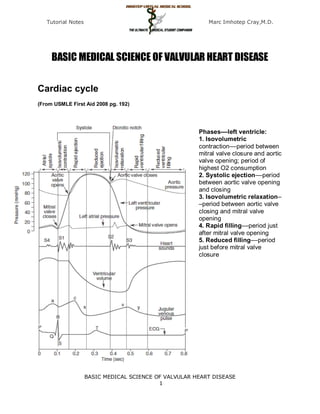
  • 1. Tutorial Notes Marc Imhotep Cray,M.D. BASIC MEDICAL SCIENCE OF VALVULAR HEART DISEASE Cardiac cycle (From USMLE First Aid 2008 pg. 192) Phases––left ventricle: 1. Isovolumetric contraction––period between mitral valve closure and aortic valve opening; period of highest O2 consumption 2. Systolic ejection––period between aortic valve opening and closing 3. Isovolumetric relaxation– –period between aortic valve closing and mitral valve opening 4. Rapid filling––period just after mitral valve opening 5. Reduced filling––period just before mitral valve closure BASIC MEDICAL SCIENCE OF VALVULAR HEART DISEASE 1
  • 2. Tutorial Notes Marc Imhotep Cray,M.D. Ventricular Pressure-Volume Loop Changes in Valve Disease Phases––left ventricle: 1. Isovolumetric contraction––period between mitral Cardiac valve disease valve closure and aortic valve opening; period of significantly alters ventricular highest O2 consumption pressure and volume 2. Systolic ejection––period between aortic valve relationships during the cardiac opening and closing cycle. A convenient way to analyze cardiac pressure and 3. Isovolumetric relaxation––period between aortic volume changes is by using valve closing and mitral valve opening ventricular pressure-volume 4. Rapid filling––period just after mitral valve loops. The links below will opening illustrate the pressure-volume 5. Reduced filling––period just before mitral valve changes that occur with the closure following valve defects:  Mitral stenosis  Aortic stenosis  Mitral regurgitation  Aortic regurgitation BASIC MEDICAL SCIENCE OF VALVULAR HEART DISEASE 2
  • 3. Tutorial Notes Marc Imhotep Cray,M.D. Cardiac Valve Disease Tutorial Aortic valve stenosis http://en.wikipedia.org/wiki/Aortic_valve_stenosis Aortic valve stenosis (AS) is a valvular heart disease caused by the incomplete opening of the aortic valve. The aortic valve controls the direction of blood flow from the left ventricle to the aorta. When in good working order, the aortic valve does not impede the flow of blood between these two spaces. Under some circumstances, the aortic valve becomes narrower than normal, impeding the flow of blood. This is known as aortic valve stenosis, or aortic stenosis, often abbreviated as AS. An aortic valve that, due to rheumatic heart disease, has a severe stenosis (centre of image). The pulmonary trunk is seen at the lower right (of the image). The proximal portion of right coronary artery and its ostium can be seen at the lower left (of the image). The proximal left main coronary artery and its ostium are seen on the right (of the image). Autopsy specimen. BASIC MEDICAL SCIENCE OF VALVULAR HEART DISEASE 3
  • 4. Tutorial Notes Marc Imhotep Cray,M.D. Simultaneous left ventricular and aortic pressure tracings demonstrate a pressure gradient between the left ventricle and aorta, suggesting aortic stenosis. The left ventricle generates higher pressures than what is transmitted to the aorta. The pressure gradient, caused by aortic stenosis, is represented by the green shaded area. (AO = ascending aorta; LV = left ventricle; ECG = electrocardiogram.) Symptoms of aortic stenosis Congestive heart failure Syncope Angina BMS TUTORIAL/ Cardiovascular Physiology Concepts Cardiovascular Physiology Concepts Cardiac Valve Disease Pathophysiology of valve disease CLINICAL READ/ Emedicine Aortic Stenosis BASIC MEDICAL SCIENCE OF VALVULAR HEART DISEASE 4
  • 5. Tutorial Notes Marc Imhotep Cray,M.D. Aortic insufficiency http://en.wikipedia.org/wiki/Aortic_insufficiency Aortic insufficiency (AI), also known as aortic regurgitation (AR), is the leaking of the aortic valve of the heart that causes blood to flow in the reverse direction during ventricular diastole, from the aorta into the left ventricle. Aortic insufficiency can be due to abnormalities of either the aortic valve or the aortic root (the beginning of the aorta). BEFORE SURGICAL REPAIR AFTER SURGICAL REPAIR Pathophysiology In aortic insufficiency, when the pressure in the left ventricle falls below the pressure in the aorta, the aortic valve is not able to completely close. This causes a leaking of blood from the aorta into the left ventricle. This means that some of the blood that was already ejected from the heart is regurgitating back into the heart. The percentage of blood that regurgitates back through the aortic valve due to AI is known as the regurgitant fraction. For instance, if an individual with AI has a stroke volume of 100 ml and during ventricular diastole 25 ml regurgitates back through the aortic valve, the regurgitant fraction is 25%. This regurgitant flow causes a decrease in the diastolic blood pressure in the aorta, and therefore an increase in the pulse pressure (systolic pressure - diastolic pressure). Since some of the blood that is ejected during systole regurgitates back into the left ventricle during diastole, there is decreased effective forward flow in AI. BASIC MEDICAL SCIENCE OF VALVULAR HEART DISEASE 5
  • 6. Tutorial Notes Marc Imhotep Cray,M.D. Note that while diastolic blood pressure is diminished and the pulse pressure widens, systolic blood pressure generally remains normal or can even be slightly elevated. This is because sympathetic nervous system and the renin-angiotensin-aldosterone axis of the kidneys compensate for the decreased cardiac output. Catecholamines will increase the heart rate and increase the strength of ventricular contraction, directly increasing cardiac output. Catecholamines will also cause peripheral vasoconstriction, which causes increased systemic vascular resistance and ensures that core organs are adequately perfused. Renin, a proteolytic enzyme, cleaves angiotensinogen to angiotensin I, which is converted to angiotensin II, which is also a potent vasoconstrictor. In the case of chronic aortic insufficiency with resultant cardiac remodeling, heart failure will develop, and it is possible to see systolic pressures diminish. Aortic insufficiency causes both volume overload (elevated preload) and pressure overload (elevated afterload) of the heart. The pressure overload (due to elevated pulse pressure and the systemic effects of neuroendocrine hormones) causes left ventricular hypertrophy (LVH). There is both concentric hypertrophy and excentric hypertrophy in AI. The concentric hypertrophy is due to the increased left ventricular systolic pressures associated with AI, while the excentric hypertrophy is due to volume overload caused by the regurgitant fraction. Hemodynamics The hemodynamic sequelae of AI are dependent on the rate of onset of AI. Acute AI and chronic AI will have different hemodynamics and individuals will have different signs and symptoms. Acute aortic insufficiency In acute AI, as may be seen with acute perforation of the aortic valve due to endocarditis, there will be a sudden increase in the volume of blood in the left ventricle. The ventricle is unable to deal with the sudden change in volume. In terms of the Frank-Starling curve, the end-diastolic volume will be very high, such that further increases in volume result in less and less efficient contraction. The filling pressure of the left ventricle will increase. This causes pressure in the left atrium to rise, and the individual will develop pulmonary edema. Severe acute aortic insufficiency is considered a medical emergency. There is a high mortality rate if the individual does not undergo immediate surgery for aortic valve replacement. If the acute AI is due to aortic valve endocarditis, there is a risk that the new valve may become seeded with bacteria. However, this risk is small. Acute AI usually presents as florid congestive heart failure, and will not have any of the signs associated with chronic AI since the left ventricle had not yet developed the eccentric hypertrophy and dilatation that allow an increased stroke volume, which in turn cause bounding peripheral pulses. On auscultation, there may be a short diastolic murmur and a soft S1. S1 is soft because the elevated filling pressures close the mitral valve in diastole (rather than the mitral valve being closed at the beginning of systole). BASIC MEDICAL SCIENCE OF VALVULAR HEART DISEASE 6
  • 7. Tutorial Notes Marc Imhotep Cray,M.D. Chronic aortic insufficiency If the individual survives the initial hemodynamic derailment that acute AI presents as, the left ventricle adapts by eccentric hypertrophy and dilatation of the left ventricle, and the volume overload is compensated for. The left ventricular filling pressures will revert to normal and the individual will no longer have overt heart failure. In this compensated phase, the individual may be totally asymptomatic and may have normal exercise tolerance. Eventually (typically after a latency period) the left ventricle will become decompensated, and filling pressures will increase. While most individuals would complain of symptoms of congestive heart failure to their physicians, some enter this decompensated phase asymptomatically. Proper treatment for AI involves aortic valve replacement prior to this decompensation phase. BMS TUTORIAL/ Cardiovascular Physiology Concepts Cardiac Valve Disease Pathophysiology of valve disease CLINICAL READ/eMEDICINE Aortic Regurgitation Mitral stenosis http://en.wikipedia.org/wiki/Mitral_valve_stenosis Mitral stenosis is a valvular heart disease characterized by the narrowing of the orifice of the mitral valve of the heart. BASIC MEDICAL SCIENCE OF VALVULAR HEART DISEASE 7
  • 8. Tutorial Notes Marc Imhotep Cray,M.D. Mitral stenosis with marked thickening of the leaflets and left atrial hypertrophy. Superior view. Autopsy preparation. Rheumatic heart disease at autopsy with characteristic findings (thickened mitral valve, thickened chordae tendineae, hypertrophied left ventricular myocardium). BASIC MEDICAL SCIENCE OF VALVULAR HEART DISEASE 8
  • 9. Tutorial Notes Marc Imhotep Cray,M.D. Intracardiac pressure measurements in an individual with severe mitral stenosis. Pressure tracings in the left atrium (LA) and the left ventricle (LV) in an individual with severe mitral stenosis. Blue areas represent the diastolic pressure gradient due to the stenotic valve. Pathophysiology The normal area of the mitral valve orifice is about 4 to 6 cm2. Under normal conditions, a normal mitral valve will not impede the flow of blood from the left atrium to the left ventricle during (ventricular) diastole, and the pressures in the left atrium and the left ventricle during ventricular diastole will be equal. The result is that the left ventricle gets filled with blood during early ventricular diastole, with only a small portion of extra blood contributed by contraction of the left atrium (the "atrial kick") during late ventricular diastole. When the mitral valve area goes below 2 cm2, the valve causes an impediment to the flow of blood into the left ventricle, creating a pressure gradient across the mitral valve. This gradient may be increased by increases in the heart rate or cardiac output. As the gradient across the mitral valve increases, the amount of time necessary to fill the left ventricle with blood increases. Eventually, the left ventricle requires the atrial kick to fill with blood. As the heart rate increases, the amount of time that the ventricle is in diastole and can fill up with blood (called the diastolic filling period) decreases. When the heart rate goes above a certain point, the diastolic filling period is insufficient to fill the ventricle with blood and pressure builds up in the left atrium, leading to pulmonary congestion. When the mitral valve area goes less than 1 cm2, there will be an increase in the left atrial pressures (required to push blood through the stenotic valve). Since the normal left ventricular diastolic pressures is about 5 mmHg, a pressure gradient across the mitral valve of 20 mmHg due to severe mitral stenosis will cause a left atrial pressure of about 25 mmHg. This left atrial pressure is transmitted to the pulmonary vasculature and causes pulmonary hypertension. Pulmonary capillary pressures in this level cause an imbalance between the hydrostatic pressure and the oncotic pressure, leading to extravasation of fluid from the vascular tree and pooling of fluid in the lungs (congestive heart failure causing pulmonary edema). The constant pressure overload of the left atrium will cause the left atrium to increase in size. As the left atrium increases in size, it becomes more prone to develop atrial fibrillation. When atrial fibrillation develops, the atrial kick is lost (since it is due to the normal atrial contraction). In individuals with severe mitral stenosis, the left ventricular filling is dependent on the atrial kick. The loss of the atrial kick due to atrial fibrillation can cause a precipitous decrease in cardiac output and sudden congestive heart failure. Patients with mitral stenosis prompts a series of hemodynamic changes that frequently cause deterioration of the patient's clinical status. A reduction in cardiac output, associated with acceleration of heart rate and shortening of the diastolic time, frequently leads to congestive heart failure. In addition, when AF sets in, systemic embolization becomes a real danger. BMS TUTORIAL/ Cardiovascular Physiology Concepts Cardiac Valve Disease Pathophysiology of valve disease CLINICAL READ/ eMEDICINE BASIC MEDICAL SCIENCE OF VALVULAR HEART DISEASE 9
  • 10. Tutorial Notes Marc Imhotep Cray,M.D. Mitral Stenosis Mitral regurgitation http://en.wikipedia.org/wiki/Mitral_insufficiency Mitral regurgitation (MR), mitral insufficiency or mitral incompetence is a disorder of the heart in which the mitral valve does not close properly when the heart pumps out blood. It is the abnormal leaking of blood from the left ventricle, through the mitral valve, and into the left atrium, when the left ventricle contracts, i.e. there is regurgitation of blood back into the left atrium. MR is the most common form of valvular heart disease. An illustration of mitral annular calcification, an aging phenomenon which can cause mitral regurgitation. It can also be associated with calcifications of aortic cusps causing stenosis and extension of calcification into cardiac skeleton causing heart block. Acute phase Acute mitral regurgitation (as may occur due to the sudden rupture of a chordae tendineae or papillary muscle) causes a sudden volume overload of both the left atrium and the left ventricle. The left ventricle develops volume overload because with every contraction it now has to pump out not only the volume of blood that goes into the aorta (the forward cardiac output or forward stroke volume), but also the blood that regurgitates into the left atrium (the regurgitant volume). The combination of the forward stroke volume and the regurgitant volume is known as the total stroke volume of the left ventricle. BASIC MEDICAL SCIENCE OF VALVULAR HEART DISEASE 10
  • 11. Tutorial Notes Marc Imhotep Cray,M.D. In the acute setting, the stroke volume of the left ventricle is increased (increased ejection fraction), this happens because of more complete emptying of heart. However, as it progresses the LV volume increases and the contractile function deteriorates and thus leading to dysfunctional LV and a decrease in ejection fraction.The mechanism by which the total stroke volume is increased is known as the Frank-Starling mechanism. The regurgitant volume causes a volume overload and a pressure overload of the left atrium. The increased pressures in the left atrium inhibit drainage of blood from the lungs via the pulmonary veins. This causes pulmonary congestion. Chronic phase Compensated If the mitral regurgitation develops slowly over months to years or if the acute phase can be managed with medical therapy, the individual will enter the chronic compensated phase of the disease. In this phase, the left ventricle develops eccentric hypertrophy in order to better manage the larger than normal stroke volume. The eccentric hypertrophy and the increased diastolic volume combine to increase the stroke volume (to levels well above normal) so that the forward stroke volume (forward cardiac output) approaches the normal levels. In the left atrium, the volume overload causes enlargement of the chamber of the left atrium, allowing the filling pressure in the left atrium to decrease. This improves the drainage from the pulmonary veins, and signs and symptoms of pulmonary congestion will decrease. These changes in the left ventricle and left atrium improve the low forward cardiac output state and the pulmonary congestion that occur in the acute phase of the disease. Individuals in the chronic compensated phase may be asymptomatic and have normal exercise tolerances. Decompensated An individual may be in the compensated phase of mitral regurgitation for years, but will eventually develop left ventricular dysfunction, the hallmark for the chronic decompensated phase of mitral regurgitation. It is currently unclear what causes an individual to enter the decompensated phase of this disease. However, the decompensated phase is characterized by calcium overload within the cardiac myocytes. In this phase, the ventricular myocardium is no longer able to contract adequately to compensate for the volume overload of mitral regurgitation, and the stroke volume of the left ventricle will decrease. The decreased stroke volume causes a decreased forward cardiac output and an increase in the end-systolic volume. The increased end-systolic volume translates to increased filling pressures of the ventricular and increased pulmonary venous congestion. The individual may again have symptoms of congestive heart failure. The left ventricle begins to dilate during this phase. This causes a dilatation of the mitral valve annulus, which may worsen the degree of mitral regurgitation. The dilated left ventricle causes an increase in the wall stress of the cardiac chamber as well. BASIC MEDICAL SCIENCE OF VALVULAR HEART DISEASE 11
  • 12. Tutorial Notes Marc Imhotep Cray,M.D. Demonstrations: Heart Sounds & Murmurs While the ejection fraction is less in the chronic decompensated phase http://depts.washington.edu/p than in the acute phase or the chronic compensated phase of mitral hysdx/heart/demo.html regurgitation, it may still be in the normal range (ie: > 50 percent), and Description Sounds may not decrease until late in the disease course. A decreased ejection fraction in an individual with mitral regurgitation and no other cardiac Normal abnormality should alert the physician that the disease may be in its heart sounds decompensated phase. Murmurs Audio examples Aortic stenosis ( early) & AS- late Mitral regurgitation Pulmonic stenosis Aortic insufficiency Mitral stenosis Benign murmur Atrial septal defect Ventricular septal defect Patent ductus arteriosus Extra heart Audio examples sounds Split S2 S3 BASIC MEDICAL SCIENCE OF VALVULAR HEART DISEASE 12