
Recomendados
Recomendados
Más contenido relacionado
La actualidad más candente
La actualidad más candente (20)
Similar a Pyramid of ANC care
Similar a Pyramid of ANC care (20)
Más de Sameer Dikshit
Último
Último (20)
Pyramid of ANC care
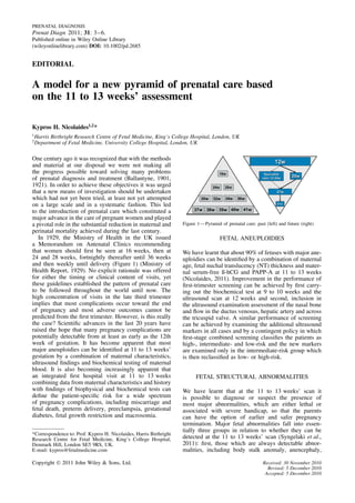
- 1. PRENATAL DIAGNOSIS Prenat Diagn 2011; 31: 3–6. Published online in Wiley Online Library (wileyonlinelibrary.com) DOI: 10.1002/pd.2685 EDITORIAL A model for a new pyramid of prenatal care based on the 11 to 13 weeks’ assessment Kypros H. Nicolaides1,2 * 1 Harris Birthright Research Centre of Fetal Medicine, King’s College Hospital, London, UK 2 Department of Fetal Medicine, University College Hospital, London, UK One century ago it was recognized that with the methods and material at our disposal we were not making all the progress possible toward solving many problems of prenatal diagnosis and treatment (Ballantyne, 1901, 1921). In order to achieve these objectives it was urged that a new means of investigation should be undertaken which had not yet been tried, at least not yet attempted on a large scale and in a systematic fashion. This led to the introduction of prenatal care which constituted a major advance in the care of pregnant women and played a pivotal role in the substantial reduction in maternal and Figure 1—Pyramid of prenatal care: past (left) and future (right) perinatal mortality achieved during the last century. In 1929, the Ministry of Health in the UK issued FETAL ANEUPLOIDIES a Memorandum on Antenatal Clinics recommending that women should first be seen at 16 weeks, then at We have learnt that about 90% of fetuses with major ane- 24 and 28 weeks, fortnightly thereafter until 36 weeks uploidies can be identified by a combination of maternal and then weekly until delivery (Figure 1) (Ministry of age, fetal nuchal translucency (NT) thickness and mater- Health Report, 1929). No explicit rationale was offered nal serum-free ß-hCG and PAPP-A at 11 to 13 weeks for either the timing or clinical content of visits, yet (Nicolaides, 2011). Improvement in the performance of these guidelines established the pattern of prenatal care first-trimester screening can be achieved by first carry- to be followed throughout the world until now. The ing out the biochemical test at 9 to 10 weeks and the high concentration of visits in the late third trimester ultrasound scan at 12 weeks and second, inclusion in implies that most complications occur toward the end the ultrasound examination assessment of the nasal bone of pregnancy and most adverse outcomes cannot be and flow in the ductus venosus, hepatic artery and across predicted from the first trimester. However, is this really the tricuspid valve. A similar performance of screening the case? Scientific advances in the last 20 years have can be achieved by examining the additional ultrasound raised the hope that many pregnancy complications are markers in all cases and by a contingent policy in which potentially detectable from at least as early as the 12th first-stage combined screening classifies the patients as week of gestation. It has become apparent that most high-, intermediate- and low-risk and the new markers major aneuploidies can be identified at 11 to 13 weeks’ are examined only in the intermediate-risk group which gestation by a combination of maternal characteristics, is then reclassified as low- or high-risk. ultrasound findings and biochemical testing of maternal blood. It is also becoming increasingly apparent that an integrated first hospital visit at 11 to 13 weeks FETAL STRUCTURAL ABNORMALITIES combining data from maternal characteristics and history with findings of biophysical and biochemical tests can We have learnt that at the 11 to 13 weeks’ scan it define the patient-specific risk for a wide spectrum is possible to diagnose or suspect the presence of of pregnancy complications, including miscarriage and most major abnormalities, which are either lethal or fetal death, preterm delivery, preeclampsia, gestational associated with severe handicap, so that the parents diabetes, fetal growth restriction and macrosomia. can have the option of earlier and safer pregnancy termination. Major fetal abnormalities fall into essen- tially three groups in relation to whether they can be *Correspondence to: Prof. Kypros H. Nicolaides, Harris Birthright Research Centre for Fetal Medicine, King’s College Hospital, detected at the 11 to 13 weeks’ scan (Syngelaki et al., Denmark Hill, London SE5 9RS, UK. 2011): first, those which are always detectable abnor- E-mail: kypros@fetalmedicine.com malities, including body stalk anomaly, anencephaly, Copyright 2011 John Wiley & Sons, Ltd. Received: 30 November 2010 Revised: 5 December 2010 Accepted: 5 December 2010
- 2. 4 EDITORIAL alobar holoprosencephaly, exomphalos, gastroschisis sonographic measurement of cervical length at 11 to and megacystis and second, undetectable abnormalities 13 weeks which is inversely related to the likelihood because sonographic signs are only manifest during the for subsequent spontaneous early delivery (Greco et al., second or third trimester of pregnancy, including some 2011). At present there are no other useful biophysical or brain abnormalities, such as microcephaly, hypoplasia of biochemical markers of spontaneous early delivery (Beta the cerebellum or vermis, hydrocephalus and agenesis et al., 2011). Future research will determine whether of the corpus callosum, achondroplasia, echogenic lung early intervention with such measures as prophylactic lesions, many renal anomalies and bowel obstruction. A use of progesterone or cervical cerclage will be effective third group includes abnormalities that are potentially in reducing spontaneous preterm delivery. detectable depending on first the objectives set for such a scan and consequently the time allocated for the fetal examination, the expertise of the sonographer and the quality of the equipment used and second, the pres- PREECLAMPSIA ence of an easily detectable marker for an underlying abnormality. Good examples of such markers in the first trimester include high NT in some fetuses with lethal We have learnt that in preeclampsia, which is a major skeletal dysplasias (Khalil et al., 2011) and diaphrag- cause of maternal and perinatal morbidity and mor- matic hernia, high NT and abnormal flow in the ductus tality, both the degree of impaired placentation and venosus and across the tricuspid valve in major cardiac the incidence of adverse fetal and maternal short-term defects (Chelemen et al., 2011) and increase in brain and long-term consequences are inversely related to stem diameter with decrease in the diameter of the fourth the gestational age at the onset of the disease. Con- ventricle-cisterna magna complex in open spina bifida sequently, in screening for preeclampsia the condition (Lachmann et al., 2011). should be subdivided according to gestational age at delivery. Algorithms which combine maternal charac- teristics, mean arterial pressure, uterine artery Doppler MISCARRIAGE AND STILLBIRTH and biochemical tests at 11 to 13 weeks could potentially identify about 90, 80 and 60% of pregnancies that sub- sequently develop early (before 34 weeks), intermediate We have learnt that the rates of miscarriage and stillbirth (34–37 weeks) and late (after 37 weeks) preeclampsia, after demonstration of a live fetus at 11 to 13 weeks are for a false positive rate of 5% (Akolekar et al., 2011b). about 1 and 0.4%, respectively. Increased risk for mis- Further investigations will determine whether in the carriage and stillbirth are associated with certain mater- high-risk group pharmacological interventions, such as nal characteristics, including increasing maternal age low-dose aspirin, starting from the first trimester could and maternal weight, previous miscarriage or stillbirth improve placentation and reduce the prevalence of the and African racial origin. Miscarriage and stillbirth are disease. also associated with abnormal results of first-trimester screening for aneuploidies, including increased fetal NT thickness, reversed a-wave in the fetal ductus veno- sus and low maternal serum PAPP-A (Akolekar et al., 2011a). At present there is no useful intervention for GESTATIONAL DIABETES MELLITUS avoidance of miscarriage and so the use of this algo- rithm in clinical practice is debatable (van Ravenswaaij We have learnt that in gestational diabetes mellitus et al., 2011). In contrast, early identification of the group (GDM), which is associated with increased risk of mater- at high-risk for stillbirth could lead to a reduction of this nal and perinatal short-term and long-term complica- complication through closer monitoring of fetal growth tions, the performance of traditional screening at the and wellbeing and appropriate timing of delivery. end of the second trimester by a series of independent maternal characteristics is poor with a detection rate of about 60%, at a false positive rate of 30 to 40% (Waugh PRETERM DELIVERY et al., 2007). Algorithms which combine maternal char- acteristics and maternal serum levels of adiponectin, an We have learnt that preterm birth is the leading cause adipocyte-derived polypeptide, and sex hormone binding of perinatal death and handicap in children and the globulin, a liver-derived glycoprotein, at 11 to 13 weeks vast majority of mortality and morbidity relates to early could potentially identify about 75% of pregnancies that delivery before 34 weeks which occurs in about 2% subsequently develop GDM, for a false positive rate of of singleton pregnancies. In two-thirds of the cases 20% (Nanda et al., 2011). Additionally, the diagnosis of this is due to spontaneous onset of labor or preterm GDM can be made in the first trimester by appropriate prelabor rupture of membranes and in the other one- adjustments to the traditional criteria of the oral glucose third it is iatrogenic, mainly due to preeclampsia. The tolerance test (Plasencia et al., 2011). Future research patient-specific risk for spontaneous delivery before will determine whether in the high-risk group appro- 34 weeks can be determined by an algorithm combining priate dietary advice and pharmacological interventions, maternal characteristics and obstetric history (Beta et al., such as metformin, could reduce the incidence of GDM 2011). This a priori risk can be modified by the and associated fetal macrosomia. Copyright 2011 John Wiley & Sons, Ltd. Prenat Diagn 2011; 31: 3–6. DOI: 10.1002/pd
- 3. EDITORIAL 5 SMALL FOR GESTATIONAL AGE FETUSES patient-specific risk based on maternal age is modi- fied by the sonographic findings and results of bio- chemical testing both in the first and second trimesters We have learnt that small for gestational age (SGA) of pregnancy. The new approach will adhere to the fetuses may be constitutionally small or growth restricted teachings of Hippocrates, that we should learn the past due to impaired placentation, genetic disease or and research the present to predict the future, and the environmental damage. In the growth-restricted group pronouncements of Galileo Galilei, that the language of the risks of perinatal death and handicap are substantially God is mathematics and that we should measure every- increased but these risks can be reduced if the condition thing that is measurable and make measurable every- is detected prenatally allowing appropriate monitoring thing that is not so. and delivery. Algorithms which combine maternal char- At 11 to 13 weeks the great majority of women acteristics, mean arterial pressure, uterine artery Doppler would be classified as being at low-risk for pregnancy and the measurement of various placental products in complications and a small proportion of women would maternal blood at 11 to 13 weeks could potentially be selected as being at high-risk (Figure 1). In the identify, at a false positive rate of 10%, about 75% low-risk group the number of medical visits can be of pregnancies without preeclampsia delivering SGA substantially reduced to perhaps three. One visit at neonates before 37 weeks and 45% of those delivering at around 20 weeks will re-evaluate fetal anatomy and term (Karagiannis et al., 2011). Since the proportion of growth and reassess risk for such complications as growth restricted to constitutional small fetuses is higher preeclampsia and preterm delivery. Another visit at in the preterm rather than term SGA, it is likely that the 37 weeks will assess maternal and fetal wellbeing and early biophysical and biochemical markers identify the determine the best time and method of delivery and this growth-restricted subgroup among the SGA. will be repeated at 41 weeks for the few that remain pregnant at this stage. The high-risk group can have close surveillance in specialist clinics both in terms of FETAL MACROSOMIA the investigations to be performed and the personnel involved in the provision of care. In each of these visits their risk will be reassessed and they will either remain We have learnt that fetal macrosomia is associated with high-risk or they will become low-risk in which case the increased risks for the mother, including cesarean section intensity of their care can be reduced. and trauma to the birth canal, and for the baby, including Future research will inevitably expand the number of shoulder dystocia and consequent brachial plexus or conditions that can be identified in early pregnancy and facial nerve injuries, fractures of the humerus or clavicle define genetic markers of disease that will improve the and birth asphyxia. Screening for macrosomia (birth accuracy of the a priori risk based on maternal charac- weight above the 90th centile for gestational age at teristics and medical history. Similarly, new biophysical delivery) by a combination of maternal characteristics and biochemical markers will be described that may and obstetric history with fetal NT and maternal serum- replace some of the current ones and modify the value free ß-hCG and PAPP-A at 11 to 13 weeks could of others. As the years pass, it will become necessary to potentially identify, at a false positive rate of 10%, re-evaluate and improve the timing and content of each about 35% of women who deliver macrosomic neonates visit and the likelihood ratios for each test. Early iden- (Poon et al., 2011). Future research would identify tification of high-risk groups will also stimulate further new biophysical and biochemical markers which could research that will define the best protocol for their fol- improve the performance of screening and determine low up and development of strategies for the prevention the extent to which early identification of the high-risk of disorders of pregnancy or their adverse consequences. group can improve prenatal surveillance and prevention It is likely that the new challenge for improvement of of macrosomia itself or the intrapartum complications pregnancy outcome will be met by inverting the pyramid related to the condition. of prenatal care (Figure 1) to introduce on a large scale and in a systematic fashion a new model of prenatal care which will be based on the results of a comprehensive THE NEW PYRAMID OF CARE assessment at 11 to 13 weeks. Early estimation of patient-specific risks for pregnancy REFERENCES complications would improve pregnancy outcome by shifting prenatal care from a series of routine visits Akolekar R, Bower S, Flack N, Bilardo CM, Nicolaides KH. 2011a. Prediction to a more individualized patient and disease-specific of miscarriage and stillbirth at 11–13 weeks and the contribution of chorionic approach both in terms of the schedule and content of villus sampling. Prenat Diagn 31(1): 38–45. such visits. Each visit would have a predefined objective Akolekar R, Syngelaki A, Sarquis R, Zvanca M, Nicolaides KH. 2011b. and the findings will generate likelihood ratios that can Prediction of early, intermediate and late pre-eclampsia from maternal factors, be used to modify the individual patient and disease- biophysical and biochemical markers at 11–13 weeks. Prenat Diagn 31(1): 66–74. specific estimated risk from the initial assessment at Ballantyne JW. 1901. A plea for a pro-maternity hospital. BMJ 2101: 813–814. 11 to 13 weeks. Such sequential screening is now well Ballantyne JW. 1921. The maternity hospital, with its antenatal and neo-natal established in screening for aneuploidies whereby the departments. BMJ 3137: 221–224. Copyright 2011 John Wiley & Sons, Ltd. Prenat Diagn 2011; 31: 3–6. DOI: 10.1002/pd
- 4. 6 EDITORIAL Beta J, Akolekar R, Ventura W, Syngelaki A, Nicolaides KH. 2011. Prediction Nanda S, Savvidou M, Syngelaki A, Akolekar R, Nicolaides KH. 2011. of spontaneous preterm delivery from maternal factors, obstetric history Prediction of gestational diabetes mellitus by maternal factors and biomarkers and placental perfusion and function at 11–13 weeks. Prenat Diagn 31(1): at 11 to 13 weeks. Prenat Diagn 31(2): In press. 75–83. Nicolaides KH. 2011. Screening for fetal aneuploidies at 11 to 13 weeks. Prenat Chelemen T, Syngelaki A, Maiz M, Allan L, Nicolaides KH. 2011. Contri- Diagn 31(1): 7–15. bution of ductus venosus Doppler in first trimester screening for major Plasencia W, Garcia R, Pereira S, Akolekar R, Nicolaides KH. 2011. Criteria cardiac defects. Fetal Diagn Ther. [Epub ahead of print]. DOI: for screening and diagnosis of gestational diabetes mellitus in the first- 10.1159/000322138. trimester of pregnancy. Fetal Diagn Ther. (In press). Greco E, Lange A, Ushakov F, Rodriguez Calvo J, Nicolaides KH. 2011. Poon LCY, Karagiannis G, Stratieva V, Syngelaki A, Nicolaides KH. 2011. Prediction of spontaneous preterm delivery from endocervical length at 11 First-trimester prediction of macrosomia. Fetal Diagn Ther. [Epub ahead of to 13 weeks. Prenat Diagn 31(1): 84–89. print]. DOI: 10.1159/000318565. Karagiannis G, Akolekar R, Sarquis R, Wright D, Nicolaides KH. 2011. Syngelaki A, Chelemen T, Dagklis T, Allan L, Nicolaides KH. 2011. Chal- Prediction of small for gestation neonates from biophysical and biochemical lenges in the diagnosis of fetal non-chromosomal abnormalities at markers at 11–13 weeks. Fetal Diagn Ther. [Epub ahead of print]. DOI: 11–13 weeks. Prenat Diagn 31(1): 90–102. 10.1159/000321694. van Ravenswaaij R, Tesselaar-van der Goot M, de Wolf S, van Leeuwen- Khalil A, Pajkrt E, Chitty LS. 2011. Early prenatal diagnosis of skeletal Spruijt M, Visser GHA, Schielen PCJI. 2011. First-trimester serum PAPP-A anomalies. Prenat Diagn 31(1): 115–124. and fβ-hCG concentrations and other maternal characteristics to establish Lachmann R, Chaoui R, Moratalla J, Picciarelli G, Nicolaides KH. 2011. logistic regression-based predictive rules for adverse pregnancy outcome. Posterior brain in fetuses with spina bifida at 11 to 13 weeks. Prenat Diagn Prenat Diagn 31(1): 50–57. 31(1): 103–106. Waugh N, Scotland G, McNamee P, et al. 2007. Screening for type 2 diabetes: Ministry of Health Report. 1929. Memorandum on antenatal clinics: their literature review and economic modelling. Health Technol Assess 11(1–125). conduct and scope. His Majesty’s Stationery Office, 1930. London. Copyright 2011 John Wiley & Sons, Ltd. Prenat Diagn 2011; 31: 3–6. DOI: 10.1002/pd