Artries, veins and lymphatics
•Descargar como PPT, PDF•
11 recomendaciones•2,588 vistas

Arteries are blood vessels that carry blood away from the heart. This blood is normally oxygenated, exceptions made for the pulmonary and umbilical arteries

Recomendados
Más contenido relacionado
La actualidad más candente
La actualidad más candente (20)
Similar a Artries, veins and lymphatics
Similar a Artries, veins and lymphatics (20)
Más de Shady Negm
Último
Último (20)
Artries, veins and lymphatics
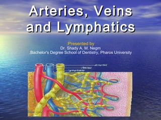
- 1. Arteries, Veins and Lymphatics :Presented by Dr. Shady A. M. Negm .Bachelor's Degree School of Dentistry, Pharos University
- 2. Arteries
- 3. • Arteries are blood vessels that carry blood away from the heart . This blood is normally oxygenated, exceptions made for the pulmonary and umbilical arteries
- 4. Description • Arterial pressure varies between the peak pressure during heart contraction, called the systolic pressure, and the minimum, or diastolic pressure between contractions, when the heart expands and refills. • Arteries carry blood away from the heart. Except for the pulmonary arteries, which carry blood to the lungs for oxygenation, all arteries carry oxygenated blood away from the heart to the tissues that require oxygen.
- 5. Anatomy ( Gross anatomy ) • The arterial system of the human body is divided into systemic arteries, carrying blood from the heart to the whole body, and pulmonary arteries, carrying blood from the heart to the lungs.
- 6. Systemic arteries • Systemic arteries are the arteries of the systemic circulation, which is the part of the cardiovascular system which carries oxygenated blood away from the heart, to the body, and returns deoxygenated blood back to the heart.
- 7. Pulmonary arteries • Pulmonary arteries are the arteries of the pulmonary circulation, which is the portion of the cardiovascular system which carries deoxygenated blood away from the heart, to the lungs, and returns oxygenated blood back to the heart.
- 8. (Anatomy (microscopic anatomy The outermost layer is known as the tunica externa formerly composed of connective tissue. Inside this layer is the tunica media, or media, which is made up of smooth muscle cells and elastic tissue. The innermost layer, which is in direct contact with the flow of blood is the tunica intima, commonly called the intima. This layer is made up of mainly endothelial cells. The hollow internal cavity in which the blood flows is called the lumen
- 10. The Aorta The aorta is the root systemic artery. It receives blood directly from the left ventricle of the heart via the aortic valve. As the aorta branches, and these arteries branch in turn, they become successively smaller in diameter, down to the arteriole. The arterioles supply capillaries which in turn empty into venules. The very first branches off of the aorta are the coronary arteries, which supply blood to the heart muscle itself. These are followed by the branches off the aortic arch, namely the brachiocephalic artery, the left common carotid and the left subclavian arteries.
- 11. Arterioles • Arterioles, the smallest of the true arteries, help regulate blood pressure by the variable contraction of the smooth muscle of their walls, and deliver blood to the capillaries.
- 12. Capillaries • The capillaries are where all of the important exchanges happen in the circulatory system. The capillaries are a single cell in diameter to aid fast and easy diffusion of gases, sugars and other nutrients to surrounding tissues.
- 13. Pathology
- 14. Blood pressure • Systemic arterial pressures, are generated by the forceful contractions of the heart's left ventricle. • Healthy resting arterial pressures, are relatively low, mean systemic pressures typically being under 100 mmHg, about 1.8 lbf/in², above surrounding atmospheric pressure (about 760 mmHg). • Over time, elevated arterial blood sugar, lipoprotein cholesterol, and pressure, smoking, and other factors are all involved in damaging both the endothelium and walls of the arteries, resulting in atherosclerosis.
- 15. Atheroma • An atheroma or plaque in the artery wall is a build up of cell debris, that contain lipids (cholesterol and fatty acids), calcium and a variable amount of fibrous connective tissue
- 16. Veins
- 17. Vein • veins (from the Latin vena) are blood vessels that carry blood towards the heart . Most veins carry deoxygenated blood from the tissues back to the heart; exceptions are the pulmonary and umbilical veins, both of which carry oxygenated blood to the heart. Veins differ from arteries in structure and function; for example, arteries are more muscular than veins, veins contain valves, and they carry blood away from the heart.
- 18. Anatomy • In general, veins function to return deoxygenated blood to the heart, and are essentially tubes that collapse when their lumens are not filled with blood. The thick outermost layer of a vein is made of connective tissue, called tunica adventitia or tunica externa. Deeper are bands of smooth muscle called tunica media, which are, in general, thin, as veins do not function primarily in a contractile manner. The interior is lined with endothelial cells called tunica intima. Most veins have one-way flaps called venous valves that prevent blood from flowing back and pooling in the lower extremities due to the effects of gravity.
- 19. Notable veins and vein systems • The pulmonary veins carry relatively oxygenated blood from the lungs to the heart. The superior and inferior venae cavae carry relatively deoxygenated blood from the upper and lower systemic circulations, respectively. • A portal venous system is a series of veins or venules that directly connect two capillary beds. Examples of such systems include the hepatic portal vein and hypophyseal portal system • The Thebesian veins within the myocardium of the heart are valveless veins that drain directly into the chambers of the heart. The coronary veins all empty into the coronary sinus which
- 20. Classification • Veins are classified in a number of ways, including superficial vs. deep, pulmonary vs. systemic, and large vs. small.
- 21. • Superficial veins – Superficial veins are those whose course is close to the surface of the body, and have no corresponding arteries. • Deep veins – Deep veins are deeper in the body and have corresponding arteries. • Pulmonary veins – The pulmonary veins are a set of veins that deliver oxygenated blood from the lungs to the heart. • Systemic veins – Systemic veins drain the tissues of the body
- 23. Phlebology • Phlebology is the medical discipline that involves the diagnosis and treatment of disorders of venous origin. Diagnostic techniques used include the history and physical examination, venous imaging techniques and laboratory evaluation related to venous thromboembolism. The American Medical Association has added phlebology to their list of self-designated practice specialties. A medical specialist in Phlebology is termed a Phlebologist. A related image is called a phlebography
- 24. Venous diseases • Deep vein thrombosis : Deep-vein thrombosis is a condition in which a blood clot forms in a deep vein, which can lead to pulmonary embolism and chronic venous insufficiency. • Thrombophlebitis : Thrombophlebitis is an inflammatory condition of the veins related to
- 25. Lymph vessel
- 26. • lymph vessels are thin walled, valved structures that carry lymph. As part of the lymphatic system, lymph vessels are complementary to the cardiovascular system. Lymph vessels are lined by endothelial cells, and deep to that have a thin layer of smooth muscles, and adventitia that bind the lymph vessel to the surroundings. Lymph vessels are devoted to propulsion of the lymph from the lymph capillaries, which are mainly concerned with absorption of interstitial fluid from the tissues. Lymph vessel that carries lymph to a lymph node are called the afferent lymph vessel, and one that carries it from a lymph node is called the efferent lymph vessel, from where the lymph may travel to another lymph node or may be returned to a vein, or may travel to a larger lymph duct. Lymph ducts drain the lymph into one of the subclavian veins and thus return it to general
- 27. Function • Lymph vessels act as a reservoir from plasma and other substances including cells that leaked from the vascular system and transport lymph fluid back from the tissues to the circulatory system. Without functioning lymph vessels, lymph cannot be effectively drained and edema typically results.
- 28. General structure of Lymphatics • There is an inner lining of single flattened cells composed of a type of epithelium that is called endothelium, and the cells are called endothelial cells. This layer functions to mechanically transport fluid and since the basement membrane on which it rests is discontinuous; it leaks easily.The next layer is that of smooth muscles that are arranged in a circular fashion around the endothelium, which by shortening (contracting) or relaxing alter the diameter (caliber) of the lumen. The outermost layer is the adventitia that consists of fibrous tissue.
- 29. • The general structure described here is seen only in larger lymphatics; smaller lymphatics have fewer layers. The smallest vessels (lymphatic or lymph capillaries) lack both the muscular layer and the outer adventitia. As they proceed forward and in their course are joined by other capillaries, they grow larger and first take on an adventitia, and then smooth muscles.
- 30. Lymph vessels • The lymph capillaries drain the lymph to larger contractile lymphatics, which have valves as well as smooth muscle walls. These are called the collecting lymphatics.As the collecting lymph vessel accumulates lymph from more and more lymph capillaries in its course, it becomes larger and is called the afferent lymph vessel as it enters a lymph node. Here the lymph percolates through the lymph node tissue and is removed by the efferent lymph vessel.
- 31. • An efferent lymph vessel may directly drain into one of the (right or thoracic) lymph ducts, or may empty into another lymph node as its afferent lymph vessel. Both the lymph ducts return the lymph to the blood stream by emptying into the subclavian veins
- 32. Head & Neck
- 33. Circulatory system • Blood circulates from the upper systemic loop originating at the aortic arch, and includes: the brachiocephalic artery, left common carotid and left subclavian artery. The head and neck are emptied of blood by the subclavian vein and jugular vein.
- 34. Blood supply
- 35. • The brachiocephalic artery or trunk is the first and largest artery that branches to form the right common carotid artery and the right subclavian artery. This artery provides blood to the right upper chest, right arm, neck, and head, through a branch called right vertebral artery. The right and left vertebral artery feed into the basilar artery and upward to the Posterior cerebral artery, which provides most of the brain with oxygenated blood. The posterior cerebral artery and the posterior communicating artery are within the circle of Willis .
- 36. • The left common carotid artery divides to form the: internal carotid artery (ICA) and an external carotid artery (ECA). The ICA supplies the brain. The ECA supplies the neck and face. • The left subclavian artery and the right subclavian artery , one on each side of the body form the internal thoracic artery, the vertebral artery, the thyrocervical trunk, and the costocervical trunk. The subclavian becomes the axiliary artery at the lateral border of the first rib. The left subclavian artery also provides blood to the left upper chest and left arm.
- 38. • The Blood-brain barrier (BBB) is semi- permeable membrane that controls the capillary leak potential of the circulatory system. In most parts of the body, the smallest blood vessels, called capillaries, are lined with endothelial cells. Endothelial tissue has small spaces between each individual cell so substances can move readily between the inside and the outside of the vessel. However, in the brain, the endothelial cells fit tightly together to create a tight junction and substances cannot pass out of the bloodstream. Some molecules, such as glucose, are transported out of the blood by active transport.
- 39. Blood return
- 40. • Blood from the brain and neck flows within the cranium via the internal jugular veins, a continuation of the sigmoid sinuses. The right and left external jugular veins drain from the parotid glands, facial muscles, scalp into the subclavian veins. The right and left vertebral veins drain the vertebrae and muscles into the right subclavian vein and into the superior vena cava, into the right atrium of the heart.
- 41. Lymphatic system
- 42. • The lymphatic system drains the head and neck of excess interstitial fluid via lymph vessels or capillaries, equally into the right lymphatic duct and the thoracic duct. • Lymph nodes line the cervical spine and neck regions as well as along the face and jaw. • The tonsils also are lymphatic tissue and help mediate the ingestion of pathogens. • Tonsils in humans include, from superior to inferior: nasopharyngeal tonsils (also known as adenoids), palatine tonsils, and lingual tonsils. • Together this set of lymphatic tissue is called the tonsillar ring or Waldeyer's ring .
- 43. Nervous system
- 44. • The nervous system is composed of a central nervous system (CNS), brain and spinal cord , and the peripheral nervous system (PNS), cranial nerves and spinal nerves. The CNS is located within the dorsal cavity, and the PNS extends through the . The central nervous system provides control and coordination of all eleven body systems and utilizes the endocrine system to form hormone chemical messengers that transport through the blood to influence the activity of individual cells of the body and their associated tissues, organs and systems.
- 45. • The CNS receives sensory (afferent) input from the PNS and directs the flow of information to association neurons (interneurons), located in the grey matter of the spinal cord and brain to create chemical synapse responses which in turn cause the formation of motor (efferent nerve) responses to stimulus. • The CNS is protected by the cranium, vertebral column, meninges, cerebrospinal fluid. The spinal cord, which is an extension of the brain, and brain stem are joined at the base of the cranium at the foramen magnum.
- 46. • The PNS has two subdivisions : A)somatic nervous system (SNS). The SNS is associated with the voluntary control of body movements through the action of skeletal muscles, and also reception of external stimuli. B) the autonomic nervous system (ANS). The ANS is divided into subsystems: the sympathetic nervous system (SNS) and the parasympathetic (PNS) nervous systems. The SNS and PNS often have opposing effects in the same organs or physiological systems, and the ANS is a major factor in maintaining homeostasis.
- 47. References