The Study of Human Heart (PDF version)
•
11 recomendaciones•11,122 vistas
The human heart is about the size of a clenched fist and is located in the chest. It is made of cardiac muscle and is divided into four chambers - two upper atria and two lower ventricles. The heart pumps blood through two circuits: the pulmonary circuit oxygenates blood in the lungs, while the systemic circuit pumps oxygenated blood to the entire body. It contracts over 70 times per minute due to electrical impulses generated by the sinoatrial node that spread through the heart and cause the chambers to contract in the correct sequence, pumping blood out of the heart.

Recomendados
Más contenido relacionado
La actualidad más candente
La actualidad más candente (20)
Destacado
Destacado (20)
Similar a The Study of Human Heart (PDF version)
Similar a The Study of Human Heart (PDF version) (20)
Último
Último (20)
The Study of Human Heart (PDF version)
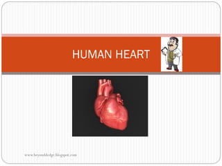
- 2. Human heart which has the size of a clenched fist is a specialised organ that pumps blood to all parts of the body. It is made up of cardiac muscles. It is divided into 2 parts (left and right) and has 4 chambers (2 atria and 2 ventricles). The wall of the atria is slightly thinner than the wall of the ventricles because atria functioned as collection chambers while the ventricles pumps blood away from the heart. The wall of the left ventricle is thicker than the right ventricle because it has to pump blood under high pressure to all parts of the body while the right ventricle only pumps blood to the lungs for re-oxygenated. www.beyonddedge.blogspot.com
- 3. Coronary arteries supply the heart muscles with food (glucose) and oxygen. The heart has valves that only allow blood to flow in one direction: Valves Location Tricuspid valve /Atrioventricular valve Between right atrium and right ventricle Bicuspid valve / Mitral valve Between left atrium and left ventricle Semilunar valve / Pulmonic valve In the wall of pulmonary artery and aorta BioInfo: Chordae tendineae anchor the valve flaps to papillary muscles in the ventricular wall. (Source: How the body works by DR Peter Abraham) www.beyonddedge.blogspot.com
- 5. PUMPING OF THE HEART Cardiac muscle is myogenic which means it contracts and relaxes without the need of receiving stimulation by the nerve impulses. The contraction of the heart is initiated by pacemaker. SINO-ATRIAL NODE (SA Node) Cluster of cells situated within the wall of right atrium. SA Node acts as heart pacemaker by contracting at a faster rate than any other cardiac muscle cells. It generates electrical impulses for each contraction which then spread rapidly to the wall of both atria, causing the contraction of the atria. ATRIOVENTRICULAR NODE (AV Node) AV Node is located within the floor of the right atrium. It passes electrical impulses originated from the SA Node to the wall of ventricles to contract. www.beyonddedge.blogspot.com
- 7. BioInfo: Cardiac muscle is made up of 3 layers: the epicardium (outer layer), the myocardium (middle layer) and the endocardium (inner layer). Myocardial layer responsible for the contraction of the heart. Thus, myocardial layer of the left ventricle is much thicker because the ventricle pumps blood to all parts of the body under high pressure. www.beyonddedge.blogspot.com
- 8. Sequence of heart contraction: The SA Node generated electrical impulses. 2. The electrical impulses spread rapidly to the wall of both atria causing the contraction of the atria. These allow the blood to be pump into the ventricles. 3. The impulses are then relayed to the AV Node down to the bundle of His, Purkinje fibres and the apex of the heart. 4. The electrical impulses spread to the ventricles allowing the ventricular contractions begins from the apex and upwards to pump blood away from the heart. 1. BioInfo: Electrical impulses from the SAN is being delayed by 0.1s upon reaching AVN, thus allowing ventricles to fill up with blood. www.beyonddedge.blogspot.com
- 9. CARDIAC CYCLE (a series of changes within the heart that causes it to pump blood) 1. Blood enters the atria. At the same time, the tricuspid valve and bicuspid valve open as the atrial pressure exceeds the ventricular pressure. The resting phase is called diastole. 2. As diastole ends and the atrial systole begins, more blood is pushed to fill the ventricles. Hence, the both atria contract. 3. During the ventricular systole (contract), the ventricular pressure increase causing the tricuspid valve and bicuspid valve to close and semilunar valve to open. Hence, pushing the blood into the aorta and pulmonary artery. At this time, the atria relax. 4. The ventricles relax (ventricular diastole) and its pressure will soon decrease causing the semilunar valve to close while blood starting to fill the atria. www.beyonddedge.blogspot.com
- 11. ELECTROCARDIOGRAM (ECG) – use to detect the sequence of the heartbeat Component Description P wave Atrial contraction (atrial depolarization) happens and excitation travels through the AV node (atrial excitation) PR interval Delay of electrical impulses from the AVN and depolarization of the atria QRS complex Ventricular contraction occurs (ventricular excitation) T wave Resting period of the ventricle (ventricular repolarization) ST segment (beginning of ventricular repolarization) PR interval www.beyonddedge.blogspot.com