
Recomendados
Recomendados
Más contenido relacionado
La actualidad más candente
La actualidad más candente (20)
Similar a Comparison of 2 Techniques of Subepithelial Connective Tissue Graft in the Treatmen of Gingival Recessions
Similar a Comparison of 2 Techniques of Subepithelial Connective Tissue Graft in the Treatmen of Gingival Recessions (20)
Comparison of 2 Techniques of Subepithelial Connective Tissue Graft in the Treatmen of Gingival Recessions
- 1. 0331_IPC_AAP_553351 11/5/01 4:49 PM Page 1470 Volume 72 • Number 11 Comparison of 2 Techniques of Subepithelial Connective Tissue Graft in the Treatment of Gingival Recessions Giampiero Cordioli,* Cinzia Mortarino,† Andrea Chierico,‡ Maria Gabriella Grusovin,§ and Zeina Majzoub*ሻ Background: The clinical outcome of connective tissue grafts in the treat- ment of gingival recessions has been documented in numerous studies. How- ever, no attempt has been made to correlate the postoperative mucogingival changes with the surgical parameters. The present retrospective clinical study was undertaken to 1) evaluate root coverage and mucogingival changes 1 to 1.5 years following treatment of Miller’s Class I and II recession defects using 2 variants of the subepithelial connective tissue graft (SCTG) procedure, and B ilaminar techniques 2) assess the effect of the surgical parameters on the postoperative gingival have been proposed width. to enhance the pre- Methods: Thirty-one recessions in 10 patients treated with the envelope tech- dictability of gingival graft- nique (E) and 31 recessions in 11 patients treated with coronally positioned flap ing procedures by improv- combined with connective tissue graft (CP) were retrospectively analyzed to ing the blood supply to the evaluate: 1) percentage of root coverage obtained with the 2 procedures and grafted tissues.1-3 In these variations in width of keratinized tissue (KT) 1 to 1.5 years postsurgery, and 2) techniques, a free connec- the effect of the surgical parameters on the postoperative gingival width. tive tissue graft is posi- Results: Results showed a mean root coverage percentage of 89.6 ± 15% tioned over the recession for the E group and 94.7 ± 11.4% for the CP group; the difference between site and covered by a pedi- groups was statistically insignificant (P = 0.1388). Mean KT increased signifi- cle flap. Variations of the cantly from 1.4 ± 1.1 mm presurgery to 4.5 ± 1.1 mm postsurgery for the E bilaminar technique include group while a minor increase in KT was observed in the CP group (2 ± 1.5 mm the use of various types of presurgery versus 2.7 ± 1.6 mm postsurgery). For both treatment groups, the primary flap and different mean postsurgical width of keratinized tissue (POSTKT) was found to be math- techniques for harvesting ematically correlated with the mean presurgical width of keratinized tissue the connective tissue graft.4 (PREKT) and the corono-apical height of the graft that remained exposed (GE) In one variant of the bilam- coronal to the flap margin in the recipient site. inar procedure, the enve- Conclusions: Treatment of human gingival recession defects by the 2 vari- lope technique, a split- ants of SCTG resulted in significant recession reduction. When SCTG is grafted thickness flap including the beneath alveolar mucosa using the combined technique (CP), transformation interdental papillae in the of the mucosa into keratinized tissue does not seem to occur, at least within 1 flap design is elevated; a to 1.5 years postsurgery. The treatment outcome in terms of keratinized tissue connective tissue graft with width seems to be correlated with the presurgical gingival dimensions and the or without an epithelial mar- height of the graft that remains exposed at the end of the surgical procedure. ginal collar is inserted into J Periodontol 2001;72:1470-1476. the recipient site; and the KEY WORDS covering flap is sutured back to its preoperative Mucogingival surgery; grafts, connective tissue; gingival recession/surgery; position, leaving the mid- gingiva/anatomy and histology; gingiva/surgery; tooth root; surgical flaps; buccal portion of the graft comparison studies; grafts, subepithelial. exposed.4 Another variant, the connective tissue graft * Department of Periodontology, University of Padova, Institute of Clinical Dentistry, Padova, Italy. combined with coronally † Department of Statistics, University of Padova. advanced flap, differs in ‡ Private practice, Verona, Italy. § Private practice, Gorizia, Italy. that 1) the split-thickness ሻ Department of Clinical Research, St. Joseph University, School of Dentistry, Beirut, Lebanon. flap at the recipient site is raised, frequently with ver- 1470
- 2. 0331_IPC_AAP_553351 11/5/01 4:49 PM Page 1471 J Periodontol • November 2001 Cordioli, Mortarino, Chierico, Grusovin, Majzoub tical releasing incisions, further apically to allow the flap of the tooth presenting the recession. A sulcular inci- to be coronally positioned without tension, and 2) the sion was made connecting the horizontal incisions. A graft, devoid of an epithelial collar, is almost or com- partial-thickness dissection was then carried out pletely covered by the primary flap.4 extending apically beyond the mucogingival junction The predictability of the multiple surgical variants and mesiodistally beyond the osseous margins of the of subepithelial connective tissue grafts (SCTGs) and bony dehiscence. Donor connective tissue with 1 to 2 the mucogingival changes associated with these pro- mm epithelial collar was harvested from the molar- cedures following treatment of human gingival reces- premolar area of the palate using 2 parallel horizontal sions have been documented in numerous studies.5-17 incisions located at least 2 mm apically to the gingi- Various clinical investigations reported an increase in val margin of the maxillary teeth. The graft dimen- gingival width following treatment of recession defects sions were determined to allow coverage of the surgi- with subepithelial connective tissue graft combined with cally exposed root surface and the adjacent periosteum coronally positioned flaps.10-17 However, no attempt of the recipient site. The donor connective tissue was has been made in these studies to justify this increase secured in position with its coronal margin in corre- in gingival width or correlate the increase to surgical spondence to the CEJ using sling or interrupted parameters. chromic gut 5-0 bioabsorbable sutures.¶ The overly- The purpose of this retrospective clinical study was ing partial-thickness flap was sutured over the graft to evaluate the differences between 2 variants of the using 5-0 monofilament interrupted sutures# into the SCTG procedure, the envelope technique and the coro- interproximal papillae with no attempt to completely nally positioned flap combined with connective tissue cover the donor tissue. graft, relative to the predictability of root coverage and In the CP group, 2 slightly oblique releasing inci- the increase in keratinized tissue width. We also sions were made starting at least 0.5 mm from the assessed the effect of the surgical parameters on the gingival margin of the adjacent teeth and extending postoperative gingival width. into the alveolar mucosa. A trapezoidal full-thickness flap was elevated 3 to 4 mm apical to the bone dehis- MATERIALS AND METHODS cence. This was followed by split-thickness sharp dis- Patient and Site Selection section further apically to allow for coronal position- Twenty-one systemically healthy patients, 13 females ing of the flap. A similar technique for harvesting the and 8 males, previously treated in private practice for palatal donor tissue was used but the collar band of isolated and multiple Class I and II (Miller’s Classifi- epithelium was excised. The flap was then repositioned cation)18 gingival recessions were included in this as coronally as possible without tension to cover the study. The patients were divided into 2 groups of 10 connective tissue graft. The embedded graft was and 11 consecutively treated patients, with no attempt secured through the flap using 5-0 monofilament to control the makeup of the groups to maintain the sutures# in the interdental papillae. Vertical releasing randomization required to perform statistical analy- incisions were closed with interrupted 5-0 monofila- sis. The first group included 31 recessions treated ment sutures.# using one variant of the SCTG procedure, the enve- The surgical procedures were performed by 3 oper- lope technique (group E). The second group included ators with similar clinical backgrounds and following 31 recessions treated with connective tissue graft com- standardized surgical protocols for both techniques. bined with coronally positioned flap (group CP). All patients were subjected to initial therapy when nec- Postoperative Care essary and instructed in adequate oral hygiene mea- Group E and CP patients were subjected to a similar sures prior to surgery. At the end of the hygienic postsurgical protocol consisting of the administration phase, none of the patients used a traumatic brush- of analgesics and the prescription of 0.12% chlorhex- ing technique or displayed full-mouth plaque scores idine 3 times daily for 3 weeks following surgery. above 15%. Sutures were removed 10 to 15 days postoperatively. The patients were asked to avoid mechanical plaque Surgical Procedures control until healing had progressed sufficiently to Following local anesthesia, the exposed root surface allow resuming normal oral hygiene measures. was carefully planed with ultrasonic and hand instru- Patients were recalled for oral hygiene reinforcement ments to remove plaque, calculus, and soft or carious and professional supragingival plaque control on a tooth structure and to flatten the root in areas of root monthly basis for the first 6 months and every 3 prominence. In group E, horizontal right-angle inci- months thereafter. sions were made into the adjacent interdental papillae mesially and distally to the defect, at or slightly coro- ¶ Johnson-Johnson International, Brussels, Belgium. nal to the level of the cemento-enamel junction (CEJ) # Ethilon, Johnson-Johnson International. 1471
- 3. 0331_IPC_AAP_553351 11/5/01 4:49 PM Page 1472 Comparison of 2 Variants of Connective Tissue Graft Volume 72 • Number 11 Clinical Measurements Table 1. The following clinical measurements were evaluated Baseline Clinical Characteristics of Patients at baseline and follow-up examination at 1 to 1.5 years postsurgery by the clinicians who performed the sur- and Recession Sites gical procedures: height of recession measured from the cemento-enamel junction to the gingival margin; E Group CP Group and width of keratinized tissue (KT) measured from Patient age* 38.0 ± 11.5 34.6 ± 8.5 the gingival margin to the mucogingival junction. At the end of the surgical procedure, the corono- Smoking habits Smokers 3 8 apical height of the graft remaining outside the coro- Non-smokers 7 3 nal border of the flap at the recipient site (GE) was recorded. All measurements were performed at the Recession classification midbuccal level using a Williams periodontal probe** Class I 19 26 and rounded to the nearest 1 mm. Class II 12 5 Root surface nature Statistical Analysis Caries 4 7 A generalized linear model was fitted to the data to Abrasion 6 14 quantify the effects of pre-operative width of kera- Normal 21 10 tinized tissue (PREKT) and corono-apical height of the * Values are expressed as mean ± standard deviation. graft exposed beyond the margin of the flap at the donor site (GE) on the postoperative width of kera- years (mean 34.6 ± 8.5 years). In this group, 4 reces- tinized tissue (POSTKT) for both techniques. The gen- sions were located at maxillary lateral incisors, 7 at eralization of a standard linear model was done maxillary canines, 6 at maxillary first premolars, 2 at because of the way data were collected; that is, mea- maxillary second premolars, 1 at a maxillary first surements of PREKT were rounded to the nearest inte- molar, 3 at mandibular canines, 7 at mandibular first ger, making normality assumption for POSTKT diffi- premolars, and 1 at a mandibular second premolar. cult to support. Therefore, a discrete Poisson distribution Twenty-six were Class I and 5 were Class II. Ten reces- concentrated only on integer numbers was fitted to sion sites exhibited a normal root surface, 12 a slight POSTKT. The mean value of POSTKT was modeled as abrasion, 2 a deep abrasion, while 7 had root caries. a function of the explanatory variables, i.e., surgical The postoperative follow-up period in this group ranged technique (TECH), PREKT, and GE (including possi- between 12 and 18 months (13.03 ± 2.06 months). ble interaction between them) to evaluate if, and to Table 1 lists the clinical characteristics of the patient what extent, these explanatory variables influenced the population and recession sites. mean value of POSTKT. The differences between the Table 2 reports the mucogingival parameters 2 techniques were analyzed using the Kruskal Wallis recorded preoperatively and at the evaluation inter- ANOVA. vals. No statistically significant differences were found RESULTS between the 2 groups relative to the baseline recession height (P = 0.6052). Both treatment groups resulted in The 10 patients treated with the envelope technique significant recession reduction (Figs. 1 through 3). The (group E) consisted of 6 women and 4 men ranging percentage of root coverage was 89.6 ± 15% for group in age between 24 and 56 years (mean 38 ± 11.5 E and 94.7 ± 11.4% for group CP; the difference years). In this group, the recession sites included 2 between the groups was statistically insignificant (P = maxillary central incisors, 2 maxillary lateral incisors, 0.1388). Sixty-four percent of the defects in group E 3 maxillary canines, 2 maxillary first premolars, 2 max- and 81% of the defects in group CP showed 100% root illary second premolars, 3 mandibular central incisors, coverage. At completion of the surgical procedure, 2.8 2 mandibular lateral incisors, 5 mandibular canines, 5 ± 1.1 mm of the graft remained exposed coronal to the mandibular first premolars, 3 mandibular second pre- flap margin in the recipient site in the E group. In the molars, 1 maxillary first molar, and 1 mandibular first CP group, the corresponding value was 0.5 ± 0.6 mm molar. Nineteen were Class I and 12 were Class II. The (Table 2). The postsurgical increase in width of kera- exposed root surface was normal in 21 sites, had slight tinized tissue was greater for group E (1.4 ± 1.1 mm abrasion in 3 recessions, deep abrasion in 3 reces- presurgery to 4.5 ± 1.1 mm postsurgery) when com- sions, and root caries in 4 sites. Patients in this group pared to group CP (2.0 ± 1.5 mm presurgery to 2.7 ± were followed postoperatively for a period ranging 1.6 mm postsurgery) (Table 2). between 12 and 18 months (mean 15.87 ± 1.61 months). The 11 patients in group CP consisted of 7 women and 4 men ranging in age between 25 and 50 ** Hu-Friedy Mfg. Co., Inc., Leimen, Germany. 1472
- 4. 0331_IPC_AAP_553351 11/5/01 4:49 PM Page 1473 J Periodontol • November 2001 Cordioli, Mortarino, Chierico, Grusovin, Majzoub Table 2. Pre- and Postsurgical Mucogingival Parameters CP Group E Group Completion of Completion of Parameter Presurgery Surgery Postsurgery Presurgery Surgery Postsurgery Recession (CEJ-GM) (mm) 3.5 ± 1.1 — 0.2 ± 0.4 3.6 ± 1.2 — 0.5 ± 0.7 Graft exposed (mm) — 0.5 ± 0.6 — — 2.8 ± 1.1 — Keratinized tissue (mm) 2.0 ± 1.5 — 2.7 ± 1.6 1.4 ± 1.1 — 4.5 ± 1.1 All values are expressed as mean ± standard deviation. CEJ = cemento-enamel junction; GM = gingival margin. Figure 1. A. Presurgical view of recessions at the buccal aspect of maxillary right canine and first premolar treated with connective tissue graft combined with coronally positioned flap (CP). B. An attempt was made to completely cover the donor tissue; however, the most coronal portion of the graft was left exposed beyond the flap margin. C. Healing at 13 months postoperatively.The observed increase in width of keratinized tissue is partly related to the presurgical dimension of KT and the corono-apical height of the donor tissue that remained exposed coronally to the flap margin.The most apical portion of the connective tissue grafted under the alveolar mucosa did not yield differentiation of the mucosa epithelium into keratinized epithelium. Statistically, a full model (including all explanatory parentheses, and the scaled deviance was equal to variables and all interactions) was initially fitted. How- 17.069 for 59 degrees of freedom. The error assump- ever, many terms were subsequently excluded due to tion used for this model was also checked with an their negligible impact on the mean value of POSTKT analysis of residuals. The normal quantile plot of stan- (the extent of impact was measured through an dardized residuals did not demonstrate any abnormal increase on scaled deviance). The final model obtained pattern, and Filliben correlation coefficient was equal was the following: to 0.9905 (P <0.25), indicating that the model used in this study was well supported by the data. POSTKT= 1.214 + 0.6193 PREKT + 0.8013 GE The statistical model applied in this study can be (0.3637) (0.1757) (0.1617) interpreted as follows: given values of PREKT and GE, The standard error of the estimates is reported within the technique (TECH) does not seem to influence the 1473
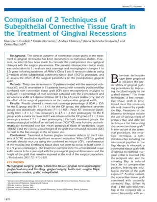
- 5. 0331_IPC_AAP_553351 11/5/01 4:49 PM Page 1474 Comparison of 2 Variants of Connective Tissue Graft Volume 72 • Number 11 Figure 2. A. Presurgical view of recession defects at the buccal aspect of mandibular right canine and first premolar treated with the CP variant of SCTG. B. At 12 months postoperatively, satisfactory root coverage is evident on both teeth.The thickness of the buccal tissues has been increased. Note the minimal increase in width of keratinized tissue, the coronal shift of the mucogingival junction, and the imprecise limits of the connective tissue graft underlying the mucosal-type soft tissue covering. mean value of POSTKT. The coefficient correspond- ing to the variable TECH could not be considered sta- tistically different from zero, and the same conclusion held for coefficients corresponding to interactions between TECH and any other variable examined. The Figure 3. mean value of POSTKT for a given recession can be A. A 4 mm deep gingival recession at a mandibular left canine with obtained by summing a constant number of 1.2 mm, minimal buccal keratinized tissue treated with the envelope technique. B. 61.93% of the recession PREKT and 80.13% of the GE The coronal portion of the connective tissue graft with its 2 mm wide during the surgical procedure. epithelial collar was left exposed beyond the flap margin. C. Note the final clinical outcome 1 year postoperatively showing a substantial DISCUSSION increase in width of keratinized tissue without displacement of the The present retrospective study compared the mucogingival junction from its presurgical position. mucogingival changes following treatment of Miller’s Class I and II recessions with subepithelial connective tissue grafts with and without coronal positioning of comparable for the 2 techniques. However, keratinized the flap. The results indicate that the treatment out- tissue increase was significantly greater in sites treated come relative to the percentage of root coverage was with the envelope technique. 1474
- 6. 0331_IPC_AAP_553351 11/5/01 4:49 PM Page 1475 J Periodontol • November 2001 Cordioli, Mortarino, Chierico, Grusovin, Majzoub In the present report, connective tissue grafts asso- tendency derive from a comparative study conducted ciated with coronally advanced flap resulted in 94.68% by Bouchard et al.,14 who obtained an increase in ker- root coverage at 1 to 1.5 years postoperatively. These atinized tissue on the buccal aspect of recession sites data in the CP group are in agreement with those pre- treated with connective tissue graft combined with viously reported in recession defects treated with a coronally positioned flap from 2.13 mm to 3.07 mm similar procedure. Comparable short- and long-term at 6 months following surgery. In the same study, a percentages of root coverage were reported by greater increase was observed in the connective tissue Trombelli et al.11 at 6 months (81%) , Caffesse et al.12 group where the coronal aspect of the graft was left at 6 months (84.33%), Bouchard et al.15 at 6 months exposed (1.73 mm to 3.8 mm). Other indications in (79.3% to 84.0%), Zucchelli et al.16 at 12 months the literature substantiating the superiority of the enve- (93.5%), and Paolantonio et al.10 at 5 years (85.23%). lope technique in terms of keratinized tissue increase When the envelope technique was used, an average can be drawn from Borghetti et al.,13 who suggest that root coverage of 89.57% was obtained in our study. attempting to completely cover the graft should be Similar techniques led to comparable results in vari- avoided when the initial height of keratinized tissue is ous studies.6,14,19 Jahnke et al.6 obtained an average poor. root coverage of 80% at 6 months, while Bouchard et While the increase in width of KT is biologically sub- al.14 reported 69.2% at the same evaluation period. stantiated when the envelope variant of SCTG is used Müller et al.,19 employing a modification of the enve- to treat gingival recessions, the connective tissue lope technique, demonstrated an average root cover- grafted under the alveolar mucosa in the combined age of 74% at 12 months postsurgery. Differences in technique should not yield differentiation of the mucosa treatment outcome among various reports may be epithelium into keratinized epithelium.20,21 The results partly attributed to the clinical characteristics of the of the histological healing studies were confirmed by patient population and recession sites. clinical data from various studies.8,11,14 Borghetti and For both treatment groups E and CP, the interpre- Louise,8 in a 1-year controlled clinical evaluation of tation of the statistical model applied in this study indi- connective tissue grafts covered by a double papilla cated a mathematical correlation between the mean full-thickness flap, reported that the position of the postsurgical width of KT and 2 clinical and surgical mucogingival junction was stable throughout the study. factors: 1) the average presurgical dimension of KT This finding indicated that only the portion of the con- and 2) the corono-apical height of the graft that nective tissue graft covered by the keratinized con- remained exposed coronal to the flap margin at the nective tissue of the sutured pedicles yielded kera- recipient site. The application of this mathematical tinized tissue postoperatively, while the more apical model in clinical situations could explain the variabil- part of the graft located beyond the mucogingival junc- ity of results reported in previous studies relative to tion and covered by alveolar mucosa did not induce the increase in width of keratinized tissue following the keratinization of the overlying mucosal epithelium. connective tissue grafting in recession sites. When The increase in KT in recessions treated with CP in reviewing reports involving the treatment of gingival the present study and all above-mentioned investiga- recessions with the envelope technique, Jahnke et al.6 tions can be attributed to a possible repositioning of obtained an increase in gingival width from 0.6 mm to the covering flap in an apical direction during wound 3.6 mm at 6 months postsurgery, whereas Müller et healing, thus exposing the connective tissue graft.14 al.19 demonstrated an augmentation from 2.1 mm to Although an attempt is routinely made to completely 3.2 mm at 12 months postoperatively. Increase in width cover the connective tissue grafts with the gingival flap of keratinized tissue was also observed in recession at completion of the surgical procedure in the com- sites treated with SCTG combined with coronal posi- bined technique, trauma to the flap during recipient tioning of the flap.10-12,15,16 Since data relative to the site preparation and manipulation, and non-passive height of the graft tissue that remained exposed coro- flap coronal positioning may result in marginal reces- nally to the flap margin at the end of the surgical pro- sion/shrinkage of the gingival flap or loss of epithe- cedure were not reported in the above-mentioned stud- lial/connective tissue integrity at the flap margin. In ies, it is impossible to establish whether the mean width this case, the inductive potential of the graft on epithe- of KT obtained postsurgically in these reports is con- lial phenotype is restricted to the exposed portion of sistent with the mathematical correlation presented in the SCTG in the short-term perspective.8,22 The ques- the present investigation. According to this mathe- tion of whether the mucogingival junction displaced matical model, a greater postsurgical increase in width coronally in recession sites treated with the combined of KT is to be expected in sites treated with the enve- technique will revert back to its more apical, geneti- lope technique; however, the mean numbers do not cally predetermined position at longer evaluation peri- seem to reflect such a tendency in the previously ref- ods23-25 cannot be answered within the short-term erenced studies. The only clinical data confirming this observation interval of the present study. 1475
- 7. 0331_IPC_AAP_553351 11/5/01 4:49 PM Page 1476 Comparison of 2 Variants of Connective Tissue Graft Volume 72 • Number 11 In conclusion, no significant differences could be subepithelial connective tissue graft in the treatment of found between the 2 variants of SCTG in terms of per- human gingival recession. J Periodontol 1999;70:123- 130. centage of root coverage. When SCTG combined with 14. Bouchard P, Etienne D, Ouhayoun J-P, Nilvéus R. Subep- coronally positioned flap is used in the treatment of ithelial connective tissue grafts in the treatment of gin- human gingival recessions, the palatal connective tis- gival recessions. A comparative study of 2 procedures. sue grafted beneath alveolar mucosa does not seem J Periodontol 1994;65:929-936. to induce the differentiation of alveolar mucosa epithe- 15. Bouchard P, Nilvéus R, Etienne D. Clinical evaluation of tetracycline HCl conditioning in the treatment of gingi- lium into keratinized epithelium, at least within the val recessions. A comparative study. J Periodontol 1997; short-term follow-up period of this study. The treat- 68:262-269. ment outcome in terms of keratinized tissue width 16. Zucchelli G, Clauser C, De Sanctis M, Calandriello M. seems to be related to the presurgical dimensions of Mucogingival versus guided tissue regeneration proce- keratinized tissue and the height of the grafted tissue dures in the treatment of deep recession type defects. J Periodontol 1998;69:138-145. that is left exposed coronal to the flap margin at the 17. Harris RJ. A comparison of 2 root coverage techniques: end of the surgical procedure. Attempts to completely Guided tissue regeneration with a bioabsorbable matrix cover the graft should be avoided if the initial gingival style membrane versus a connective tissue graft com- width is poor and if augmentation of keratinized tissue bined with a coronally positioned pedicle graft without is required. vertical incisions. Results of a series of consecutive cases. J Periodontol 1998;69:1426-1434. 18. Miller PD. A classification of marginal tissue recession. REFERENCES Int J Periodontics Restorative Dent 1985;5(2):8-13. 1. Langer B, Langer L. Subepithelial connective tissue graft 19. Müller HP, Eger T, Schorb A. Gingival dimensions after technique for root coverage. J Periodontol 1985;56:715- root coverage with free connective tissue grafts. J Clin 720. Periodontol 1998;25:424-430. 2. Raetzke PB. Covering localized areas of root exposure 20. Karring T, Lang NP, Löe H. The role of gingival con- employing the “envelope” technique. J Periodontol 1985; nective tissue in determining epithelial differentiation. J 56:397-402. Periodont Res 1975;10:1-11. 3. Nelson SW. The subepithelial connective tissue graft. A 21. Karring T, Östergaard E, Löe H. Conservation of tissue bilaminar reconstructive procedure for the coverage of specificity after heterotopic transplantation of gingiva denuded root surfaces. J Periodontol 1987;58:95-102. and alveolar mucosa. J Periodont Res 1971;6:282-293. 4. De Sanctis M, Zucchelli G. Bilaminar techniques. In: De 22. Trombelli L. Periodontal regeneration in gingival reces- Sanctis M, Zucchelli G, eds. Soft Tissue Plastic Surgery, sion defects. Periodontol 2000 1998;19:138-150. 2nd ed. Bologna: Martina;1997:132-192. 23. Ainamo A, Bergenholtz A, Hugoson A, Ainamo J. Loca- 5. Harris RJ. The connective tissue and partial thickness tion of the mucogingival junction 18 years after apically double pedicle graft: A predictable method for obtaining repositioned flap surgery. J Clin Periodontol 1992;19:49- root coverage. J Periodontol 1992;63:477-486. 52. 6. Jahnke PV, Sandifer JB, Gher ME, Gray JL, Richardson 24. Wennström JL, Zucchelli G. Increased gingival dimen- AC. Thick free gingival and connective tissue autografts sions: A significant factor for successful outcome of root for root coverage. J Periodontol 1993;64:315-322. coverage procedures? J Clin Periodontol 1996;23:770- 7. Harris RJ. The connective tissue with partial thickness 777. double pedicle graft. The results of 100 consecutively 25. Romanos GE, Bernimoulin J-P, Marggraf E. The double treated defects. J Periodontol 1994;65:448-461. lateral bridging flap for coverage of denuded root sur- 8. Borghetti A, Louise F. Controlled clinical evaluation of face: Longitudinal study and clinical evaluation after 5 the subpedicle connective tissue graft for the coverage to 8 years. J Periodontol 1993;64:683-688. of gingival recession. J Periodontol 1994;65:1107-1112. 9. Ricci G, Silvestri M, Tinti C, Rasperini G. A clinical/sta- Send reprint requests to: Dr. Zeina Majzoub, Università degli tistical comparison between the subpedicle connective Studi di Padova, Istituto di Clinica Odontoiatrica, via Gius- tissue graft method and the guided tissue regeneration tiniani, 2, 35100 Padova, Italy. Fax: 39 049 8218229; e- technique in root coverage. Int J Periodontics Restorative mail: majzoub@libero.it. Dent 1996;16:539-545. 10. Paolantonio M, di Murro C, Cattabriga A, Cattabriga M. Accepted for publication May 4, 2001. Subpedicle connective tissue graft versus free gingival graft in the coverage of exposed root surfaces. A 5-year clinical study. J Clin Periodontol 1997;24:51-56. 11. Trombelli L, Scabbia A, Tatakis DN, Calura G. Sub- pedicle connective tissue graft versus guided tissue regeneration with bioabsorbable membrane in the treat- ment of human gingival recession defects. J Periodon- tol 1998;69:1271-1277. 12. Caffesse RG, De LaRosa M, Garza M, Munne-Travers A, Mondragon JC, Weltman R. Citric acid demineralization and subepithelial connective tissue grafts. J Periodontol 2000;71:568-572. 13. Borghetti A, Glise J-M, Monnet-Corti V, Dejou J. Com- parative clinical study of a bioabsorbable membrane and 1476