Case Review #14: 16 year old female with progressive adolescent scoliosis
•
1 recomendación•826 vistas

A 16-year-old female presented with progressive adolescent idiopathic scoliosis. Her curve had increased from 40 degrees to 46 degrees over two years while wearing a brace. Pre-op x-rays showed a 46 degree thoracic curve. The indications for surgery were a progressive curve, pain, and deformity. The surgical strategy involved segmental spinal instrumentation from T2 to L1 using pedicle screws, multiple osteotomies from T5 to T10 including Smith-Peterson osteotomies, and posterior spinal fusion from T3 to L1. Post-op films showed the patient was well-balanced in the coronal and sagittal planes, and her symptoms resolved following surgery.

Recomendados
Recomendados
Más contenido relacionado
La actualidad más candente
La actualidad más candente (20)
Similar a Case Review #14: 16 year old female with progressive adolescent scoliosis
Similar a Case Review #14: 16 year old female with progressive adolescent scoliosis (20)
Más de Robert Pashman
Más de Robert Pashman (15)
Último
Último (20)
Case Review #14: 16 year old female with progressive adolescent scoliosis
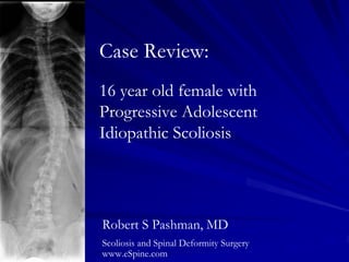
- 1. Case Review: 16 year old female with Progressive Adolescent Idiopathic Scoliosis Robert S Pashman, MD Scoliosis and Spinal Deformity Surgery www.eSpine.com
- 2. Patient History 14 year old female presented with progressive Adolescent Idiopathic Scoliosis. Her curve was thought to have increased 20° over the duration of her treatment. The patient wore a brace. When she presented at my office, her curve was 40°. Curve progressed to 46° two years from initial consultation. The patient has 2-cm right rib hump, minimal left lumbar fullness and what appears to be a gross thoracic shift to the right.
- 3. Pre-op X-rays 46° 46° 1AN curve with apex at T10, which is fully flexible. The patient has a significant right rib hump.
- 5. Indications for Surgery 1. Progressive adolescent idiopathic scoliosis, 1AN, 46 degree, right thoracic curve. 2. Significant pain, thoracic and lumbar spine. 3. Cosmetic deformity of thoracic spine. 4. Failed conservative therapy.
- 6. Surgical Strategy Segmental spinal instrumentation, T2 to L1 using 5.5 stainless steel pedicle screw rod construct. Multiple level osteotomies, T5 to T10, including Smith-Peterson osteotomies for induction of flexibility for improvement of adolescent idiopathic scoliosis. Posterior spinal fusion, T3 to L1, using locally harvested autogenic bone and allograft. Removal of fractured facet under loupe magnification, T7 on the left. Plastic closure of the wound. Intraoperative SSEP management. Intraoperative fluoroscopy management.
- 7. Post-Op Films The patient is well balanced in the coronal and sagittal planes.
- 9. Pre-Op/Post-op Comparison The patient’s symptoms resolved following surgery, and she is very happy with her cosmetic outcome.