SlideShare una empresa de Scribd logo
1 de 45
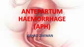
ANTEPARTUM
HAEMORRHAGE
(APH)
FAHAD ZAKWAN
INTRODUCTION
Antepartum haemorrhage (APH),
also called prepartum hemorrhage,
is bleeding from the genital tract
during pregnancy from 28 weeks
gestational age to term
CLASSIFICATION
(A) Placental site bleeding : (62%)
• Placenta praevia (22%): Bleeding from separation of a
placenta wholly or partially implanted in the lower
uterine segment.
• Abruptio placentae (30%): Premature separation of a
normally implanted placenta.
• Marginal separation (10%): Bleeding from the edge of
a normally implanted placenta.
(B) Non-placental site bleeding :
(28%)
•Vasa praevia : Bleeding from ruptured
foetal vessels.
•Rupture uterus: 3-Bloody show.
•Cervical ectopy , polyp or cancer.
•Vaginal varicosity.
It should be considered as an obstetric
emergency (regardless of whether there
is pain or not) and medical attention
should be sought immediately, because
if it is left untreated it can lead to death
of the mother and/or featus.
NOTE
Avoid doing vaginal examination in
patients with antepartum haemorrhage
until placenta praevia has been excluded
either clinically or by USS
EPIDEMIOLOGY
•It Affects 3-5% of all pregnancies.
•It is 3 times more common in
multiparous than primiparous
women
DIFFERENTIAL DIAGNOSIS OF APH
•Bloody show - most common benign cause of
APH
•Uterine rupture
•Bleeding from the lower genital tract
•Cervical bleeding - cervicitis, cervical neoplasm,
cervical polyp
•Bleeding from the vagina itself - trauma, neoplasm
PLACENTA PRAEVIA
• Abnormally implanted placenta in the lower
segment of the uterus
• Although almost all the blood loss from placental site
is maternal, some fetal loss is possible particularly if
the substance of the placenta is traumatized.
• Bleeding from the vasa praevia is the only cause of
pure fetal haemorrhage.
•Bleeding is thought to occur in association with the
development of the lower uterine segment in the
third trimester.
•Placental attachment is disrupted as this area
gradually thins in preparation for the onset of
labor.
•When this occurs, bleeding occurs at the
implantation site as the uterus is unable to contract
adequately and stop the flow of blood from the
open vessels.
RISK FACTORS
•Previous placenta praevia
•Multiple pregnancies
•Multiparity
•Previous c/s scar
•Advanced age
•History of threatened abortion in index
pregnancy
•Deficient endometrium due to pre-existent
•Uterine scar (previous CS)
•Endometritis
•Manual removal of placenta
•Curettage (especially for miscarriage or
termination of pregnancy)
•Submucous fibroid
Typically:
•First episode of bleeding occurs:
•After 36th week: in 60% of cases
•32-36th week: in 30% of cases
•Before 32nd week: in 10% of cases
CLINICAL PRESENTATION
Painless vaginal bleeding, usually bright red, but variable amount.
Soft non tender uterus.
Fetal malpresentation
unusually high and mobile presenting part
Fetal kicks are usually present.
Normal foetal heart tones – foetal distress if blood loss is severe
ABDOMINAL EXAMINATION
Uterus is soft and non-tender
The presenting part is usually high
Fetal malpresentation may be found.
Fetal heart heard
No contraction.
Vaginal examination should be avoided, if
done should, be in theatre under GA with
preparation for emergency c/s in case
profuse bleeding occurs. Explore the fornices
first then the head. Passing the finger
through the cx should be avoided.
GRADING PLACENTA PRAEVIA
Placenta praevia is classified
according to the placement of
the placenta:
GRADE 1 – low lying
•Placenta
encroaches
lower segment
but does not
reach the
cervical os.
GRADE 2 - marginal
•Placenta
reaches
cervical os
but does not
cover it
GRADE 3 - partial
•Placenta covers
part of the cervical
os.
•The placenta
covers the internal
os when it is
closed or partially
dilated but not
when it is fully
dilated
GRADE 4 - total
• Placenta completely
covers the os, even when
the cervix is dilated.
• The placenta covers the
internal os completely
whether the cervix is
partially or fully dilated.
LOCALIZATION OF THE PLACENTA
Ultrasound
i. Transabdominal
ii. Transvaginal.
iii. Transperineal done if instrumentation of
the vagina is likely to cause problems.
The transvaginal US is the most
accurate route to locate the
placenta but the probe should
not touch the cx or the placental
placental site.
Management
•Depends on
-Gestation age
-Severity of bleeding
-Viability of fetus.
IMMEDIATE MEASURES
Call for help!!! Involve the senior staff on call
put in 2 large bore (14 gauge) IV cannulas.
Keep patient flat and worm.
Send blood for group, cross matching diagnostic tests and ask
for at least 2 units of blood
Infuse rapidly 2 litres of normal saline (crystalloids) to re-
expand the vascular bed.
Give oxygen by mask 10 – 15L/min
Insert Foley catheter to empty the bladder
and monitor urine output.
Monitor the following:
pulse
BP
RR
urine output (continuous cathetarization)
type and amount of fluid the patient received
The management includes
• Restore blood volume by giving IV fluids – normal
saline or ringers lactate
• Assess blood loss:
• If bleeding is heavy or continuous, arrange or do
caesarean section delivery irrespective of foetal maturity
within 6 – 8 hrs.
• If bleeding is light and stopped and the foetus alive and
premature consider expectant management until 37/38
wks.
•Keep the woman in hospital for bed rest
until delivery
•Correct anaemia
•Give iron/ folic acid, vit. B12
•Blood transfusion
•Packed cells
•Whole blood
NOTE
•Decide management after weighing benefits and risks
to the woman and fetus.
•PRIORITY IS SAVING THE MOTHER!!
•ALL GRADES OF PLACENTA PRAEVIA ARE DELIVERED
BY C/S EXCEPT GRADE IA WHERE THE PLACENTA JUST
DIPS IN THE LOWER SEGMENT AND ITS ANTERIORLY
LOCATED
Bloods Tests
• Complete blood picture and D-Dimer
• Group and cross match 4 units
• Consider need for Anti-D if Rh negative.
Ultrasound
• Confirm diagnosis.
Corticosteroids
• Consider corticosteroid prophylaxis if < 34 weeks
ABRUPTIO PLACENTA
•Refers to bleeding due to the premature
separation of a normally sited placenta from its
attachment to the uterus.
May be described as
•Concealed ( if no external bleeding is seen)
•Revealed (if there is obvious external bleeding)
RISK FACTORS
• Hypertension
• Maternal thrombophilias
• Increasing maternal age, parity
• Cigarette smoking
• Abdominal trauma e.g. motor vehicle accident
• Substance abuse (crack, cocaine, amphetamines)
• Sudden decrease in uterine volume (e.g. SROM in the presence of
polyhydramnios, or after delivery of a first twin)
• External cephalic version (ECV) or instrumental delivery.
CLASSIFICATION
•Classification of placental abruption is based on:
•extent of separation (ie, partial vs complete) and
•location of separation (ie, marginal vs central).
Clinical characteristics include the following:
Class 0 is asymptomatic.
Diagnosis is made
retrospectively by finding an
organized blood clot or a
depressed area on a delivered
placenta.
Class 1 is mild
• represents approximately 48% of all cases.
• Characteristics include the following:
• No vaginal bleeding to mild vaginal bleeding
• Slightly tender uterus
• Normal maternal BP and heart rate
• No coagulopathy
• No fetal distress
Class 2 is moderate
• represents approximately 27% of all cases.
• Characteristics include the following:
• No vaginal bleeding to moderate vaginal bleeding
• Moderate-to-severe uterine tenderness with possible
tetanic contractions
• Maternal tachycardia with orthostatic changes in BP and
heart rate
• Fetal distress
• Hypofibrinogenaemia (i.e. 50-250 mg/dL)
Class 3 is severe
•represents approximately 24% of all cases.
Characteristics include the following:
•No vaginal bleeding to heavy vaginal bleeding
•Very painful tetanic uterus
•Maternal shock
•Hypofibrinogenemia (ie, <150 mg/dL)
•Coagulopathy
•Fetal death
Clinical features
• Vaginal bleeding is usually associated with abdominal pain,
uterine contractions, tenderness and / or irritability
• May be faint and / or collapse
• Signs of haemorrhagic shock
• Consider concealed abruption if abdominal or back pain is
present
• Fetal moments are usually absent unless it is an early
abruption or partial
•Contractions/uterine tenderness
•Uterine contractions are a common
finding with placental abruption.
•Contractions progress as the abruption
expands, and uterine hypertonus may be
noted.
•Contractions are painful and palpable.
Principles of early management
• Intravenous (IV) access (2 large bore iv lines must be put in
place)
• Indwelling catheter
• Start intensive care list (fluid balance, blood pressure, pulse
rate etc)
• The cornerstone of appropriate management is adequate
resuscitation with intravenous fluids (plasma volume
expanders, blood)
• Consider need for early delivery dependent on maternal and
fetal condition
• Achieving adequate perfusion is essential to allow the
reticulo-endothelial system (Kupfer cells, spleen) to clear
fibrinogen / fibrin degradation products (FDPs). Acceptable
perfusion is signalled by urine output of > 30 mL per hour
• Cross match blood
• Urgent complete blood picture, D-dimer, thrombin time,
fibrinogen levels, creatinine
• Bed side clotting time (NORMAL – 7mins)
• Consider giving FFP if there is prolonged bed side clotting time
or abnormal bleeding indices.
Later on
•Monitor in-put and output
•Monitor maternal vital signs.
•Continuous CTG to assess for signs of fetal
compromise if the fetus was alive at the time of
diagnosis.
•Anti D prophylaxis for Rh negative women
Active management:
• Consultation with anaesthetist as indicated
• Consider central venous pressure monitoring
• Ensure adequate blood, blood products (6-8 packed cells to start with in
case of total abruption), and fresh frozen plasma (FFP) guided by fibrinogen
levels and mostly also 1 FFP unit required for each 4-6 units of blood
transfused. Specific replacement of coagulation factors is very rarely
required - only in consultation with laboratory / haematologist
• If fetal death is confirmed
• Aim for vaginal delivery with ARM and Syntocinon® if condition allow.
Delivery
• Consider vaginal delivery with continuous electronic fetal monitoring if
fetus is alive and rapid recourse to caesarean section depending on
fetal/maternal condition.
• Caesarean section if acute fetal compromise occurs (if it was alive at
diagnosis) or if other obstetric indications dictate so.
Following delivery:
• Recognize increased risk of PPH
After delivery do placental examination for:
• Completeness
• Any area of abruption
• Associated pathological features e.g. abnormal degree of calcification
• Send for histopathology

Más contenido relacionado

La actualidad más candente (20)

Normal labour
Normal labourNormal labour
Normal labour
 
POLYHYDRAMINOS
POLYHYDRAMINOSPOLYHYDRAMINOS
POLYHYDRAMINOS
 
1st stage managment
1st stage managment1st stage managment
1st stage managment
 
Oligohydramnios
OligohydramniosOligohydramnios
Oligohydramnios
 
Antepartum haemorrhage
Antepartum haemorrhageAntepartum haemorrhage
Antepartum haemorrhage
 
Premature labour
Premature labourPremature labour
Premature labour
 
Pph
PphPph
Pph
 
Ectopic Pregnancy
Ectopic PregnancyEctopic Pregnancy
Ectopic Pregnancy
 
Hyperemesis gravidarum
Hyperemesis gravidarumHyperemesis gravidarum
Hyperemesis gravidarum
 
Breech presentation
Breech presentationBreech presentation
Breech presentation
 
Cord Prolapse
Cord ProlapseCord Prolapse
Cord Prolapse
 
Induction of labor
Induction of laborInduction of labor
Induction of labor
 
Amniotic fluid embolism
Amniotic fluid embolismAmniotic fluid embolism
Amniotic fluid embolism
 
Eclampsia ppt
Eclampsia pptEclampsia ppt
Eclampsia ppt
 
Normal puerperium
Normal puerperiumNormal puerperium
Normal puerperium
 
Rupture of the uterus
Rupture of the uterusRupture of the uterus
Rupture of the uterus
 
Placenta previa
Placenta previaPlacenta previa
Placenta previa
 
Preterm labour
Preterm labourPreterm labour
Preterm labour
 
HIV In Pregnancy
HIV In Pregnancy HIV In Pregnancy
HIV In Pregnancy
 
Abruptio placentae
Abruptio placentae Abruptio placentae
Abruptio placentae
 

Destacado

Aph Antepartum hemorrhage
Aph Antepartum hemorrhageAph Antepartum hemorrhage
Aph Antepartum hemorrhageHuzaifaMD
 
Antepartum haemorrhage s
Antepartum haemorrhage  sAntepartum haemorrhage  s
Antepartum haemorrhage sDr E.W Ugboma
 
Placenta ultrasound
Placenta ultrasoundPlacenta ultrasound
Placenta ultrasoundDoaa Gadalla
 
Antepartum Hemorrhage
Antepartum Hemorrhage Antepartum Hemorrhage
Antepartum Hemorrhage Nooriya Afghan
 
ABNORMALITY IN STRUCTURE OF UTERUS
ABNORMALITY IN STRUCTURE OF UTERUSABNORMALITY IN STRUCTURE OF UTERUS
ABNORMALITY IN STRUCTURE OF UTERUSEmmanuel Masih
 
Antepartum Haemorrhage
Antepartum HaemorrhageAntepartum Haemorrhage
Antepartum Haemorrhagelimgengyan
 
Antepartum haemorhage
Antepartum haemorhageAntepartum haemorhage
Antepartum haemorhageDrNadir Khan
 
Medical Complications in Pregnancy
Medical Complications in PregnancyMedical Complications in Pregnancy
Medical Complications in PregnancyDJ CrissCross
 
Medical Complications In Pregnancy
Medical Complications In PregnancyMedical Complications In Pregnancy
Medical Complications In PregnancyDJ CrissCross
 
Pregnancy Complications.ppt
Pregnancy Complications.pptPregnancy Complications.ppt
Pregnancy Complications.pptShama
 
antepartum haemorrhage
antepartum haemorrhageantepartum haemorrhage
antepartum haemorrhageEddie Lim
 
Overview management of postpartum haemorrhage
Overview management of postpartum haemorrhageOverview management of postpartum haemorrhage
Overview management of postpartum haemorrhageAhmed Almumtin
 
Haemorrhage during late pregnancy
Haemorrhage during late pregnancyHaemorrhage during late pregnancy
Haemorrhage during late pregnancyKripa Susan
 

Destacado (19)

Aph Antepartum hemorrhage
Aph Antepartum hemorrhageAph Antepartum hemorrhage
Aph Antepartum hemorrhage
 
Antepartum haemorrhage s
Antepartum haemorrhage  sAntepartum haemorrhage  s
Antepartum haemorrhage s
 
Antepartum hemorrhage
Antepartum hemorrhageAntepartum hemorrhage
Antepartum hemorrhage
 
Placenta ultrasound
Placenta ultrasoundPlacenta ultrasound
Placenta ultrasound
 
Placental grading
Placental gradingPlacental grading
Placental grading
 
Antepartum Hemorrhage
Antepartum Hemorrhage Antepartum Hemorrhage
Antepartum Hemorrhage
 
Bleeding in late px
Bleeding in late pxBleeding in late px
Bleeding in late px
 
ABNORMALITY IN STRUCTURE OF UTERUS
ABNORMALITY IN STRUCTURE OF UTERUSABNORMALITY IN STRUCTURE OF UTERUS
ABNORMALITY IN STRUCTURE OF UTERUS
 
Antepartum Haemorrhage
Antepartum HaemorrhageAntepartum Haemorrhage
Antepartum Haemorrhage
 
Antepartum haemorhage
Antepartum haemorhageAntepartum haemorhage
Antepartum haemorhage
 
Pregnancy Complications
Pregnancy ComplicationsPregnancy Complications
Pregnancy Complications
 
Medical Complications in Pregnancy
Medical Complications in PregnancyMedical Complications in Pregnancy
Medical Complications in Pregnancy
 
Medical Complications In Pregnancy
Medical Complications In PregnancyMedical Complications In Pregnancy
Medical Complications In Pregnancy
 
Placenta Praevia
Placenta PraeviaPlacenta Praevia
Placenta Praevia
 
Obg seminar
Obg seminarObg seminar
Obg seminar
 
Pregnancy Complications.ppt
Pregnancy Complications.pptPregnancy Complications.ppt
Pregnancy Complications.ppt
 
antepartum haemorrhage
antepartum haemorrhageantepartum haemorrhage
antepartum haemorrhage
 
Overview management of postpartum haemorrhage
Overview management of postpartum haemorrhageOverview management of postpartum haemorrhage
Overview management of postpartum haemorrhage
 
Haemorrhage during late pregnancy
Haemorrhage during late pregnancyHaemorrhage during late pregnancy
Haemorrhage during late pregnancy
 

Similar a Aph

APH.pptx
APH.pptxAPH.pptx
APH.pptxMrsP6
 
Vaginal bleeding in early pregnancy
Vaginal bleeding in early pregnancyVaginal bleeding in early pregnancy
Vaginal bleeding in early pregnancyAmit Poudel
 
Antepartumhaemorrhage 121128013531-phpapp02
Antepartumhaemorrhage 121128013531-phpapp02Antepartumhaemorrhage 121128013531-phpapp02
Antepartumhaemorrhage 121128013531-phpapp02Ahmed Farrasyah
 
Antepartum Hemorrhage
Antepartum HemorrhageAntepartum Hemorrhage
Antepartum HemorrhageZana Hossam
 
ANTEPARTUM HAEMORRHAGE (APH).pptx
ANTEPARTUM        HAEMORRHAGE (APH).pptxANTEPARTUM        HAEMORRHAGE (APH).pptx
ANTEPARTUM HAEMORRHAGE (APH).pptxAnthonyMatu1
 
Late Pregnancy Bleeding.pptx
Late Pregnancy Bleeding.pptxLate Pregnancy Bleeding.pptx
Late Pregnancy Bleeding.pptxLara Masri
 
POSTPARTUM HAEMORRHAGE IN MIDWIFERY .ppt
POSTPARTUM HAEMORRHAGE IN MIDWIFERY .pptPOSTPARTUM HAEMORRHAGE IN MIDWIFERY .ppt
POSTPARTUM HAEMORRHAGE IN MIDWIFERY .pptJuma675663
 
Antepartum Haemorrage.pdf
Antepartum Haemorrage.pdfAntepartum Haemorrage.pdf
Antepartum Haemorrage.pdfChantal Settley
 
anti partum hemorrhage and its complication
anti partum hemorrhage and its complicationanti partum hemorrhage and its complication
anti partum hemorrhage and its complicationdevtesfaye77
 
Third trimester bleeding
Third trimester bleedingThird trimester bleeding
Third trimester bleedingHasan Ismail
 
ANTE PARTUM HAEMORRHAGE (APH).pptx
ANTE PARTUM HAEMORRHAGE (APH).pptxANTE PARTUM HAEMORRHAGE (APH).pptx
ANTE PARTUM HAEMORRHAGE (APH).pptxAFSALA18
 
OBSTETRIC HAEMORRHAGE.pptx
OBSTETRIC HAEMORRHAGE.pptxOBSTETRIC HAEMORRHAGE.pptx
OBSTETRIC HAEMORRHAGE.pptxHuda800869
 
OBSTETRIC EMERGENCIES.pptx
OBSTETRIC EMERGENCIES.pptxOBSTETRIC EMERGENCIES.pptx
OBSTETRIC EMERGENCIES.pptxSwizzyKhalfa
 
Ectopic Pregnancy-1.pptx
Ectopic Pregnancy-1.pptxEctopic Pregnancy-1.pptx
Ectopic Pregnancy-1.pptxImranKhan127540
 

Similar a Aph (20)

Antepartum hemorrhage
Antepartum hemorrhageAntepartum hemorrhage
Antepartum hemorrhage
 
Antepartum haemorrhage
Antepartum haemorrhageAntepartum haemorrhage
Antepartum haemorrhage
 
APH.pptx
APH.pptxAPH.pptx
APH.pptx
 
ABRUPTIO PLACENTAE
ABRUPTIO PLACENTAEABRUPTIO PLACENTAE
ABRUPTIO PLACENTAE
 
Vaginal bleeding in early pregnancy
Vaginal bleeding in early pregnancyVaginal bleeding in early pregnancy
Vaginal bleeding in early pregnancy
 
Antepartumhaemorrhage 121128013531-phpapp02
Antepartumhaemorrhage 121128013531-phpapp02Antepartumhaemorrhage 121128013531-phpapp02
Antepartumhaemorrhage 121128013531-phpapp02
 
Antepartum Hemorrhage
Antepartum HemorrhageAntepartum Hemorrhage
Antepartum Hemorrhage
 
ANTEPARTUM HAEMORRHAGE (APH).pptx
ANTEPARTUM        HAEMORRHAGE (APH).pptxANTEPARTUM        HAEMORRHAGE (APH).pptx
ANTEPARTUM HAEMORRHAGE (APH).pptx
 
Late Pregnancy Bleeding.pptx
Late Pregnancy Bleeding.pptxLate Pregnancy Bleeding.pptx
Late Pregnancy Bleeding.pptx
 
POSTPARTUM HAEMORRHAGE IN MIDWIFERY .ppt
POSTPARTUM HAEMORRHAGE IN MIDWIFERY .pptPOSTPARTUM HAEMORRHAGE IN MIDWIFERY .ppt
POSTPARTUM HAEMORRHAGE IN MIDWIFERY .ppt
 
APH.pdf
APH.pdfAPH.pdf
APH.pdf
 
APH-PPT.pptx
APH-PPT.pptxAPH-PPT.pptx
APH-PPT.pptx
 
Antepartum Haemorrage.pdf
Antepartum Haemorrage.pdfAntepartum Haemorrage.pdf
Antepartum Haemorrage.pdf
 
anti partum hemorrhage and its complication
anti partum hemorrhage and its complicationanti partum hemorrhage and its complication
anti partum hemorrhage and its complication
 
Third trimester bleeding
Third trimester bleedingThird trimester bleeding
Third trimester bleeding
 
ANTE PARTUM HAEMORRHAGE (APH).pptx
ANTE PARTUM HAEMORRHAGE (APH).pptxANTE PARTUM HAEMORRHAGE (APH).pptx
ANTE PARTUM HAEMORRHAGE (APH).pptx
 
OBSTETRIC HAEMORRHAGE.pptx
OBSTETRIC HAEMORRHAGE.pptxOBSTETRIC HAEMORRHAGE.pptx
OBSTETRIC HAEMORRHAGE.pptx
 
OBSTETRIC EMERGENCIES.pptx
OBSTETRIC EMERGENCIES.pptxOBSTETRIC EMERGENCIES.pptx
OBSTETRIC EMERGENCIES.pptx
 
Ectopic Pregnancy-1.pptx
Ectopic Pregnancy-1.pptxEctopic Pregnancy-1.pptx
Ectopic Pregnancy-1.pptx
 
Obstetric emergencies
Obstetric  emergencies Obstetric  emergencies
Obstetric emergencies
 

Más de Fahad Zakwan (20)

Version
VersionVersion
Version
 
Us in obstretics
Us in obstreticsUs in obstretics
Us in obstretics
 
Rpl
RplRpl
Rpl
 
Pueperium
PueperiumPueperium
Pueperium
 
Pueperal sepsis
Pueperal sepsisPueperal sepsis
Pueperal sepsis
 
Ptl
PtlPtl
Ptl
 
Prom
PromProm
Prom
 
Prolonged labour
Prolonged labourProlonged labour
Prolonged labour
 
Pph
PphPph
Pph
 
Placenta praevia
Placenta praeviaPlacenta praevia
Placenta praevia
 
Pih
PihPih
Pih
 
Pid
PidPid
Pid
 
Ovarian tumors
Ovarian tumorsOvarian tumors
Ovarian tumors
 
Ovarian cysts
Ovarian cystsOvarian cysts
Ovarian cysts
 
Obtetrics terms
Obtetrics termsObtetrics terms
Obtetrics terms
 
Multiple pregnancy
Multiple pregnancyMultiple pregnancy
Multiple pregnancy
 
Malaria in prgnancy
Malaria in prgnancyMalaria in prgnancy
Malaria in prgnancy
 
Labour and delivery
Labour and deliveryLabour and delivery
Labour and delivery
 
Iugr
IugrIugr
Iugr
 
Iufd
IufdIufd
Iufd
 

Aph

  • 2. INTRODUCTION Antepartum haemorrhage (APH), also called prepartum hemorrhage, is bleeding from the genital tract during pregnancy from 28 weeks gestational age to term
  • 4. (A) Placental site bleeding : (62%) • Placenta praevia (22%): Bleeding from separation of a placenta wholly or partially implanted in the lower uterine segment. • Abruptio placentae (30%): Premature separation of a normally implanted placenta. • Marginal separation (10%): Bleeding from the edge of a normally implanted placenta.
  • 5. (B) Non-placental site bleeding : (28%) •Vasa praevia : Bleeding from ruptured foetal vessels. •Rupture uterus: 3-Bloody show. •Cervical ectopy , polyp or cancer. •Vaginal varicosity.
  • 6. It should be considered as an obstetric emergency (regardless of whether there is pain or not) and medical attention should be sought immediately, because if it is left untreated it can lead to death of the mother and/or featus.
  • 7. NOTE Avoid doing vaginal examination in patients with antepartum haemorrhage until placenta praevia has been excluded either clinically or by USS
  • 8. EPIDEMIOLOGY •It Affects 3-5% of all pregnancies. •It is 3 times more common in multiparous than primiparous women
  • 9. DIFFERENTIAL DIAGNOSIS OF APH •Bloody show - most common benign cause of APH •Uterine rupture •Bleeding from the lower genital tract •Cervical bleeding - cervicitis, cervical neoplasm, cervical polyp •Bleeding from the vagina itself - trauma, neoplasm
  • 10. PLACENTA PRAEVIA • Abnormally implanted placenta in the lower segment of the uterus • Although almost all the blood loss from placental site is maternal, some fetal loss is possible particularly if the substance of the placenta is traumatized. • Bleeding from the vasa praevia is the only cause of pure fetal haemorrhage.
  • 11. •Bleeding is thought to occur in association with the development of the lower uterine segment in the third trimester. •Placental attachment is disrupted as this area gradually thins in preparation for the onset of labor. •When this occurs, bleeding occurs at the implantation site as the uterus is unable to contract adequately and stop the flow of blood from the open vessels.
  • 12. RISK FACTORS •Previous placenta praevia •Multiple pregnancies •Multiparity •Previous c/s scar •Advanced age •History of threatened abortion in index pregnancy
  • 13. •Deficient endometrium due to pre-existent •Uterine scar (previous CS) •Endometritis •Manual removal of placenta •Curettage (especially for miscarriage or termination of pregnancy) •Submucous fibroid
  • 14. Typically: •First episode of bleeding occurs: •After 36th week: in 60% of cases •32-36th week: in 30% of cases •Before 32nd week: in 10% of cases
  • 15. CLINICAL PRESENTATION Painless vaginal bleeding, usually bright red, but variable amount. Soft non tender uterus. Fetal malpresentation unusually high and mobile presenting part Fetal kicks are usually present. Normal foetal heart tones – foetal distress if blood loss is severe
  • 16. ABDOMINAL EXAMINATION Uterus is soft and non-tender The presenting part is usually high Fetal malpresentation may be found. Fetal heart heard No contraction.
  • 17. Vaginal examination should be avoided, if done should, be in theatre under GA with preparation for emergency c/s in case profuse bleeding occurs. Explore the fornices first then the head. Passing the finger through the cx should be avoided.
  • 18. GRADING PLACENTA PRAEVIA Placenta praevia is classified according to the placement of the placenta:
  • 19. GRADE 1 – low lying •Placenta encroaches lower segment but does not reach the cervical os.
  • 20. GRADE 2 - marginal •Placenta reaches cervical os but does not cover it
  • 21. GRADE 3 - partial •Placenta covers part of the cervical os. •The placenta covers the internal os when it is closed or partially dilated but not when it is fully dilated
  • 22. GRADE 4 - total • Placenta completely covers the os, even when the cervix is dilated. • The placenta covers the internal os completely whether the cervix is partially or fully dilated.
  • 23. LOCALIZATION OF THE PLACENTA Ultrasound i. Transabdominal ii. Transvaginal. iii. Transperineal done if instrumentation of the vagina is likely to cause problems.
  • 24. The transvaginal US is the most accurate route to locate the placenta but the probe should not touch the cx or the placental placental site.
  • 25. Management •Depends on -Gestation age -Severity of bleeding -Viability of fetus.
  • 26. IMMEDIATE MEASURES Call for help!!! Involve the senior staff on call put in 2 large bore (14 gauge) IV cannulas. Keep patient flat and worm. Send blood for group, cross matching diagnostic tests and ask for at least 2 units of blood Infuse rapidly 2 litres of normal saline (crystalloids) to re- expand the vascular bed. Give oxygen by mask 10 – 15L/min
  • 27. Insert Foley catheter to empty the bladder and monitor urine output. Monitor the following: pulse BP RR urine output (continuous cathetarization) type and amount of fluid the patient received
  • 28. The management includes • Restore blood volume by giving IV fluids – normal saline or ringers lactate • Assess blood loss: • If bleeding is heavy or continuous, arrange or do caesarean section delivery irrespective of foetal maturity within 6 – 8 hrs. • If bleeding is light and stopped and the foetus alive and premature consider expectant management until 37/38 wks.
  • 29. •Keep the woman in hospital for bed rest until delivery •Correct anaemia •Give iron/ folic acid, vit. B12 •Blood transfusion •Packed cells •Whole blood
  • 30. NOTE •Decide management after weighing benefits and risks to the woman and fetus. •PRIORITY IS SAVING THE MOTHER!! •ALL GRADES OF PLACENTA PRAEVIA ARE DELIVERED BY C/S EXCEPT GRADE IA WHERE THE PLACENTA JUST DIPS IN THE LOWER SEGMENT AND ITS ANTERIORLY LOCATED
  • 31. Bloods Tests • Complete blood picture and D-Dimer • Group and cross match 4 units • Consider need for Anti-D if Rh negative. Ultrasound • Confirm diagnosis. Corticosteroids • Consider corticosteroid prophylaxis if < 34 weeks
  • 32. ABRUPTIO PLACENTA •Refers to bleeding due to the premature separation of a normally sited placenta from its attachment to the uterus. May be described as •Concealed ( if no external bleeding is seen) •Revealed (if there is obvious external bleeding)
  • 33. RISK FACTORS • Hypertension • Maternal thrombophilias • Increasing maternal age, parity • Cigarette smoking • Abdominal trauma e.g. motor vehicle accident • Substance abuse (crack, cocaine, amphetamines) • Sudden decrease in uterine volume (e.g. SROM in the presence of polyhydramnios, or after delivery of a first twin) • External cephalic version (ECV) or instrumental delivery.
  • 34. CLASSIFICATION •Classification of placental abruption is based on: •extent of separation (ie, partial vs complete) and •location of separation (ie, marginal vs central). Clinical characteristics include the following:
  • 35. Class 0 is asymptomatic. Diagnosis is made retrospectively by finding an organized blood clot or a depressed area on a delivered placenta.
  • 36. Class 1 is mild • represents approximately 48% of all cases. • Characteristics include the following: • No vaginal bleeding to mild vaginal bleeding • Slightly tender uterus • Normal maternal BP and heart rate • No coagulopathy • No fetal distress
  • 37. Class 2 is moderate • represents approximately 27% of all cases. • Characteristics include the following: • No vaginal bleeding to moderate vaginal bleeding • Moderate-to-severe uterine tenderness with possible tetanic contractions • Maternal tachycardia with orthostatic changes in BP and heart rate • Fetal distress • Hypofibrinogenaemia (i.e. 50-250 mg/dL)
  • 38. Class 3 is severe •represents approximately 24% of all cases. Characteristics include the following: •No vaginal bleeding to heavy vaginal bleeding •Very painful tetanic uterus •Maternal shock •Hypofibrinogenemia (ie, <150 mg/dL) •Coagulopathy •Fetal death
  • 39. Clinical features • Vaginal bleeding is usually associated with abdominal pain, uterine contractions, tenderness and / or irritability • May be faint and / or collapse • Signs of haemorrhagic shock • Consider concealed abruption if abdominal or back pain is present • Fetal moments are usually absent unless it is an early abruption or partial
  • 40. •Contractions/uterine tenderness •Uterine contractions are a common finding with placental abruption. •Contractions progress as the abruption expands, and uterine hypertonus may be noted. •Contractions are painful and palpable.
  • 41. Principles of early management • Intravenous (IV) access (2 large bore iv lines must be put in place) • Indwelling catheter • Start intensive care list (fluid balance, blood pressure, pulse rate etc) • The cornerstone of appropriate management is adequate resuscitation with intravenous fluids (plasma volume expanders, blood) • Consider need for early delivery dependent on maternal and fetal condition
  • 42. • Achieving adequate perfusion is essential to allow the reticulo-endothelial system (Kupfer cells, spleen) to clear fibrinogen / fibrin degradation products (FDPs). Acceptable perfusion is signalled by urine output of > 30 mL per hour • Cross match blood • Urgent complete blood picture, D-dimer, thrombin time, fibrinogen levels, creatinine • Bed side clotting time (NORMAL – 7mins) • Consider giving FFP if there is prolonged bed side clotting time or abnormal bleeding indices.
  • 43. Later on •Monitor in-put and output •Monitor maternal vital signs. •Continuous CTG to assess for signs of fetal compromise if the fetus was alive at the time of diagnosis. •Anti D prophylaxis for Rh negative women
  • 44. Active management: • Consultation with anaesthetist as indicated • Consider central venous pressure monitoring • Ensure adequate blood, blood products (6-8 packed cells to start with in case of total abruption), and fresh frozen plasma (FFP) guided by fibrinogen levels and mostly also 1 FFP unit required for each 4-6 units of blood transfused. Specific replacement of coagulation factors is very rarely required - only in consultation with laboratory / haematologist • If fetal death is confirmed • Aim for vaginal delivery with ARM and Syntocinon® if condition allow.
  • 45. Delivery • Consider vaginal delivery with continuous electronic fetal monitoring if fetus is alive and rapid recourse to caesarean section depending on fetal/maternal condition. • Caesarean section if acute fetal compromise occurs (if it was alive at diagnosis) or if other obstetric indications dictate so. Following delivery: • Recognize increased risk of PPH After delivery do placental examination for: • Completeness • Any area of abruption • Associated pathological features e.g. abnormal degree of calcification • Send for histopathology