Lessons Learned in Teleradiology for Global Health: Providing Imaging Services to Rwanda, Africa
•
1 recomendación•410 vistas

Access to medical imaging expertise in the developing world is limited. Radiography & ultrasound has emerged as a diagnostic tool in certain resource-poor settings in developing nations. This exhibit aims to demonstrate how a pro-bono teleradiology service can be developed, executed, & evaluated via the example of our experience in Rwanda, Africa
Recomendados

Recomendados
Más contenido relacionado
Último
Último (20)
Destacado
Destacado (20)
Lessons Learned in Teleradiology for Global Health: Providing Imaging Services to Rwanda, Africa
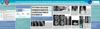
- 1. Lessons Learned in Teleradiology for Global Health: Providing Imaging Services to Rwanda, Africa Sung H Kim, MD; Subba R Digumarthy, MD; Sjirk J Westra, MD; Randheer Shailam, MD; Parul Penkar, MBBS; Garry Choy, MD Purpose/Aim Summary Access to medical imaging expertise in the developing world is limited. In Rwanda, nearly all cases are referred to our service via Partners in Health, but Radiology can play a signi cant role in Radiography & ultrasound has emerged as a diagnostic tool in certain resource- we also provide radiology services to other NGOs and organizations including: global health. poor settings in developing nations. This exhibit aims to demonstrate how a pro-bono teleradiology service can be developed, executed, & evaluated via the Case 4. Patient SG is a example of our experience in Rwanda, Africa. 45-year-old male who While diagnostic radiology remains a presented with vague abdominal pain. Plain lm key component in practice of medicine Content Organization of the abdomen revealed even in resource-poor settings of the mass pneumoperitoneum. developing world, radiologists are Case 2. Patient MM is a 18-year-old female with 6-month history of nonpainful mass in the distal lower Case 3. Patient DM is a 12-year-old male with back The case was interpreted In our exhibit, we will review key elements in teleradiology for resource-poor pain for months who also developed left hip pain seldom involved when compared to within 5 minutes of settings: radiographs of the tibia and bula showed aggressive sunburst periosteal reaction in the mid to distal tibia so that he was unable to walk. Plain lm of the receipt from Rwanda, the e orts of other medical specialties. Low-cost Teleradiology Informatics Solutions pelvis was obtained which showed joint space Africa. Communication narrowing and erosion of the left sacroiliac joint. was made to the referring consistent with metastatic disease. Unfortunately the patient expired within the year following diagnotic Concern for septic joint was raised based on the physician and patient was Numerous non-pro t organizations work-up. lm and the clinical team performed aspiration. immediately transferred for surgery to evaluate for perforated ulcer One liter of purulent material was obtained from and volunteer physicians in elds the left SI joint. Although no organism was isolated, pending but surgeons found a perforated bowel from a large obstructing outside of imaging are looking to tuberculosis was presumed at last update. small bowel tumor. radiologists to participate and play a critical role in providing much needed teleradiology services. Review of Teleradiology Cases from Rwanda, Africa Cases are typically sent for Informatics tools are low cost and Recruiting Volunteer Radiologists, Technologists, ubiquitous, enabling access to high and Imaging Experts: consultation quality medical imaging globally for r own secure image sharing platform those in underserved regions. Radiologists and technologists from various private practices and academic institutions. Policy and Legal issues Recent members come from various areas including: Massachusetts General Hospital, Boston, MA; Legal and policy issues in global health work often center around two Mount Auburn Hospital, Cambridge, MA; Case 1. Patient VJ is a 14-year-old male with one major areas: medicolegal risk and institutional research compliance. While Mayo Clinic, Rochester, MN; For more information on our month history of leg pain. The pain started in Figure 8. Images taken on digital x-ray units can minimal, there is still a low risk for malpractice liability. However, fortunately Columbia University Medical Center, New York, NY; issues of jurisdiction and good Samaritan laws will often protect healthcare Brown University, Providence, RI; the knee and migrated to the distal thigh. The often be sent via direct export to DICOM or JPEG. work and if you are interested in However, majority of volume for teleradiology are professionals in volunteer activities for global health. and National Jewish Medical and Research Center, Denver, CO. the left femur showed a large area of permeative non-digital lms. As a result digital photography participating, please contact – lytic lesion extending from mid diaphysis to distal using cameras is heavily utilized. It is important to check with your insurance provider. Our insurance provider 8A. Viewboxes are not always available but Garry Choy, MD has provided additional coverage for outreach and charitable medical Informatics professionals in industry and in academic settings. Case 6. NB is a 3-month-old status Case 7. CN is a 45-year-old patient with practices. Case 5. Patient NG is a 4-year-old patient presenting with fever and post NG tube placement but referring history of HIV with fevers presents with referring physicians do their best. Mass General Imaging the diaphysis of the femur with Codman's triangle seizures. Patient received a CT scan from the nearest major hospital of 8B. CT scan images are also not always available Academic and industry collaboration helpful for providing resources and Kigali. Abscess, parasitic infection, and malignancy were considered. Case physicians could not nd the tip and heterogeneous lesion in liver. Ultrasound via DICOM but certain questions can be answered gchoy@partners.org unclear if patient was successful. sweep images and static images provided Additionally, needs assessments and research activities that arise from work expertise. malignancies such as osteosarcoma, primary lymphoma of bone, Ewing's sarcoma as well as osteomyelitis with a large associated infected was sent from neurosurgeon for second opinion regarding full range of via digital photography of the lm. In this case, 617-383-9729 (mobile) Radiologists via teleradiology demonstrating loculated intrahepatic abroad need to also be actively cleared not only by our local IRB but also the collection. As primary malignancy was inthe di erential diagnosis, MRI was recommended to look for any skipped lesions and the patient di erential diagnostic considerations. Due to signi cant mass e ect, patient has di use metastatic prostate cancer. identi ed tip was coiled in mouth collection. Surgical aspiration con rmed was sent to Kigali for the study. No skipped lesion was seen and a biopsy was performed which showed focal myositis without evidence of patient was taken to surgery for treatment/further diagnosis. Diagnosis 8C. Digital photograph of a chest radiograph Educational E orts in Rwanda, Africa liver abscess. was neurocysticercosis at the time of surgery. hanging on a viewbox.