Temporomandibular joint. dr. gaurav salunkhe
•Descargar como PPTX, PDF•
14 recomendaciones•2,417 vistas

temporomandibular joint

Recomendados
Más contenido relacionado
La actualidad más candente
La actualidad más candente (20)
Destacado
Similar a Temporomandibular joint. dr. gaurav salunkhe
Similar a Temporomandibular joint. dr. gaurav salunkhe (20)
Más de Gaurav Salunkhe
Más de Gaurav Salunkhe (10)
Último
Último (20)
Temporomandibular joint. dr. gaurav salunkhe
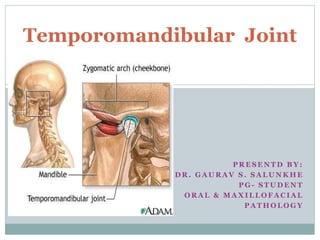
- 1. P R E S E N T D B Y : D R . G A U R A V S . S A L U N K H E P G - S T U D E N T O R A L & M A X I L L O F A C I A L P A T H O L O G Y Temporomandibular Joint
- 2. INTRODUCTION The temporomandibular joint is the joint of the jaw and is frequently referred to as TMJ. ALSO KNOWN AS MANDIBULAR JOINT. TYPE-SYNOVIAL JOINT SUBTYPE-BICONDYLAR The name is derived from the two bones which form the joint: the upper temporal bone which is part of the cranium (skull), and the lower jaw bone called the mandible. The unique feature of the TMJs is the articular disc. The part of the mandible which mates to the under-surface of the disc is the condyle and the part of the temporal bone which mates to the upper surface of the disk is the glenoid (or mandibular) fossa.
- 4. Components There are six main components of the TMJ. Mandibular condyles Articular surface of the temporal bone Fibrous Capsule Articular disc Ligaments Lateral pterygoid
- 7. RELATIONS INFRONT LATERAL PTERYGOID,TEMPORALIS, MASSETERIC NERVE & VESSELS BEHIND PAROTID GLAND, SUPERFICIAL TEMPORAL VESSELS, AURICULOTEMPORAL NERVE LAERALLY SKIN, FASCIA MEDIALLY LATERAL PTERYGOID, MIDDLE MENINGEAL
- 9. MOVEMENTS 1. ELEVATION & DEPRESSION 2. PROTRACION & RETRACTION 3. SIDE TO SIDE MOVEMENS
- 11. MOVEMENTS Depression- lateral pterygoid mainly Elevation- masster, temporalis, medial petygoid of both sides. Protrusion- lateral and medial pterygoid. Retraction- posterior fibres of temporalis. Lateral or side to side movement eg. turning chin to left side- left lateral pterygoid and right medial pterygoid.
- 12. INNERVATION & VASCULARIZATION Sensory innervation of the temporomandibular joint is derived from the auriculotemporal and masseteric branches of TRIGEMINAL NERVE. Its arterial blood supply is provided by branches of the EXTERNAL CAROTID ARTERY, predominately the superficial temporal branch. Other branches: deep auricular artery, anterior tympanic artery, ascending pharyngeal artery, and maxillary artery- may also contribute.
- 13. HISTOLOGY Bony structures Articular fibrous covering Articular disk Synovial membrane
- 15. CLINICAL CONSIDERATIONS Most common disorder- Disc displacement. Most common cause of pain- Myofascial pain dysfunction syndrome. Temporomandibular joint disorder/ syndrome. Conditions that affect joint. 1) Ankylosis 2) Arthritis 3) Trauma 4) Developmental anomalies 5) Neoplasia
- 16. THANK YOU