Er36881887
•
0 recomendaciones•328 vistas

International Journal of Engineering Research and Applications (IJERA) is an open access online peer reviewed international journal that publishes research and review articles in the fields of Computer Science, Neural Networks, Electrical Engineering, Software Engineering, Information Technology, Mechanical Engineering, Chemical Engineering, Plastic Engineering, Food Technology, Textile Engineering, Nano Technology & science, Power Electronics, Electronics & Communication Engineering, Computational mathematics, Image processing, Civil Engineering, Structural Engineering, Environmental Engineering, VLSI Testing & Low Power VLSI Design etc.

Recomendados
Recomendados
Más contenido relacionado
La actualidad más candente
La actualidad más candente (20)
Destacado
Similar a Er36881887
Similar a Er36881887 (20)
Último
Último (20)
Er36881887
- 1. E. SIVASANKARI et al Int. Journal of Engineering Research and Applications ISSN : 2248-9622, Vol. 3, Issue 6, Nov-Dec 2013, pp.881-887 RESEARCH ARTICLE www.ijera.com OPEN ACCESS Retinal Image Compression In Favour Of Hasty Transmission Using Region of Interest E. Sivasankari1 D.Gowthami2 and Prof.R.Jayanthi3 1 (PG Scholar, Department of Electronics and Communication Engineering, Nandha College of Technology, Erode-52) 2 (PG Scholar, Department of Electronics and Communication Engineering, Nandha College of Technology, Erode-52) 3 (Assosiative Professor,Department of Electronics and Communication Engineering, Nandha College of Technology, Erode-638052) ABSTRACT Teleophthalmology illustrated by transmission of retinal images and data between users is one of the promising fields in medicine. Colossal bandwidth is essential for transmitting retinal images over the wireless network. So the most essential aspiration of proposed system is to provide an efficient tool for defining to maximize compression and reconstruct image portions lossless for high speed, efficient transmission and diminution in storage space. This paper is proposed to investigate multiple compression techniques based on Region of Interest (ROI). In the diagnosis of retinal images, the significant part is separated out from the rest of the image using improved adaptive fuzzy C means algorithm and Integer multi wavelet transform is applied to enhance the visual quality in significant part. The region of less significance are compressed using SPIHT algorithm and finally modified embedded zero tree wavelet algorithm is applied which uses six symbols was applied whole image then Huffman coding is applied to get the compressed image for transmission. The proposed algorithm would give better quality, if the images used ROI compared to that of the other methods. The proposed techniques can be evaluated for performance using Compression Ratio (CR), Peak Signal to Noise Ratio (PSNR), Normalized Average Error (NAE), Average Difference (AD), Maximum Difference (MD), Mean Square Error (MSE), Root Mean Square Error (RMSE), Signal to Noise Ratio (SNR), Normalized Cross Correlation (NCC), Structural Content (SC), Encoding and Decoding time. The consequence scrutinized using MATLAB and realized in hardware. As the conclusion, the inaccessible patients used a low cost access to specialist’s eye checkups at crucial healthcare clinics, and at the same time, diminish unnecessary face-to-face consultation at the hospital specialist’s center. Keywords: Retinal Images, Image Compression, Region of Interest, Integer Multi wavelet Transform, SHIPT, and Modified Embedded Zero Trees. I. Introduction One of the most important organs of human body is eye, which gives the sensation of vision including color differentiation and perception of depth due to the presence of rods and cones in the retina. Definitely, the blindness result produces in both physical and emotional disturbance for every patient. This situation is most likely due to an acute scarcity of ophthalmology specialists in many areas and the distribution of ophthalmologists is remote from uniform such that there are much more eye disease patients in the rural regions, e.g., in India, 79% eye patients reside in rural areas. To resolve this trouble, as a promising technology-based solution, telemedicine, enables the doctor to discuss with the patient remotely through video conferencing, share data, and images the patient so as to reduce the unnecessary referrals and travel cost. Teleophthalmology is the branch of the telemedicine. In conventional ophthalmology, most of the analytic ophthalmic instrumentations are adapted www.ijera.com to accumulate still and/or video cameras to acquire images so that ophthalmologists create analytic inferences. Retinal images are acquired by a specialized camera called fundus camera. Mydriatic and non-mydriatic fundus cameras are used for retinal photography.Mydriatic camera requires dilation of pupils. It provides good quality fundus images than non mydriatic camera. Teleophthalmology system will provide eye consultation by delivering high-quality eye images and videos over a public broadband network, so as to utilize the use of communication technologies to give ophthalmology services, share and optimize medical expertise locally, as well as globally. Teleophthalmology was focused on particular eye problem, such as diabetic retinopathy and macular degeneration. Diabetic retinopathy (DR) occurs in patients suffering from diabetes, which causes damage to the retina of the eye. This finally leads to total vision loss. Diabetes is caused due to the body’s inability to store 881 | P a g e
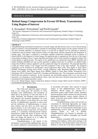
- 2. E. SIVASANKARI et al Int. Journal of Engineering Research and Applications ISSN : 2248-9622, Vol. 3, Issue 6, Nov-Dec 2013, pp.881-887 and make use of the sugar level in the blood. Diabetic retinopathy causes damage to the blood vessels in the retina, and this cause fluid to leak to leak into the macula region of the retina causing it to enlarge and leading to blurred vision. Diabetic retinopathy has two types namely non proliferative and proliferative type. Age related macular degeneration (AMD) occurs in the older group peoples, which affects the macula and degeneration of capillaries in fundus and in the central part of retina. This occurs due to the non functionality of the Bruch’s membrane, which passes the waste products and the nutrients to the retina from the choroid. Fluid leaks out from the damaged vessels and deposited at the center of macula, which results blurring, obscuring or distorting vision. These liquid deposits are called exudates. Huge amount of image is produced in the field of medical image, which can be stored in picture archiving and communication system (PACS). So, it is really hard for hospitals to manage the storing facilities for the same. Moreover, such high data demand for high end network especially for transmitting the images over the network such as in telemedicine. So compression is used and can be categorized into two categories: lossless and lossy compressions. Lossless image compression is achieved if original input image is recovered perfectly from compressed data while lossy image compression cannot regenerate original image data. Lossy image compression, can maintain most details of original image, useful for diagnosis. Precise image detail preservation is not strictly required as an image’s degraded portion is usually often not visible to humans. But lossy image compression is not commonly used in clinical practice/diagnosis because even with slight loss of data, it is possible for physicians/radiologists to miss critical diagnostic information that is needed for diagnosis of a patient. Historically, medical image compression was investigated by researchers working in the image fields. Consequently, technological growth in this field is a by-product of progress in the more general field of natural image. There is no golden rule: different coding algorithms fit different types of images and scenarios best. Depending on imaging modalities and applications, some are better suited than others to fulfill certain targets in terms of compression factor or with respect to a desired functionality. A challenging requirement concerns the visually lossless mode. Compression is not about storage costs alone. It is also about transmission time, imaging apparatus utilization and the patient’s convenience/comfort. Compression techniques by reducing file size and transmission time can thereby improve overall care. Image compression techniques take advantage of any occurring redundancy. There are different redundancy types and each compression methodology exploits www.ijera.com one of these. The different redundancy types are spatial, temporal and spectral. Present compression schemes have high compression rates when quality loss can be afforded. But physicians cannot afford deficiencies in image regions which are important, known as Regions of Interest (ROIs). An approach which brings high compression rates accompanied by good quality in ROIs is needed. A common idea is to preserve quality in diagnostically critical regions while allowing lossy encoding of other regions. The research’s aim focuses on ROI coding to ensure use of multiple and arbitrarily shaped ROIs in images, with arbitrary weights describing the importance for each ROI including background (i.e. image regions not of ROI) so that latter regions can be represented by varying levels of quality. In medical images, some structures in the data are of interest. These structures typically occupy a small percentage of the data, but their analysis requires contextual information like locations within a specific organ or adjacency to sensitive structures. Therefore, while focusing on a particular region of the data, designated as a Region of Interest (ROI), contextual information surrounding that region is important. However, the same amount of detail is not required for the context and the ROI. Improved Adaptive Fuzzy C-means logic is used to separate out ROI by extracting image features. After performing segmentation lossless compression is applied to significant region i.e., Integer Multi Wavelet Transform and lossy is applied to rest of the image i.e., SPIHT. The lossless and lossy compression part is combined and whole image is applied modified embedded zero wavelet for improve the PSNR and Huffman coding is applied for file transmission. The specialist hospital will receive compressed image then it will decompress with better quality of image. II. Methodology The proposed system is a novel telemedicine application, which meets the special requirements of tele-ophthalmologic field using secure image transmission via public Internet networks. It assuages the network bandwidth bottleneck by modeling the network bandwidth usage. Fig.1 Block Diagram of Proposed System www.ijera.com 882 | P a g e
- 3. E. SIVASANKARI et al Int. Journal of Engineering Research and Applications ISSN : 2248-9622, Vol. 3, Issue 6, Nov-Dec 2013, pp.881-887 The Teleophthalmology system has two kinds of locations. Without loss of generality, one location is called clinic and another is called hospital. The patient’s medical data will be captured by instruments (e.g., camera and ophthalmoscope) in the clinic. At the other end, the ophthalmologist will receive the patient’s medical data and provide consultation advice in the hospital. The clinic system is responsible for patient registration, medical examination, and patient consultation. It is equipped with three monitors. The first monitor enables a clinic physician to register the patient bio data and capture the retinal image in real time. The proposed system accepts input image and produces segmented image as output. It consists of various modules namely preprocessing unit, segmentation, compression and decompression unit. The proposed system starts with the input image, preprocessing of the image is done for removing the noise for a better segmentation. After preprocessing, segmentation and tracking are performed. A model fitting technique is to be proposed after tracking the borders. The tracked borders are to be decomposed into meaningful regional parameters. The original image can be reconstructed from the compressed image using inverse transforms to the above proposed algorithm model. 2.1. Noise Removal In order to make the image noise free, preprocessing should be performed as the first step. Preprocessing phase of the images is necessary to improve the quality of the images and make the images more reliable for further processing. Preprocessing is always a necessity whenever the image to be compressed in noisy, inconsistent or incomplete and it significantly improves the effectiveness of the image compression techniques. Wiener filtering is a method of restoring images in the presence of noise and blur. 2.2. Extraction of ROI To separate out ROI from the diagnosis image, Improved Adaptive Fuzzy C-means Clustering Algorithm (IAFCM) segmentation has to be performed which plays a dominant role in image analysis. IAFCM improves the sensitivity, segmentation and classification accuracy in the existing system. The concept of Improved Adaptive Fuzzy C-means Clustering Algorithm (IAFCM) is it uses a new objective function with a different regulation term, which appears to be more effective in controlling the shape of the gain field. Improved Adaptive Fuzzy C-means Clustering Algorithm (IAFCM) avoids solving large differential equation and gives much faster computational speed. Improved Adaptive Fuzzy C-means Clustering Algorithm (IAFCM) with a new objective function yields better background compensation and results in improved segmentation and classification Image segmentation www.ijera.com www.ijera.com can be used in medical analysis where it is possible to identify and analyze the defects and in compressing some segments communication can be made possible by saving network resource. Clustering is a way to separate groups of objects. C-means clustering treats each object as having a location in space. C-means clustering requires that you specify the number of clusters to be partitioned and a distance metric to quantify how close two objects are to each other. Color Base Segmentation Using C-Means Clustering. The solution of the above equation gives the optimum values of ( , , ), which lead to the algorithm described as IAFCM. Initialize with 1(i=1...N) and cluster centers (k=1….NC) with random values within the image intensity, Where NC is the number of clusters. Update the membership function Update the cluster centers Calculate the gain field Update the gain field Using the above equation the segmentation of retinal images is done. The suggested approach at first divide the image into sub regions according to the distribution of various textural descriptors which belongs to two main categories. First, co occurrence matrices based features and second, coherence analysis based measures are compared at this stage of the proposed methodology. Each sub region is then classified as texturally important or not utilizing fuzzy logic unsupervised techniques. The textural features for the sliding window size of M=8 for a 256 x 256 images are considered by calculating co occurrence matrices and statistical metrics like Correlation, entropy, Inverse difference moment, energy-angular moment are calculated and coherence analysis is performed for it. From the measures significant and insignificant regions are determined using fuzzy logic unsupervised techniques. After determination of co occurrence matrices, second method for deriving textural features is the coherence analysis of the original image. The coherence measures takes on low values in regions of the textures with similar pixels and the variation is higher in those points that are between the regions with different textural structure. Then clustering technique is performed to group pixels of similar textures. Texturally significant and insignificant patterns are grouped into labeling of two logic levels “1” and “0”. A black and white image results for the significant and insignificant partitions. In short, at 883 | P a g e
- 4. E. SIVASANKARI et al Int. Journal of Engineering Research and Applications ISSN : 2248-9622, Vol. 3, Issue 6, Nov-Dec 2013, pp.881-887 first texture properties are transformed in to fuzzy set. Appropriate fuzzy membership functions are determined and values corresponding to it are assigned. 2.3. Lossless Compression Technique The Integer Multi Wavelet Transform (IMWT) is used to have lossless processing. The IMWT is proposed for an integer implementation of a multi wavelet system, based on the simple multi – scalar function.. Multi wavelet transform is implemented by multi filter with vector sequences as its input and output. The wavelet transform (WT), in general, produces floating point coefficients. Although these coefficients can be used to reconstruct an original image perfectly in theory, the use of finite precision arithmetic and quantization results in a lossy scheme. The advantages of IMWT are Higher order of approximation High energy compaction capability Symmetry Dynamic range of the coefficients will not be largely amplified Faster calculation 2.4. Lossy Compression Technique SPIHT is an extension of the Embedded Zero Tree Algorithm (EZW) and was developed by Amir Said and William Pearlman in 1996. It has been known to give significantly impressive results in image compression as compared to other techniques. The SPIHT algorithm offers significantly improved quality over other image compression techniques such as vector quantization, JPEG and wavelets combined with quantization. It offers characteristics such as: Good image quality with a high PSNR Optimized for progressive image transmission Fast coding and decoding Can be used for lossless compression Can be efficiently combined with error protection The SPIHT Algorithm works in two phases: First the wavelet transform of the input image is computed and then the wavelet coefficients are transmitted to the SPIHT coding engine. After the discrete wavelet transform of the image has been computed, SPIHT divides the wavelets in to spatial orientation trees. Each node in the tree corresponds to an individual pixel. Each pixel in the transformed image is coded on basis of its significance by comparing with a threshold value at each level. If the value of a pixel or any of its offspring is below the threshold, we can conclude that all of its descendants are insignificant at that level and need not be passed. After each pass, the threshold is divided by two and the algorithm proceeds further. This way information about the most significant bits of the wavelet coefficients will always precede information on lower-order significant bits, which is referred to as bit-plane ordering. www.ijera.com www.ijera.com The set partition in hierarchical trees (SPIHT) coding algorithm is best in terms of compression performance. Previously, the SPIHT was designed for lossy data compression. By combining the IMWT with the SPIHT, both the lossy and lossless compression modes are now supported. The major advantage of using SPIHT coding technique is that, it supports embedded coding along with progressive transmission, which is suitable for telemedicine. 2.5. Modified EZW and Huffman Coding In Shapiro’s EZW algorithm, a “zero tree” consists of a parent and its off springs are insignificant, then the ancestor is coded as zero trees. If the value of the coefficient is lower than the threshold and has one or more significant descendants with respect to ‘j’th level, then they are coded as “isolated zero”. The significant coefficients of the last sub-bands, which do not accept descendants and are not themselves descendants of a zero tree are also considered to be zero trees. The significance symbols of the image coefficients are then placed in the dominant list. The amplitudes of the significant coefficients are placed in the subordinate list. Their values in the transformed image are set to zero in order not to undergo the next step. Finally to the above coefficients, Huffman coding is applied. A bit corresponding to 2j-1 is emitted for all the significant values in the refinement list S in order to increase the precision of those values transmitted. The coefficients are then converted into binary by the coding technique. This process is repeated by dividing the threshold by 2. The process is reiterated until the desired quality of the reconstructed image is reached or until the number of transferable bit required is exceeded. The modified algorithm works in the following way: Symbols were added to the significance test stage to allow a better redistribution of the entropy. The coding of the dominant elements and the subordinate list quantization bits was optimized. If a coefficient is tested and found to be significant, its off springs are also tested. If at least one coefficient is significant, then the descendants are coded according to the doing rules of the Shapiro’s algorithm, which is the case for the coefficients. If a coefficient is tested and found to be significant, its off springs are also tested. If all the coefficients are significant, then the descendants are coded with symbols Pt, for positive coefficient and with symbol Nt. Performing the above steps for the two possibilities of coefficient values reduction in code symbol of four results for the both the cases. In Shapiro’s EZW algorithm, the dominant list D is composed of four symbols, each one coded into binary on two bits; these symbols are coded Huffman coded before transmission. Huffman coding assigns less codes for coefficients whose probabilities of occurrence is high and vice versa for coefficients 884 | P a g e
- 5. E. SIVASANKARI et al Int. Journal of Engineering Research and Applications ISSN : 2248-9622, Vol. 3, Issue 6, Nov-Dec 2013, pp.881-887 www.ijera.com whose probabilities of occurrence is low. The significant size is obtained by binary regrouping of several symbols. After the compression, the compressed image is transmitted over TCP at bit-rate 587 kb/s. Then the hospital system received compressed image and was decompressed image. The performance of proposed system had evaluated based on Compression Ratio (CR), peak signal to noise ratio (PSNR), Normalized Average Error (NAE), Average Difference (AD), Maximum Difference (MD), Mean square error (MSE),Root Mean Square error(RMSE),Signal to Noise Ratio (SNR),Normalized cross correlation (NCC), Structural Content (SC),Encoding and Decoding time. Surely this will give better result compared with other compression techniques within limited computational complexity. III. Result and Discussion Teleophthalmology targets to provide a virtual platform for eye consultation. Hence, it should select suitable network bandwidth for balancing the cost and QoS, and tolerate the network bandwidth fluctuation. ROI-based compression computational complexity is also one of the important issues to be considered, while addressing real time applications. But the method of separate transforms to the two regions proves better results compared to the ordinary way of applying only single transforms to the whole image. The proposed method evaluated in MATLAB platform. Fig. 3 Preprocessing Output Fig.4 Segmented Output Fig.2 Input Image Fig. 2 shows the input of the original retinal images where the mask for the ROI between the important space and the non-important space is computed. Fig.3 shows preprocessing output and Fig.4 shows segmented output. Using EZW method, the medical image is segmented into ROI and non ROI, it is seen that the significantly improved reconstruction image obtained. The figure 5 shows analysis of compression ratio with respect to level. The expected PSNR value is 80.The following formulas is used to find performance of reconstructed image. www.ijera.com Fig.5 Analysis of compression ratio with respect to level 885 | P a g e
- 6. E. SIVASANKARI et al Int. Journal of Engineering Research and Applications ISSN : 2248-9622, Vol. 3, Issue 6, Nov-Dec 2013, pp.881-887 [3] [4] IV. Conclusion This paper takes a detailed analysis of image compression. With the growing demand for better ROI-based Procedures for progressive transmission of digital images: comparison bandwidth utilization, efficient image data compression techniques have emerged as an important factor for image data transmission and storage. Different attributes of compression such as compression ratio, peak signal to noise ratio, bits per pixel can be calculated. Image compression technique is used to reduce the number of bits required in representing image, which helps to reduce the storage space and transmission cost. The proposed algorithm can compare the image quality using different image attributes. We can conclude that the system will provide the better visibility after the image restoration. The implementation of ROI based compression is providing better results as compared with lossless algorithms, along with preservation of diagnostically important information. Such image compression system recommended for telemedicine. Hence, it offers great potential of cutting down the overall treatment costs, as it is able to maximize the specialist productivity by having a specialist handling many cases without having to commute to be physically with the patients. Our real trials demonstrate that the telemedicine via teleophthalmology is promising. V. Acknowledge The authors would like to thank Aravinth and Arasan Eye Hospital for their contributions in providing the eye samples. The authors are grateful to the reviewers for their constructive comments to improve the manuscript. [5] [6] [7] [8] [9] [10] [11] [12] References Conference Proceedings: [1] Lavanya. M, M. Sureshkumar, “ Intelligent Compression Of Medical Images Based On Multi ROI”, International Conference on Information Systems and Computing, Vol 3, no.1, January 2013. [2] Malay Kumar, Kundu Sudeb ,Das “Lossless ROI Medical Image Watermarking Technique with Enhanced Security and High Payload Embedding” International Conference on Pattern Recognition, IEEE DOI 10.1109,2010 Journals/Periodicals: www.ijera.com [13] [14] www.ijera.com Ahmed Abu-Hajar and Ravi Shankar, “Region of Interest Coding using Partial SPIHT”, IEEE Int.Symp.Image processes vol.9, 2004, no.3. Ahmet M., Paul S., "Image quality measures and their performance", IEEE Transactions on communications, vol.1, no. 43, 1995, pp.2959-2965. Ali T J and Akhtar P “Significance of region of interest applied on MRI and CT images in teleradiology telemedicine” Heidelberg: Springer-Verlag Berlin, 2008 pp.151–159. [4] Doukas C and Maglogiannis I “Region of Interest Coding Techniques for Medical Image Compression”, IEEE Eng. Med. Biol. Mag. Vol.26 no.5, 2007, pp.29–35. Gunpreet Kaur, Mandeep Kaur “Performance Evaluation of ROI-based Image Compression Techniques” International Journal of Scientific & Engineering Research, (ISSN 2229-5518), Vol 2, no.10, 2011. J. C. Wei, D. J. Valentino, D. S. Bell, and R. S. Baker, “A web-based telemedicine system for diabetic retinopathy screening using digital fundus photography,” Telemed. EHealth, vol. 12, no. 1, 2006, pp. 50–57. Janaki. R, Tamilarasi. A, “Enhanced ROI for Medical Image Compression”, International Journal of Computer Applications (0975 – 8887), Vol 38, No.2, 2012,pp.38-43. Maglogiannis I and Kormentzas G “Wavelet-based compression with ROI coding support for mobile access to DICOM images over heterogeneous radio networks”, Trans. Inform. Technol. Biomed. Vol.13, no.4, 2009,pp.458–466. Michael B.Martin and Amy E.Bell, “New Image Compression Techniques using Multiwavelets and Multiwavelet pascket”, IEEE Transactions on Image Processing, vol.10, no.4, 2001 pp.500-511. Ngoh LH, Zhao Z, Yao H, Wei Z, Wu Y, Deng RH & Yu S. “TeleOph: a secure realtime teleophthalmology system”, IEEE Trans Inf Technol Biomed vol.14,no.5, 2010, pp.1259-66. P.G. Tahoces, J.R. Varela, M.J. Lado, M. Souto, “Image compression: Maxshift ROI encoding options in JPEG2000”, Computer Vision and Image Understanding, vol.109 no.2, 2008,pp.139-145. Pervez Akhtar, Muhammad Iqbal Bhatti2, Tariq Javid Ali1, & Muhammad Abdul Muqeet “Significance of ROI Coding using MAXSHIFT Scaling applied on MRI Images in Teleradiology-Telemedicine” J. Biomedical Science and Engineering, vol.1,2008, pp.110-115. 886 | P a g e
- 7. E. SIVASANKARI et al Int. Journal of Engineering Research and Applications ISSN : 2248-9622, Vol. 3, Issue 6, Nov-Dec 2013, pp.881-887 [15] [16] [17] [18] [19] [20] www.ijera.com R.Sumalatha, M.V.Subramanyam Medical Image Compression Using Multiwavelets for Telemedicine Applications International Journal of Scientific & Engineering Research, (ISSN 2229-5518),Vol 2, no. 9, 2013 S.Kumar, M. Tay-Kearneyw, F.Chavesz, I. J Constablew, andK.Yogesan, “Remote ophthalmology services: Cost comparison of telemedicine and alternative service delivery options,” J. Telemed. Telecare, vol. 12, no. 1, 2006, pp. 19–22. Tilak Mukherjee, M.Koteswara Rao, “Efficient Performance of Lifting Scheme Along With Integer Wavelet Transform In Image Compression”, IJERA, Vol. 3,no.4, 2013,pp.1950-1953,. V. ThulasiBai,V. Murali, R. Kim, and S.K. Srivatsa, “Teleophthalmology based rural eye care in India,” Telemed. E-Health, vol. 13, no. 3, pp. 313– 322, 2007. Verma M, Raman R, Mohan RE. “Application of tele-ophthalmology in remote diagnosis and management of ad nexal and orbital diseases” Indian J Ophthalmol, vol.57,no.5, 2009, pp.381-384. Vikash Kumar Jitu Sharma Shahanaz Ayub “Image Compression using FFN for ROI and SPIHT for background” International Journal of Computer Applications (0975 – 8887) Vol 46, No.18, 2012. Books: [21] J.Janet, Divya Mohandass and S.Meenalosini Lossless Compression Techniques for Medical Images In Telemedicine, ISBN: 978-953-307-161-9, InTech, 2011. [22] Manoj Kulshrestha, Simon Kelly and Usman Mahmood, Teleophthalmology in Practice, ISBN: 978-953-307-161-9, InTech, 2011. [23] P. J. Kertes and T. M. Johnson, Eds., “Evidence Based Eye Care. Philadelphia”, PA: Lippincott Williams & Wilkins, 2007. www.ijera.com 887 | P a g e